Metastatic adenocarcinoma of unknown primary site
Applied Radiology — Vol. 37 , Issue 6 , pp. 40A -40B
DOI: 10.37549/AR1624
Published: June 1, 2008
Categories
CASE SUMMARY
A 58-year-old woman was referred to the general surgery clinic with a 3-month history of abdominal masses. She had not undergoneabdominal surgery or trauma previously. On physical examination, she had nontender masses in the right lower abdomen. The results of labor atory studies were unremarkable. Computed tomography (CT) of the ab domen and pelvis was performed for further evaluation.
IMAGING FINDINGS
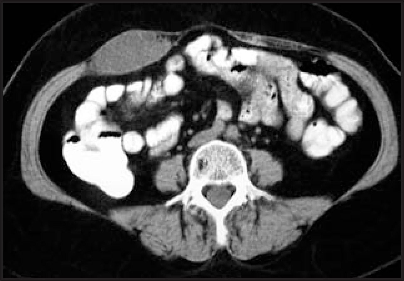
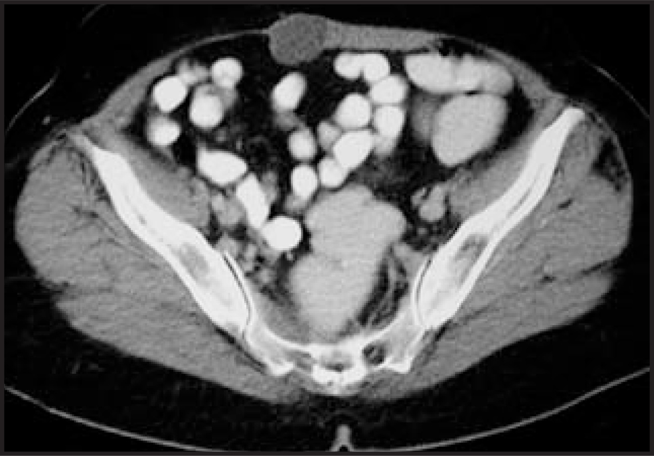
Abdominal CT revealed well-defined multiloculated cystic lesions within the right rectus muscle. The lesions measured 6 × 4 cm (Figure 1) and 3 × 3 cm (Figure 2). The rest of the imaging exam ination was unremarkable.
SURGICAL AND PATHOLOGIC FINDINGS
The patient underwent surgery during which the abdominal wall was partially removed and reconstructed using mesh. The histologic diagnosis was adenocarcinoma. The patient’s medical history, physical examination, imaging studies and pathologic evaluations did not indicate theprimary site of the disease.
DIAGNOSIS
Metastatic adenocarcinoma of unknown primary site
CASE FOLLOW-UP
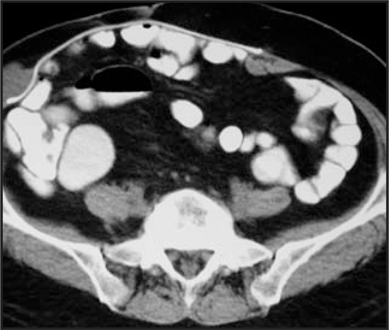
The patient received adjuvant chemo therapy. Twelve months later, the patient presented with similar clinical findings. At this time, CTshowed a 3 × 3-cm recurrent cystic mass on the mesh (Figure 3). The patient underwent repeat abdominal wall resection. A year later, thepatient’s condition worsened, and CT revealed metastases to the peritoneum, liver, subcutaneous abdominal wall, and inguinal lymph node.The patient died 32 months after diagnosis.
DISCUSSION
Primary malignant tumors of the ab dominal wall are extremely rare. The most frequent tumors include lymphoma, melanoma, andmalignant fibrous histiocytoma in adults, and sarcoma in children.1Metastatic lesions involving the abdominal wall can be frequentlyencountered in clinical practice. Metastases can develop in this region in a hematogenous or lymphatic dissemination.
In female patients with metastatic tumor, the most common primary site is the ovary, followed by the colon and breast. In male patients,the most common primary tumor is malignant melanoma.2In most metastases to the anterior abdominal wall, the patient’s primary tumor isalready known and/or metastases to the anterior abdominal wall arise together with other metastases.3To our knowledge, there are few reported cases of cancer of unknown primary site that were found to have metastasized to the cutaneous-subcutaneous anterior abdominal wall andno cases presenting as an isolated intramuscular abdominal wall mass. An interesting characteristic of this case is that the lesions wererestricted to the abdominal wall for a long period and metastasis (such as to the liver, peritoneum, and lymph nodes) emerged months afterthe anterior abdominal wall metastasis.
CONCLUSION
The occurrence of abdominal wall metastases from an unknown primary adenocarcinoma is very rare. But, as shown in this case, it shouldbe considered in the differential diagnosis of abdominal wall masses.
References
- Meuwly J, Gudinchet F. Sonography of the thoracic and abdominal walls. J Clin Ultrasound. 2004;32:500-510.
- David O, Kluskens L, Reddy V, Gattuso P. Malignant cutaneous and subcutaneous abdominal wall lesions: A fine-needle aspiration study. Diagn Cytopathol. 1998;19:267-269.
- Srinivasan R, Ray R, Nijhawan R. Metastatic cutaneous and subcutaneous deposits from internal carcinoma. An analysis of cases diagnosed by fine needle aspiration. Acta Cytol. 1993;37:894-898.
Citation
. Metastatic adenocarcinoma of unknown primary site. Applied Radiology. 2008;37(6):40A-40B. doi:10.37549/AR1624.