MR evaluation of acute abdominal pain in pregnant patients
Applied Radiology — Vol. 36 , Issue 9 , pp. 20 -25
DOI: 10.37549/AR1550
Published: September 1, 2007
Categories
The evaluation of abdominal pain in the pregnant patient can be a difficult task that is magnified by related sociologic issues and complicated by the anatomic and physiologic adaptations of the pregnant state. Evaluation is also limited by the need to avoid ionizing radiation. Pathologies directly related to pregnancy are generally evaluated by ultrasound, which usually provides for accurate assessment of the uterus, fetus, ovaries, kidneys, and gallbladder.1,2 However, ultrasound provides limited and inconsistent imaging of the bowel, pancreas, and other deep structures and may be rendered ineffective by overlying bowel gas or anatomic alteration or variation. In addition, fetal exposure to ionizing radiation limits theuse of computed tomography (CT) during pregnancy.
Serious fetal risk has not been shown to occur with radiation doses below 10 rad,3 which is less than any single commonly ordered diagnostic study. However, the International Commission on Radiological Protection has recommended that one should question whether a diagnosis can be obtained without using ionizing radiation if the expected fetal dose is high.4 The Safety Committee of the Society for Magnetic Resonance Imaging has issued a report stating that “MR may be used in pregnant women if other nonionizing forms of diagnostic imaging are inadequate or if the examination provides important information that would otherwise require exposure to ionizing radiation.”5 While fetal harm has not been reported as a result of magnetic resonance imaging (MRI), the effects of fetal exposure have not yet been fully determined, which suggests cautious use, especially during the first trimester. Particular care should be taken with the use of intravenous contrast, as gadolinium-based agents cross the placenta. Although adequate controlled studies of gadolinium use during pregnancy have not been performed, animal studies have shown an increase in skeletal malformations.6 It is suggested that these contrast agents be used only in the second and third trimester and when potential benefit outweighs the risk.
At our institution, images are obtained with a Signa EXCITE LX 1.5T system (GE Healthcare, Milwaukee, WI) using a phased-array surface coil when possible. Toward the end of pregnancy, a body coil may be required. Informed consent is obtained from each pregnant patient prior to imaging. The backbone of our protocol is T2-weighted (T2W) imaging in 3 orthogonal planes with single-shot fast spin-echo (SSFSE) or half-Fourier acquisition single-shot spin-echo (HASTE) imaging (effective repetition time [TR] = infinite; echo time [TE] = 80 msec; slice thickness = 6 mm). Typically, a 35-cm field of view is used, with a 160–192 × 256 matrix. A fat-saturated T2W sequence (SSFSE/ HASTE or fast spin-echo [FSE]) improves detection of inflammation and characterization of pelvic masses. An axial T1-weighted (T1W) sequence is also included, which may be either a breath-hold spoiled gradient-echo or a respiratory-gated fast spin-echo (FSE) sequence; TR = 700 msec; TE = minimum; number of excitation (NEX) = 3; echo-train length (ETL) = 2; matrix = 256 × 192, slice thickness = 6 mm with 1 mm spacing). The FSE sequence provides better resolution than gradient echo and may be useful for detecting small structures, such as a normal appendix. After review of the noncontrast images, the radiologist may request contrast-enhanced imaging in selected situations. If required, gadolinium may be administered at a dose of 0.1 mmol/kg, and 2-dimensional or 3-dimensional gradient-refocused echo (GRE) T1W images may subsequently be acquired.
The evaluation of abdominal pain in the pregnant patient is complicated by the body’s adaptation to the pregnant state. Physiologic changes include mild leukocytosis, “physiologic” anemia, mildly elevated alkaline phosphatase, dilatation of the renal pelvis and ureter, and a relative hypercoagulable state. In addition, the gravid uterus compresses and displaces abdominal contents, and abdominal wall laxity during late pregnancy may diminish signs of peritonitis.7,8 Diagnosis may also be confounded by other conditions common in pregnancy, such as round ligament pain, constipation, and Braxton Hicks contractions. This article presents a brief summary of the use of MRI in the evaluation of abdominal pain in the pregnant patient.
Appendicitis
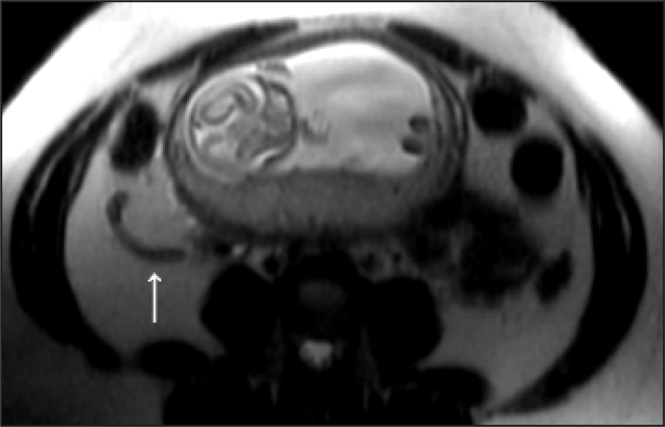
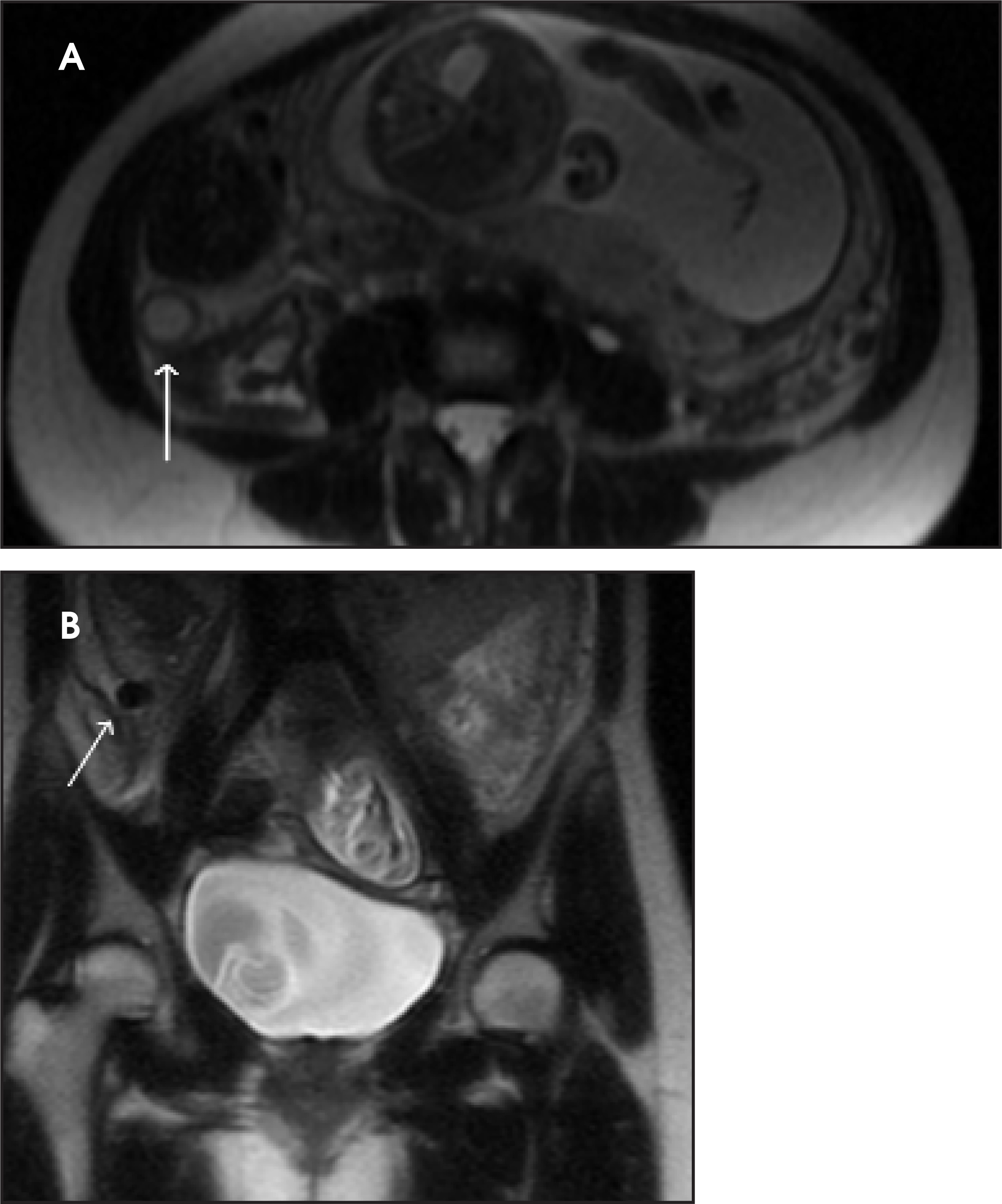
Appendicitis is the most common cause of acute abdomen in the pregnant patient, affecting in 1 in 1500 pregnancies. Ruptured appendicitis is associated with a 30% chance of fetal loss, versus a 3% to 5% risk without rupture.9 Moreover, >40% of patients who undergo appendectomy during the second or third trimesters have a normal appendix, and up to 83% of all pregnant patients who have an appendectomy experience preterm contractions10 (Figure 1). Diagnosis is confounded by “normal” causes of abdominopelvic pain during pregnancy, physiologic leukocytosis, and the fact that the gravid uterus may cause the appendix to shift superiorly,11 which distorts the classic clinical presentation. Studies have proven the ability of MRI to diagnose appendicitis and differentiate other causes of acute abdominal pain.12-15 In a series of 50 patients, the overall sensitivity, specificity, and accuracy of MRI for the diagnosis of acute appendicitis in the pregnant patient were 100%, 93.6%, and 94%, respectively15 (Figure 2). The majority of these examinations were obtained without intravenous contrast.
Small bowel obstruction
Small bowel obstruction is the second most common nonobstetric surgical indication in pregnancy, complicating approximately 1 in 3000 gestations.9,16 Adhesions are the cause in 60% to 70% of cases,9 with volvulus being the second most common etiology at 25%.17 When surgery is required, fetal and maternal mortality rates are high, approaching 25% and 6%, respectively.18 The high mortality may be partly explained by delays and errors that result from the physical and diagnostic difficulties created by the pregnant state. MRI provides an effective alternative to CT, yielding multiplanar imaging of the entire bowel without the comparatively high radiation dose. Single-shot fast spin-echo sequences are particularly effective for bowel imaging,19 minimizing artifacts caused by magnetic susceptibility and peristaltic motion.
Cholecystitis/cholelithiasis
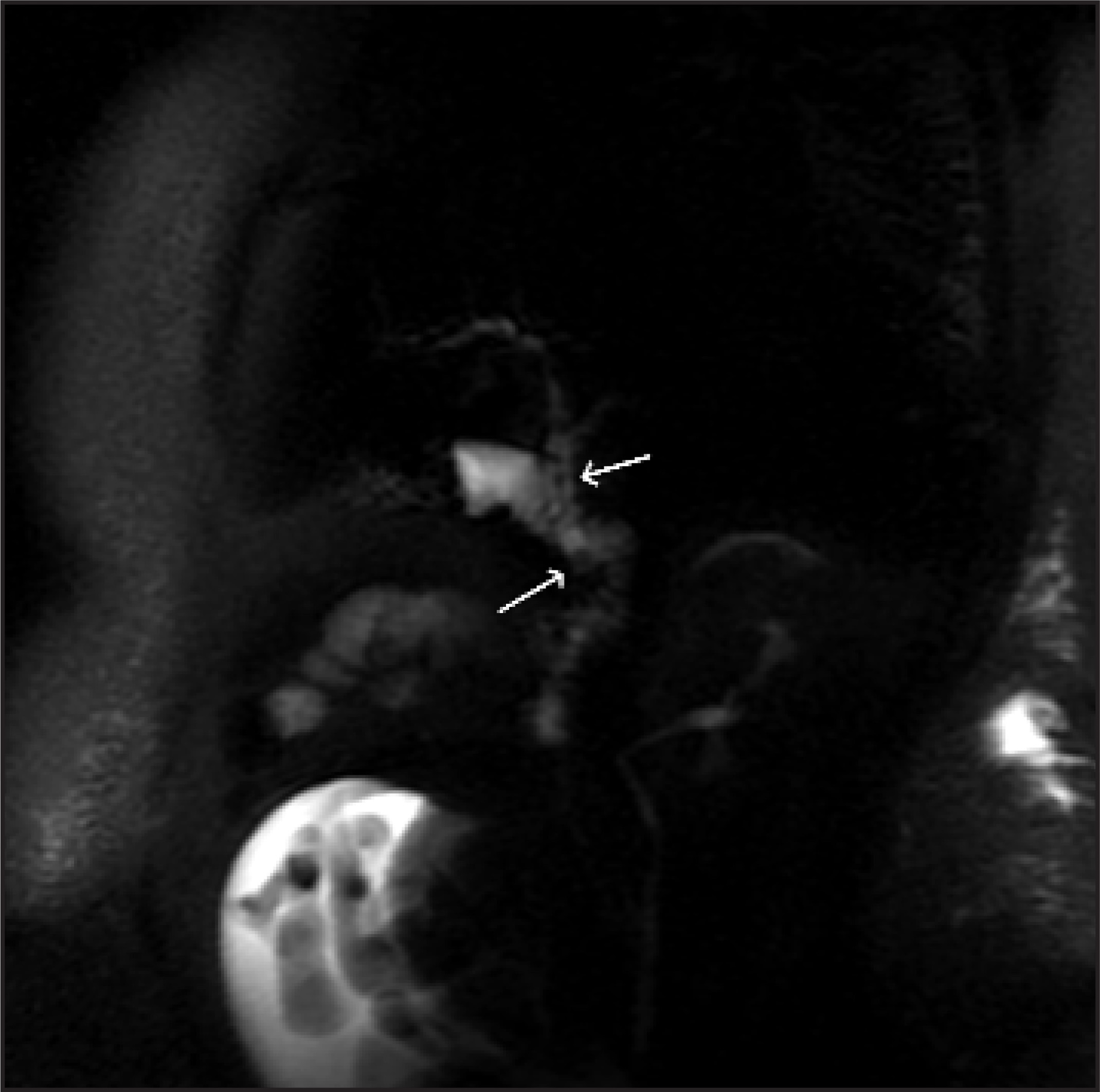
The third most common nonobstetric surgical emergency in pregnancy is cholecystitis, occurring in 1 to 8 in 10,000 pregnancies.16 The lithogenicity of bile is increased during pregnancy; in 1 large Chilean study, 12% of pregnant women had cholelithiasis versus 1.3% of nonpregnant women.20 While pregnancy has not been shown to result in an increase in cholecystitis or symptomatic cholelithiasis, cholecystectomy is associated with miscarriage during the first trimester and premature labor during the third trimester.20-22 Although signs and symptoms are not significantly altered by pregnancy,21,22 the diagnosis may again be complicated by physiologic changes related to the state. Ultrasound is obviously the initial study of choice and is generally accurate. However, if overlying soft tissues or bowel gas limits evaluation, then MRI may provide further information. MRI is of particular use if choledocholithiasis is suspected, as this modality most effectively reveals findings of biliary obstruction (Figure 3).
Urolithiasis
Renal stones cause the largest number of nonobstetric hospitalizations during pregnancy,16 with most patients presenting late in gestation. Diagnosis may be complicated by “normal” dilatation of the renal collecting system, which results from ureteral compression by the enlarged uterus and a progesterone-induced decrease in smooth muscle tone within the urinary tract. “Physiologic” hydronephrosis is usually asymptomatic but may occasionally result in abdominal pain. While ultrasound effectively reveals hydronephrosis, it inadequately evaluates the lower urinary tract and ureteral stones may go undetected. MR urography is an alternative diagnostic tool. Spencer et al23 described physiologic hydronephrosis as compression of the middle third of the ureter with distal collapse but without an intrinsic ureteral filling defect. The presence of perirenal fluid, ureteral dilatation, and an associated ureteral filling defect have been reported as MRI signs of pathological hydronephrosis, most likely secondary to a stone.23,24 In these cases, the possibility of clot, an air bubble (resulting from instrumentation), and flow artifacts must be excluded.
Pyelonephritis
Acute pyelonephritis occurs in 1% to 2% of pregnancies. It is generally related to asymptomatic bacteriuria or a history of recurrent lower urinary tract infections.25 The clinical presentation is similar to the general population, but it is associated with preterm labor. Diagnosis is based on urinalysis, and possible complications are generally shown by ultrasound. However, if a diagnostic dilemma occurs, MRI may provide additional evaluation, particularly with regard to the development of intra- or perirenal abscesses, although the diagnosis of abscess remains limited without the administration of intravenous contrast.13
Acute pancreatitis
While pregnancy does not cause or significantly alter the clinical presentation of acute pancreatitis, it occurs in approximately 1 of 3300 gestations, most commonly during the third trimester and postpartum period.26 Ultrasound accurately portrays cholelithiasis and intrahepatic biliary dilatation but provides limited and inconsistent evaluation of the common bile duct and pancreas as well as of potential complications such as pseudocyst. MRI can effectively assess all of these. Fat-suppressed T1W gradient-echo sequences best evaluate the pancreas, particularly if pre- and postgadolinium imaging is obtained. T2-weighted SSFSE imaging accurately shows the biliary and pancreatic ducts, peripancreatic inflammation, pseudocysts, and other fluid collections without the administration of intravenous contrast.27
Inflammatory bowel disease
Inflammatory bowel disease and its complications may be assessed by MRI, avoiding the comparatively large radiation doses that result from the usual barium studies and CT. Some reports have suggested that as many as one third of patients with inflammatory bowel disease will relapse during pregnancy; this high percentage may partly be related to a discontinuation of medications.28,29 Maccioni et al30 have shown a correlation between disease activity and the intensity of T2 signal within bowel wall. In addition to identifying bowel wall inflammation, MRI can effectively show other complications, such as obstruction, abscess, and possibly fistula formation.31 Single-shot fast spin-echo sequences image the bowel ideally, limiting deleterious artifacts. T2-weighted/T1W imaging with steady state acquisition (fast imaging employing steady state acquistion [FIESTA] or fast imaging with steady-state precession [True FISP]) also provides useful information.
Deep venous thrombosis
The risk of deep venous thrombosis is increased by the hypercoagulable state of pregnancy.32 Although ultrasound effectively assesses for deep venous thrombosis in the lower extremities, evaluation of the pelvic veins is limited by overlying soft tissue, bowel gas, and the gravid uterus. The lack of normal flow voids on noncontrast MRI can suggest a deep venous thrombosis, and time-of-flight MR venography can confirm its presence.33
Pelvic pathology
Most pelvic abnormalities (including leiomyomas, ectopic pregnancy, adnexal torsion, hemorrhagic cysts, and masses) are initially evaluated by ultrasound, but MRI may be useful when atypical presentation or ultrasound limitation is encountered.34 Uterine leiomyomas are seen in approximately 2% of pregnant women and 1 in 10 develops related complications during pregnancy,35 such as pain resulting from degeneration, rapid growth, or torsion. The major complication is hemorrhagic infarction that is caused by venous obstruction at the lesion’s periphery, which generally presents as second- or third-trimester pain, low-grade fever, and occasional bleeding.35 On MRI, degenerating leiomyomas usually have increased T1 and T2 signal, which may be diffuse or peripheral.6,36,37 A peripheral rim of high T1/low T2 signal may correspond to obstructed veins.37 If contrast is administered, the entire lesion fails to enhance.
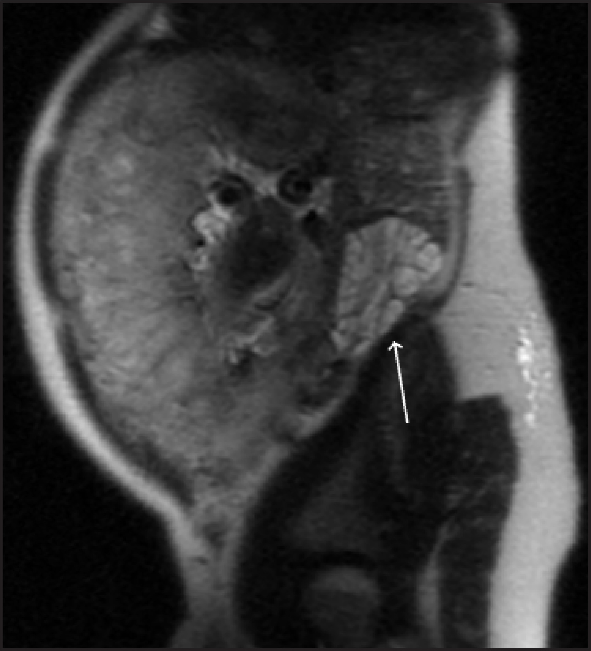
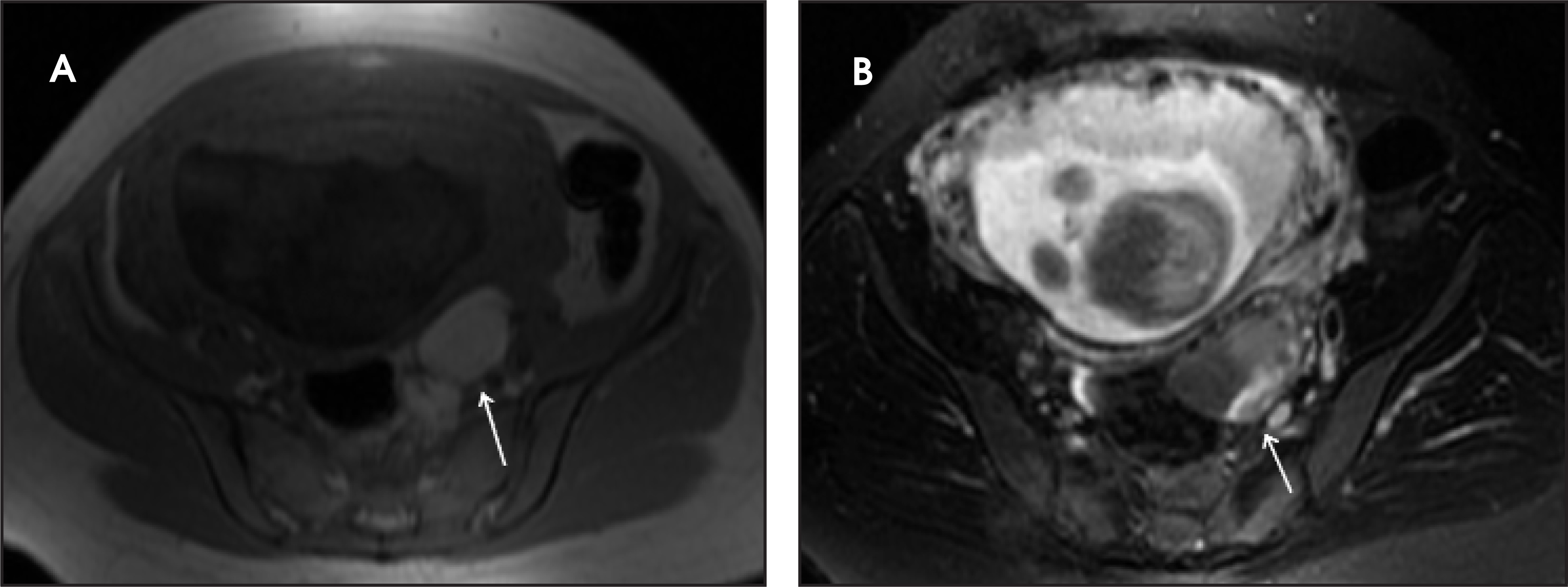
MRI may also be of use in unusual or complicated forms of ectopic pregnancy (abdominal, interstitial, myometrial, or cervical pregnancy), potentially revealing an associated gestational sac, mass, hematoma, or hemoperitoneum.38 Furthermore, Kataoka et al39 have reported that MRI allowed the early diagnosis of ectopic pregnancy, helping to decide the utility of early conservative therapy with methotrexate. In addition, adnexal torsion is one of the few causes of acute abdomen that is more common during pregnancy, usually occurring between the 6th and 14th weeks.9,40 It accounts for 3% of gynecological emergencies, but 20% to 25% of cases occur in pregnant women.41 When ultrasound fails, as may occur when limited by the gravid uterus, MRI offers an alternative diagnostic venue, potentially showing an enlarged ovary, thick and edematous pedicle, signal compatible with hemorrhage, and smooth wall thickening around a cystic ovarian mass40,42 (Figure 4). Finally, MRI provides a means of differentiating hemorrhagic cysts (which have been referred to as “the great imitator”) from a list of differential diagnoses that includes ectopic pregnancy, adnexal torsion, neoplasm, and pelvic inflammatory disease (Figure 5). Such cysts may result from rupture of the corpus luteum, and MRI provides sensitive evaluation for blood within the cysts or the peritoneal cavity, allowing differentiation from solid masses.43
Conclusion
Diagnosis of abdominal pain in the pregnant patient is fraught with multiple pitfalls, as discussed above, including a radiation-induced constraint of most imaging modalities. Ultrasound remains the initial study of choice, but its limitations necessitate consideration of alternative technology. While the evidential role of MRI utilization during pregnancy is currently being investigated, fetal harm has not been shown, and MRI shows clear potential to advance patient care.
References
- Lim H, Bae S, Seo G. Diagnosis of acute appendicitis in pregnant women: Value of sonography. AJR Am J Roentgenol. 1992;159:539-542.
- Barloon T, Brown B, Abu-Yousef M. Sonography of acute appendicitis in pregnancy. Abdom Imaging. 1995;20:149-151.
- Swartz H, Reichling B. Hazards of radiation exposure for pregnant women. JAMA. 1978;239:1907-1908.
- Pregnancy and medical radiation. Publication 84. Annals ICRP. 2000;30(1):1-43.
- Shellock F, Kanal E. Policies, guidelines, and recommendations for MR imaging safety and patient management. SMRI Safety Committee. J Magn Reson Imaging. 1991;1:97-101.
- Brown M, Birchard K, Semelka R. Magnetic resonance evaluation of pregnant patients with acute abdominal pain. Semin Ultrasound CT MR. 2005;26:206-211.
- DeVore G. Acute abdominal pain in the pregnant patient due to pancreatitis, acute appendicitis, cholecystitis, or peptic ulcer disease. Clin Perinatol. 1980;7:349-369.
- Tracey M, Fletcher H. Appendicitis in pregnancy. Am Surg. 2000;66:555-559.
- Sharp H. The acute abdomen during pregnancy. Clin Obstet Gynecol. 2002;45:405-413.
- Firstenberg M, Malangoni M. Gastrointestinal surgery during pregnancy. Gastroenterol Clin North Am. 1998;27:73-88.
- Baer J, Reis R, Arens R. Appendicitis in pregnancy with changes in position and axis of normal appendix in pregnancy. JAMA. 1932;98:1359.
- Cobben L, Groot I, Haans L. MRI for clinically suspected appendicitis during pregnancy. AJR Am J Roentgenol. 2004;183:671-675.
- Birchard K, Brown M, Hyslop W. MRI of acute abdominal and pelvic pain in pregnant patients. AJR Am J Roentgenol. 2005;184:452-458.
- Oto A, Ernst R, Shah R. Right-lower-quadrant pain and suspected appendicitis in pregnant women: Evaluation with MR imaging—Initial experience. Radiology. 2005;234:445-451.
- Pedrosa I, Levine D, Eyvazzadeh A. MR imaging evaluation of acute appendicitis in pregnancy. Radiology. 2006;238:891-899.
- Cappell M, Friedel D. Abdominal pain during pregnancy. Gastroenterol Clin North Am. 2003;32:1-58.
- Wanetick L, Roschen F, Dunn J. Volvulus of the small bowel complicating pregnancy. J Reprod Med. 1975;14:82-83.
- Sharp H. Gastrointestinal surgical conditions during pregnancy. Clin Obstet Gynecol. 1994;37:306-315.
- Regan F, Beall D, Bohlman M. Fast MR imaging and the detection of small-bowel obstruction. AJR Am J Roentgenol. 1998;170:1465-1469.
- Valdivieso V, Covarrubias C, Siegel F, Cruz F. Pregnancy and cholelithiasis: Pathogenesis and natural course of gallstones diagnosed in early puerperium. Hepatology. 1993;17:1-4.
- Davis A, Katz V, Cox R. Gallbladder disease in pregnancy. J Reprod Med. 1995;40:759-762.
- El-Shawarby S, Hendeson A, Mossa M. Ovarian cysts during pregnancy: Dilemmas in diagnosis and management. J Obstet Gynaecol. 2005;25:669-775.
- Spencer J, Chahal R, Kelly A. Evaluation of painful hydronephrosis in pregnancy: Magnetic resonance urographic patterns in physiological dilatation versus calculous obstruction. J Urol. 2004;171:256-260.
- Roy C, Saussine C, LeBras Y. Assessment of painful ureterohydronephrosis during pregnancy by MR urography. Eur Radiol. 1996;6:334-338.
- Millar L, Cox S. Urinary tract infections complicating pregnancy. Infect Dis Clin North Am. 1997;11(1):13-26.
- Ramin K, Ramin S, Richey S, Cunningham F. Acute pancreatitis in pregnancy. Am J Obstet Gynecol. 1995;173:187-191.
- Miller F, Keppke A, Dalal K. MRI of pancreatitis and its complications: Part 1, acute pancreatitis. AJR Am J Roentgenol. 2004;183:1637-1644.
- Hanan I. Inflammatory bowel disease in the pregnant woman. Compr Ther. 1993;19:91-95.
- Ferrero S, Ragni N. Inflammatory bowel disease: Management issues during pregnancy. Arch Gynecol Obstet. 2004;270:79-85.
- Maccioni F, Viscido A, Broglia L. Evaluation of Crohn disease activity with magnetic resonance imaging. Abdom Imaging. 2000;25:219-228.
- Horsthuis K, Lavini Mphil C, Stoker J. MRI in Crohn’s disease. J Magn Reson Imaging. 2005;22:1-12.
- Stone K. Acute abdominal emergencies associated with pregnancy. Clin Obstet Gynecol. 2002;45:553-561.
- Spritzer C, Evans A, Kay H. Magnetic resonance imaging of deep venous thrombosis in pregnant women with lower extremity edema. Obstet Gynecol. 1995;85:603-607.
- Weinreb J, Brown C, Lowe T. Pelvic masses in pregnant patients: MR and US imaging. Radiology. 1986;159:717-724.
- Katz V, Dotters D, Droegemeuller W. Complications of uterine leiomyomas in pregnancy. Obstet Gynecol. 1989;73:593-596.
- Ueda H, Togashi K, Konishi I. Unusual appearances of uterine leiomyomas: MR imaging findings and their histopathologic backgrounds. RadioGraphics. 1999;19(suppl 1):s131-s145.
- Kawakami S, Togashi K, Konishi I. Red degeneration of uterine leiomyoma: MR appearance. J Comput Assist Tomogr. 1994;18:925-928.
- Nagayama M, Watanabe Y, Okumura A. Fast MR imaging in obstetrics. RadioGraphics. 2002;22:563-580.
- Kataoka M, Togashi K, Kobayashi H. Evaluation of ectopic pregnancy by magnetic resonance imaging. Hum Reprod. 1999;14:2644-2650.
- Rha S, Byun J, Jung S. CT and MR imaging features of adnexal torsion. RadioGraphics. 2002;22:283-294.
- McGowan L. Surgical diseases of the ovary in pregnancy. Clin Obstet Gynecol. 1983;26:843-852.
- Born C, Wirth S, Stäbler A, Reiser M. Diagnosis of adnexal torsion in the third trimester of pregnancy: A case report. Abdom Imaging. 2004;29:123-127.
- Imaoka I, Wada A, Kaji Y. Developing an MR imaging strategy for diagnosis of ovarian masses. RadioGraphics. 2006;26:1431-1448.
Citation
. MR evaluation of acute abdominal pain in pregnant patients. Applied Radiology. 2007;36(9):20-25. doi:10.37549/AR1550.