MR imaging in the evaluation of benign pathologies of urinary bladder
Applied Radiology — Vol. 33 , Issue 8 , pp. 26 -33
DOI: 10.37549/AR1270
Published: August 1, 2004
Categories
Pathology of the urinary bladder spans congenital abnormalities, inflammatory and infectious diseases, diverticulosis, lithiasis, tumorlike conditions, and malignancies.1 Traditionally, imaging of the urinary bladder has been performed with cystoscopy, antegrade or retrograde cystography, ultrasonography, and computed tomography (CT). The role of magnetic resonance (MR) imaging in the assessment of bladder pathology is evolving. As the most common disease of the urinary bladder that requires further imaging is bladder cancer,2 the most frequent indications for MR imaging of the bladder in clinical practice are staging of a known bladder tumor and follow-up of a treated cancer patient. However, the use of MR imaging has been shown to be effective in the assessment of benign congenital bladder anomalies, such as bladder exstrophy, or acquired adult bladder pathology, such as cystocele associated with pelvic organ prolapse. The excellent soft-tissue contrast, multiplanar imaging capability, noninvasiveness, and lack of ionizing radiation make MR imaging an attractive alternative modality in the evaluation of bladder pathology in the pediatric population and in women with pelvic floor defects.
This article reviews the applications of MR imaging in the assessment of benign urinary bladder pathology. The authors describe the imaging techniques used for bladder evaluation and imaging findings for selected benign bladder pathologies.
Imaging techniques
The phased-array pelvic coil is a preferred receiver coil for high-resolution imaging of the bladder. The typical urinary bladder protocol for 1.5T MR scanner includes T1-weighted spin-echo images (repetition time [TR]/echo time [TE]: 400 to 550/minimum) obtained in the axial plane and T2-weighted fast spin-echo (TR/TE: 4000 to 5500/80 to 120) images also obtained in the axial plane. Dynamic postcontrast imaging is performed with fast multiplanar spoiled gradient-recalled echo (FMPSPGR) images with fat suppression (TR/TE: 180 to 300/1.7 to 4.2) obtained in the axial plane before, and at 20 seconds (arterial phase) and 70 to 115 seconds (venous phase) after gadopentate dimeglumine injection (0.1 mmol/kg). Sagittal or coronal T2-weighted images may be obtained, if the anteroposterior or inferosuperior extent of disease needs to be evaluated. Typical parameters for optimal bladder imaging are: 20- to 30-cm field-of-view (FOV), 6-mm slice thickness, and 2-mm intersection gap. Ideally, the urinary bladder should be moderately distended during imaging.
The protocol for imaging patients with bladder exstrophy includes T1-weighted and T2-weighted sequences obtained in 3 planes: 1) axial T1weighted images (TR/TE 600 to 650/9, slice/space 4 to 6 mm/0.5 to 1 mm, FOV 16 to 18 cm), axial T2-weighted images (TR/TE 3200 to 3500/99, slice/space 4 to 6 mm/0.5 to 1 mm, FOV 16 to 18); 2) sagittal T1-weighted images (TR/TE 600 to 650/9, slice/space 5 mm/0 mm, FOV 16 to 18 cm), sagittal T2-weighted images (TR/TE 3200 to 3500/99, slice/space 5 mm/0 mm, FOV 16 to 18 cm); and 3) coronal T1-weighted images (TR/TE 600 to 650/9, slice/space 5 mm/0 mm, FOV 16 to 18 cm), and coronal T2-weighted images (TR/TE 3200 to 3500/99, slice/space 5 mm/0 mm, FOV 16 to 18 cm). The imaging matrix is 512 × 512. The extremity or cardiac coil may be used as receiver coils, as they allow for high-resolution imaging with adequate spatial coverage. Infant patients are sedated for the procedure.
For the dynamic bladder imaging at rest and during valsalva/strain in patients with pelvic organ prolapse, fast imaging techniques are used, such as a single-shot fast spin echo (SSFSE). Typical image acquisition parameters include: TR: infinite, TE: 60 msec, slice thickness: 5 mm, and slice gap: 1 mm. Images are obtained during rest and straining in axial, sagittal, and coronal planes. No contrast administration is needed. The high-signal urine in the bladder on SSFSE images and high signal of the pelvic fat allow for detailed examination of the bladder during motion between rest and strain. Patients usually receive endovaginal and endorectal gel for simultaneous evaluation of other pelvic floor com-partments.3
Congenital abnormalities and acquired benign conditions of the urinary bladder
The two most common congenital abnormalities of the urinary bladder are the urachus abnormalities and exstrophy. The urachus is an extraperitoneal structure located in the space of Retzius (which is bounded by the transversalis fascia ventrally and the parietal peritoneum dorsally), between the dome of the bladder and umbilicus. It retracts from the bladder at birth, but its lumen may persist within the bladder wall and remains continuous with the bladder cavity. Congenital abnormalities that involve the urachus include: patent urachus (50%), urachal sinus (15%), vesicourachal diverticulum (5%), and urachal cyst (30%). A patent urachus is a fistula between the bladder and the umbilicus, through which the urine may pass into the umbilicus (50%). A urachal sinus is patent only from the side of urachus toward the umbilicus and has a blind ending. A vesicourachal diverticulum is a communication only between the bladder dome and the urachus. A urachal cyst in the anterior abdominal wall does not communicate with either the bladder or the umbilicus, and can be complicated by abscess or granulomatous omphalitis. The acquired conditions complicating the urachal anomalies include infection, which is the most common complication, as well as benign and malignant neoplasms.4-9 The urachal remnant anomalies are usually radiologically evaluated with ultrasound (US) or CT (Figure 1), with fistulography or cystography performed to illustrate contiguity of the fistulous tract with the urachus. There is a limited role for MR imaging in the evaluation of benign urachus disease.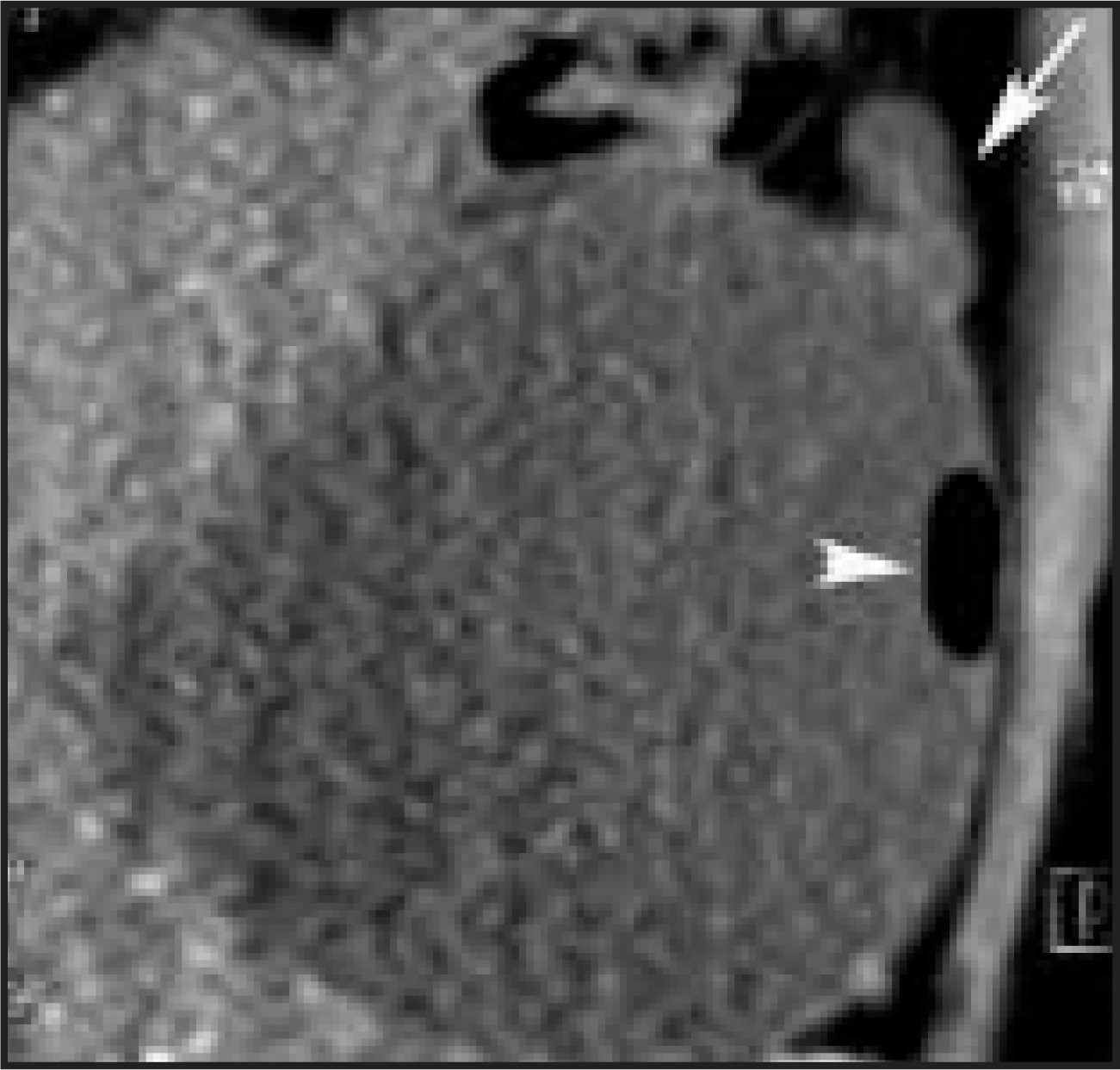
Bladder exstrophy is a complex defect involving skin, abdominal wall musculature, bone, bladder, and external genitalia. It is characterized by the absence of anterior vesical and lower abdominal wall, with the eversion of the posterior bladder wall (Figure 2). These changes may be complete or partial and are often associated with other anomalies of the urogenital tract. The exstrophy-epispadias complex comprises a spectrum of congenital abnormalities that includes classic bladder exstrophy, epispadias, cloacal exstrophy, and several variants. The embryologic defect is due to lack of separation of the primitive cloaca into the urogenital sinus and hindgut during the first trimester when the maturation of the anterior abdominal wall also occurs. In classic bladder exstrophy, the lower urinary tract, genitalia, and musculoskeletal system are affected. In cloacal exstrophy, a much more severe abnormality, there is significant involvement of the gastrointestinal tract and central nervous system. In cloacal exstrophy, up to 95% of patients have myelodysplasia, which may include myelomeningocele, lipomeningocele, and meningocele. In classic exstrophy, the bladder is open on the lower abdomen, with mucosa fully exposed through a fascial defect. In the classic bladder exstrophy male patient, the phallus is short and broad with upward angulation (dorsal chordee). The glans lies open, and the dorsal component of the foreskin is absent. The anus is anteriorly displaced. In the classic bladder exstrophy female patient, the clitoris is bifid with divergent labia, and the vagina and the anus are anteriorly displaced. In both males and females, the pubic symphysis is widened and the rectus muscles are divergent distally. In cloacal exstrophy, nearly all patients have an associated omphalocele and the bladder is open and separated into 2 halves. In the male patient, the penis is generally quite small and bifid, with a hemiglans located just caudal to each hemibladder. Infrequently, the phallus may be intact in the midline. In females, the clitoris is bifid and two vaginas are present. The anus is absent.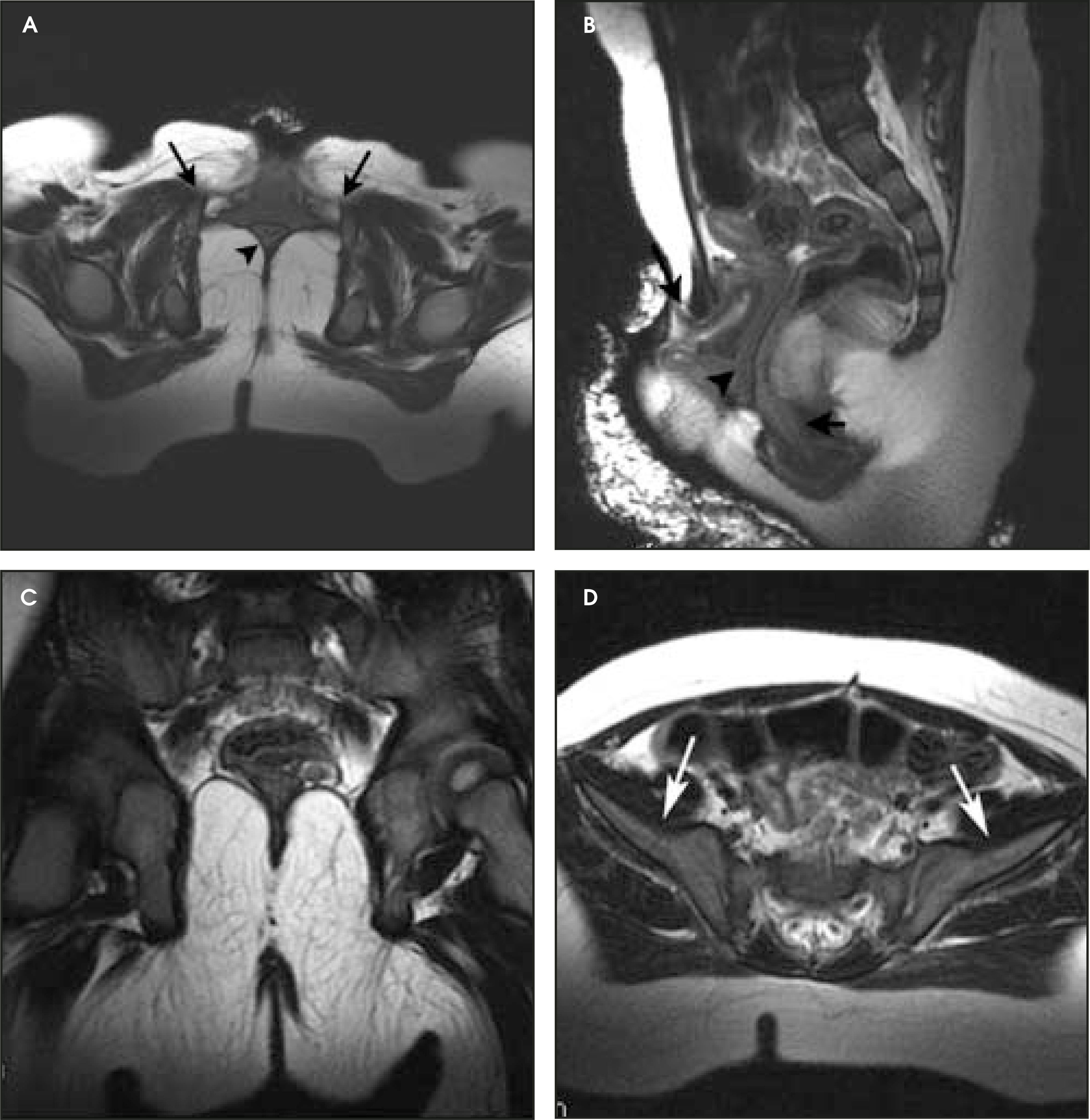
The role of MR imaging in the evaluation of patients with bladder exstrophy includes presurgical assessment (Figure 2) of the pelvic organs and evaluation for possible associated genitourinary, gastrointestinal, and spinal anomalies, as well as postoperative follow-up (Figure 3). The common findings in the simple bladder exstrophy include: lack of the fluid-filled urinary bladder in the pelvis with eversion of the bladder wall through the anterior low abdominal wall defect (Figure 2A); widening of the distance between the pubic symphysis; widening of the angle of divergence of the levator ani from midline; anterior displacement of the anus (Figure 2B); flattening of the pelvic diaphragm on the sagittal and coronal views (Figure 2C); and widening of the iliac wing angle due to external rotation of the iliac bones (Figure 2D). Malignant complications of bladder exstrophy may occur; adenocarcinoma is the most common type,1 followed by a squamous-type carcinoma.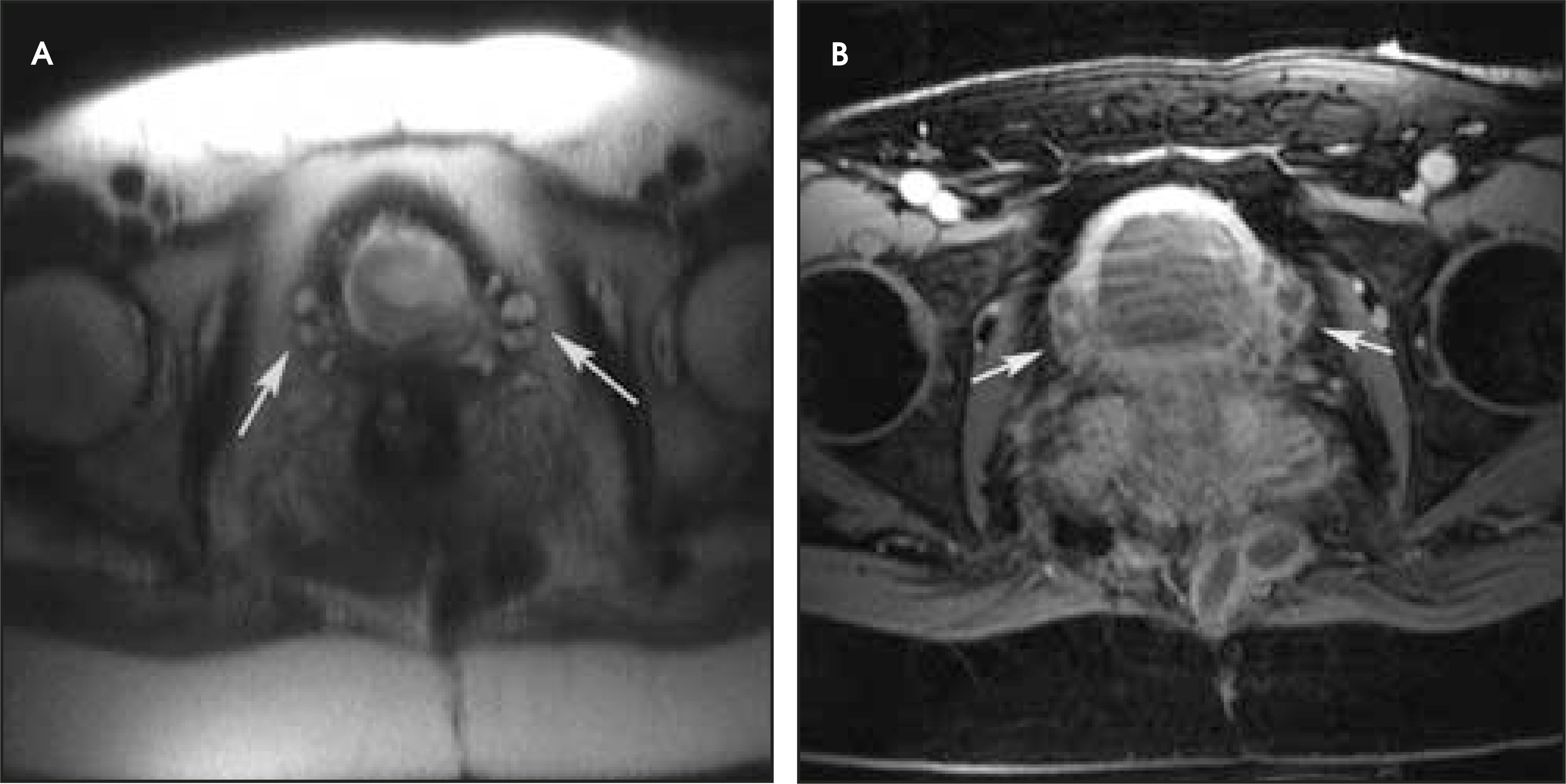
Bladder diverticuli may be congenital or acquired. Congenital diverticuli are uncommon and occur in boys. Congenital diverticuli arise as a result of herniation of the bladder mucosa through the detrusor muscle near the ureteral orifice, the so-called Hutch diverticulum. These diverticuli most often result in vesicoureteric reflux, but may also produce ureteric obstruction. Most bladder diverticuli are acquired in association with longstanding bladder outlet obstruction. They are most commonly seen in older men with benign prostatic hypertrophy, prostatitis, or carcinoma of the prostate, and in women with urethral carcinoma or urinary dysfunction. These diverticuli may present as focal wide-mouth herniation or multiple small bladder wall outpouchings (Figure 4). Bladder diverticuli are well-demonstrated by intravenous urography or cystography, but may also be seen on US, CT, or MR imaging. They are usually an incidental finding on MR imaging and rarely are the primary indication for MR, unless the cause of the bladder outlet obstruction is sought.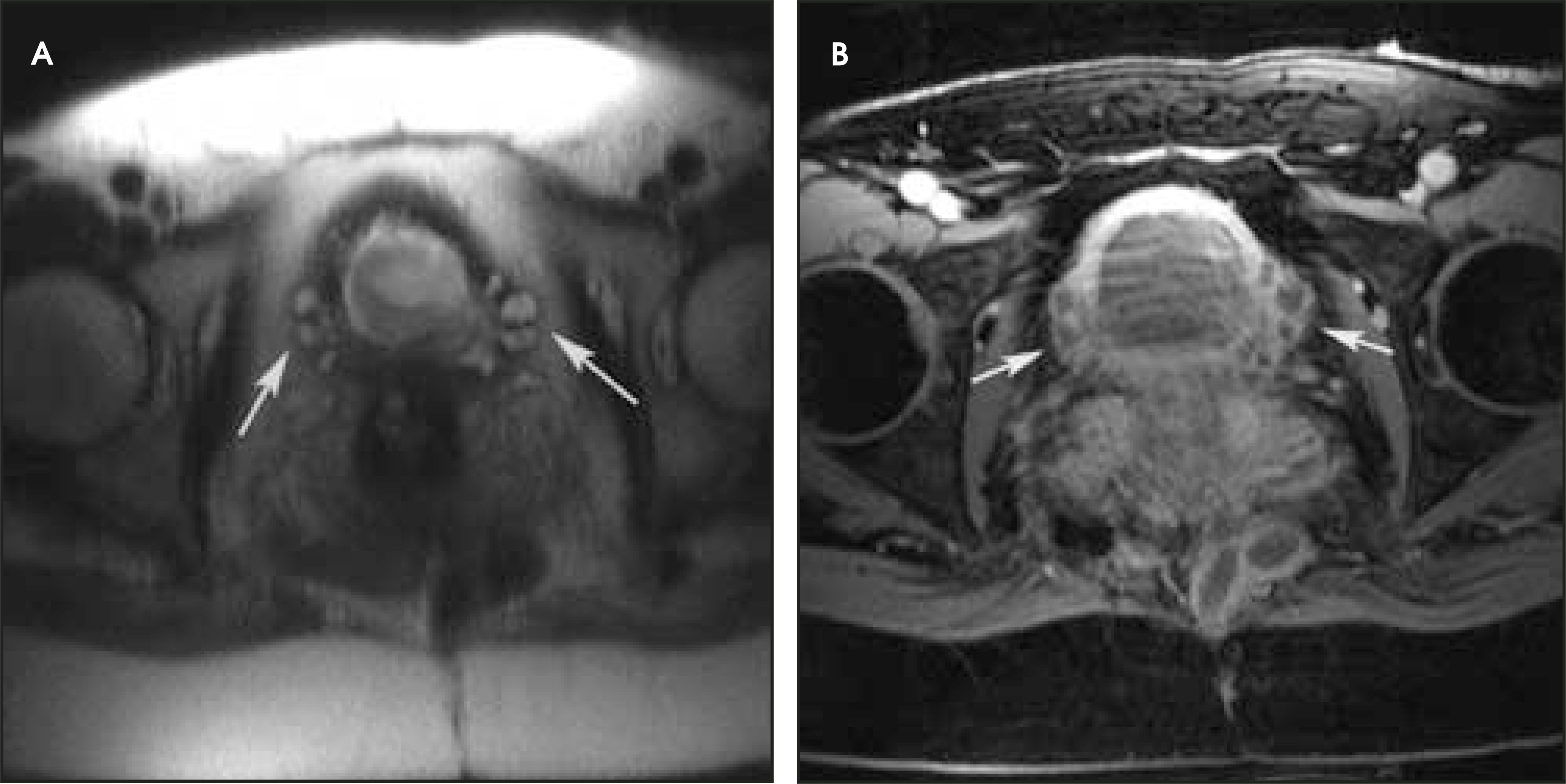
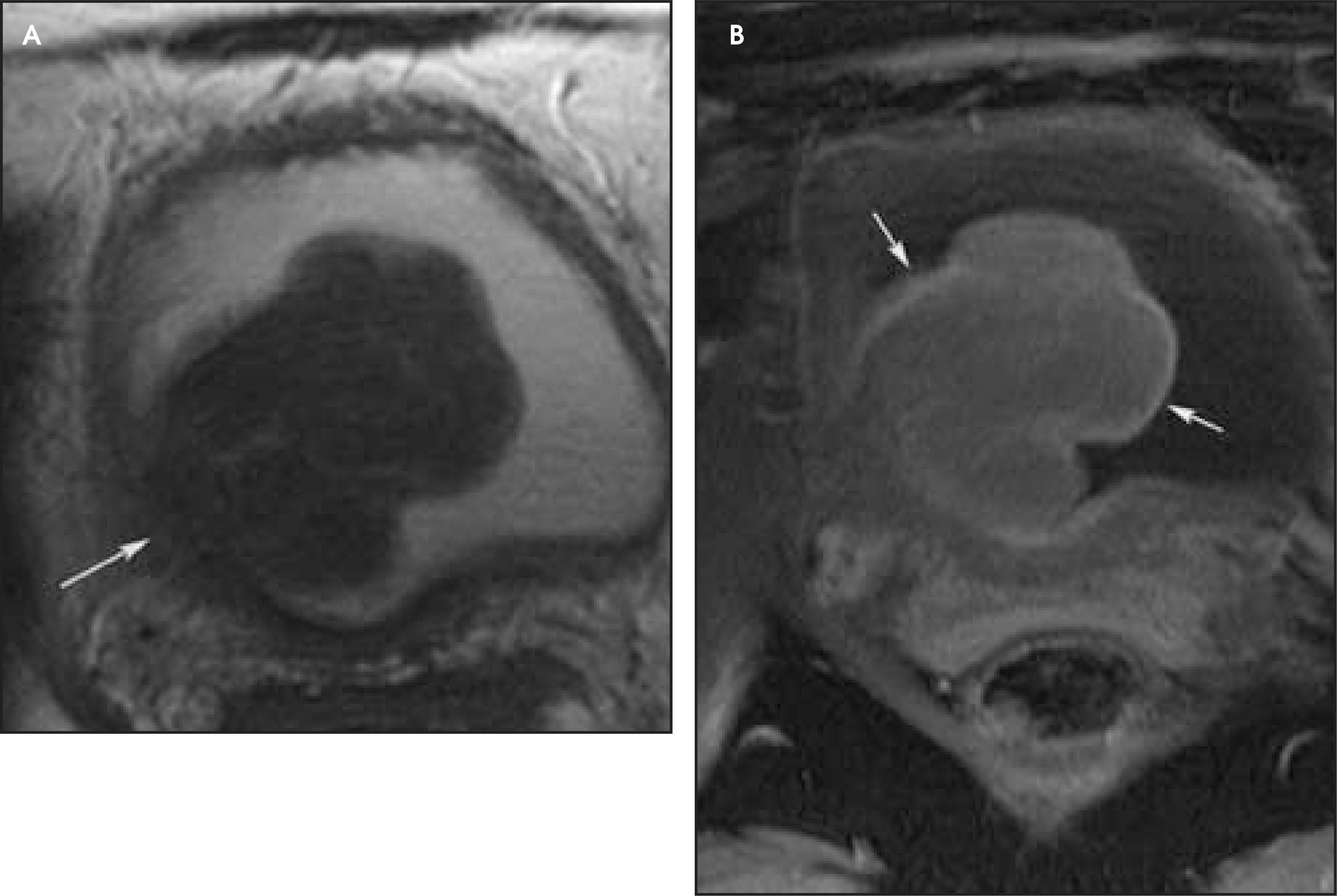
Other benign urinary bladder abnormalities include: 1) lithiasis, for which US or CT are the modalities of choice; 2) endo-metriosis, for which MR imaging can provide a specific diagnosis by demonstration of hemorrhagic implants; 3) cystitis, which usually is not evaluated by imaging (however, hemorrhagic cystitis and bladder hematoma have distinct MR appearance) (Figure 5); and 4) benign tumors (papilloma, leiomyoma [Figure 6], villous adenoma, paraganglioma, hemangioma, and neurofibroma), which can be well evaluated by MR imaging (however, specific diagnosis requires surgical excision).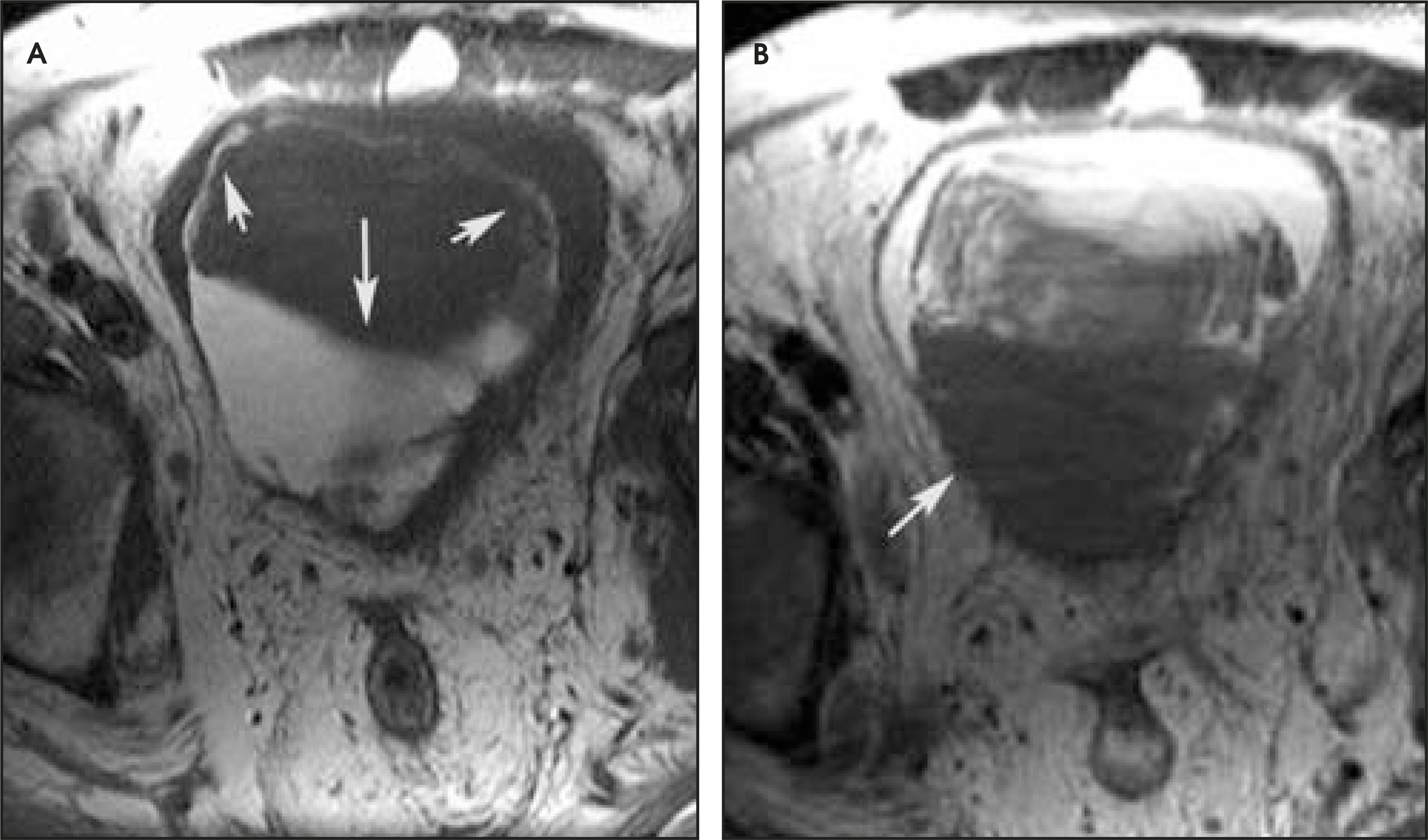
The levator ani muscles and endopelvic fascia normally support the urinary bladder. Defects in the fascia and the pelvic floor muscles result in abnormal bladder descent and formation of a cystocele, defined as caudal displacement of the bladder base below the inferior margin of the pubic bone, either at rest or during strain (Figure 7). This may lead to pain, vaginal bulging, vaginal pressure, dyspareunia, urinary tract infections, obstructive voiding symptoms and urinary retention, and urinary incontinence. MR imaging has been shown to be an excellent modality for the assessment of patients with cystoceles prior to surgical correction.3 It has been shown that when patients present with symptoms isolated to the anterior pelvic floor compartment, 42% of these patients have concomitant defects in other compartments, and those defects should also be corrected surgically to prevent recurrence and re-operations.10 MR imaging has been also used for follow-up after surgical treatment of cystoceles.3,11 A cystocele is measured as a vertical distance between the inferior aspect of the symphysis pubis or pubococcygeal line and the most inferior portion of the bladder.12 The normal vertical distance from the pubococcygeal line to the bladder base at strain should be no more than 1 cm below the line.13 Cystoceles are usually associated with urethral hypermobility and stress urinary incontinence. Higher-grade cystoceles are commonly associated with vaginal bulging, vaginal pressure, dyspareunia, urinary tract infections, obstructive voiding symptoms, and urinary retention. It can be difficult to differentiate clinically a high-grade cystocele from an enterocele, a vaginal vault prolapse or a high rectocele. When evaluating for cystocele, Gousse et al14 found that MR imaging had a sensitivity of 100%, a specificity of 83%, and a positive predictive value of 97% compared with intraoperative findings. MR imaging was shown to have a high degree of correlation with lateral cystourethrography in diagnosing cys-tocele.15 The advantage of MR imaging over other imaging modalities is simultaneous visualization of the entire pelvic floor and detection of the coexisting abnormalities involving other compartments. This is essential in planning reconstructive procedures so that the risks of surgical failure, recurrent prolapse, and reoperation can be minimized.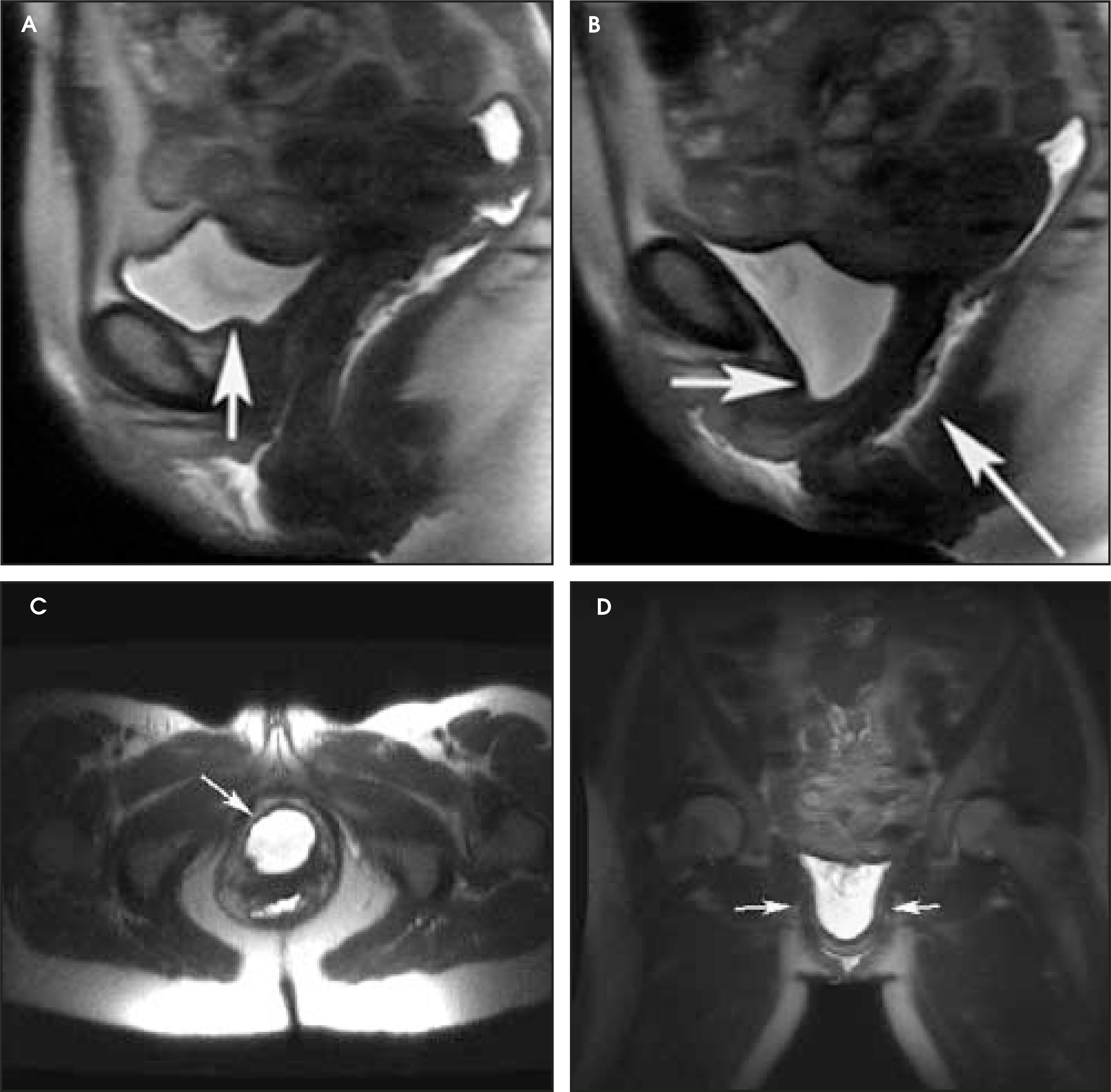
Conclusion
Due to superior soft-tissue contrast and lack of ionizing radiation, MR imaging is a compelling modality for the assessment of congenital anomalies in the pediatric population. Another patient group for which MR provides an excellent imaging alternative is adult women. The treatment for the female pelvic organ prolapse involves surgical repair of site-specific defects in pelvic floor support. MR imaging allows for a thorough preoperative evaluation of the entire female pelvis and for adequate diagnosis and staging of pelvic floor dysfunction. Although many benign bladder conditions can be diagnosed with MR imaging, currently, the most important indication for bladder MR in the adult population is staging of bladder cancer.
References
- Rosai J, Rosai J. Ackerman’s Surgical Pathology. 1996:1195-1196.
- Barentz J, Jafri S, Amendola M, Diokno A. Genitourinary Radiology: Diagnostic and Therapeutic. 1997:138-158.
- Pannu H, Kaufman H, Cundifl G. Dynamic MR imaging of pelvic organ prolapse: Spectrum of abnormalities. RadioGraphics. 2000;20:1567-1582.
- Ravi R, Shrivastava B, Chandrasekhar G. Adenocarcinoma of the urachus. J Surg Oncol. 1992;50:201-203.
- Cornil C, Reynolds C, Kickham C. Carcinoma of the urachus. J Urol. 1967;98:93-95.
- Chow Y, Lin W, Tzen C. Squamous cell carcinoma of the urachus. J Urol. 2000;163:903-904.
- Rubin J, Kasznica J, Davis C. Transitional cell carcinoma in a urachal cyst. J Urol. 1999;162:1687-1688.
- Defabiani N, Iselin C, Khan H. Benign teratoma of the urachus. Br J Urol. 1998;81:760-761.
- Yokoyama S, Hayashida Y, Nagahama J. Rhabdomyosarcoma of the urachus. A case report. Acta Cytol. 1997;41(4 suppl):1293-1298.
- Ng C, Rackley R, Appell R. Incidence of concomitant procedures for pelvic organ prolapse and reconstruction in women who undergo surgery for stress urinary incontinence. Urology. 2001;57:911-913.
- Stoker J, Halligan S, Bartram C. Pelvic floor imaging. Radiology. 2001;218:621-641.
- Lawler L. MR imaging of the bladder. Radiol Clin North Am. 2003;41(1):161-177.
- Yang A, Mostwin J, Rosenshein N. Pelvic floor descent in women: Dynamic evaluation with fast MR imaging and cinematic display. Radiology. 1991;179:25-33.
- Gousse A, Barbaric Z, Safir M. Dynamic half Fourier acquisition, single shot turbo spin-echo magnetic resonance imaging for evaluating the female pelvis. J Urol. 2000;164:1606-1613.
- Gufler H, DeGregorio G, Allamn K. Comparison of cystourethrography and dynamic MRI in bladder neck descent. J Comput Assist Tomogr. 2000;24:382-388.
Citation
. MR imaging in the evaluation of benign pathologies of urinary bladder. Applied Radiology. 2004;33(8):26-33. doi:10.37549/AR1270.