MR imaging in the evaluation of bladder cancer
Applied Radiology — Vol. 33 , Issue 10 , pp. 8 -17
DOI: 10.37549/AR1287
Published: October 1, 2004
Categories
Bladder cancer is a common tumor of the urinary tract, accounting for 6% to 8% of male malignancies and 2% to 3% of female malignancies.1 The most common histologic cell type is transitional cell carcinoma, accounting for 90% of all primary tumors of the urinary bladder. The remaining 5% to 10% of malignant bladder tumors are nontransitional and consist of squamous cell carcinomas, adenocarcinomas, small cell carcinomas, and, rarely, sarcomas. Cross-sectional imaging aids in determining tumor stage, including detection of local extension of the tumor, lymph node involvement, and distant metastasis.2 Presently, magnetic resonance (MR) imaging is the modality of choice in imaging of urinary bladder neoplasms, and staging accuracy of MR imaging in bladder cancer ranges from 73% to 96%. These values are 10% to 33% higher than those obtained with computed tomography (CT).3 Because the most common disease of the urinary bladder that requires further imaging is bladder cancer,4 the most frequent indications for MR imaging of the bladder are staging of a known bladder tumor and follow-up of a treated cancer patient. The excellent soft-tissue contrast and multiplanar capability of MR imaging improve the evaluation of the location and extent of bladder tumors.5
Technique
Currently, a 1.5T MR scanner with a phased-array pelvic coil is commonly used for imaging of the urinary bladder. The typical urinary bladder protocol includes T1-weighted spin-echo images (repetition time [TR]: 400 to 550 msec; echo time [TE]: minimum) obtained in the axial plane and T2-weighted fast spin-echo (TR: 4000 to 5500 msec; TE: 80 to 120 msec) images also obtained in the axial plane. Subsequently, fast multiplanar spoiled gradient-echo images with fat suppression (TR: 180 to 300 msec; TE:1.7 to 4.2 msec) are obtained in the axial plane before, at 20 seconds (arterial phase), and at 70 to 115 seconds (venous phase) after gadopentate dimeglumine injection (0.1 mmol/kg). Sagittal or coronal T2-weighted images may be obtained, if the anteroposterior or inferosuperior extent of disease needs to be evaluated. Sagittal and coronal gadolinium-enhanced images are also recommended for the evaluation of the bladder tumors, when the tumor is located in the base or the dome of the bladder. Typical parameters for optimal bladder imaging are: 20- to 30-cm field-of-view (FOV), 6-mm slice thickness, and 2-mm intersection gap. Ideally, the urinary bladder should be moderately distended during imaging. The low distension may cause misevaluation of the bladder wall thickening, and overdistension may obscure small tumors.
Bladder cancer
Bladder cancer is more common in men (male/female: 3:1) than in women. It is the fourth most common cancer in men, after prostate, lung, and colorectal cancer, and it accounts for 10% of all cancer cases in men. In women, it is the eighth most common cause of cancer and accounts for 4% of all cancer cases in women.6 It is generally a disease of the elderly, and its incidence peaks at the sixth to seventh decade.7
Several factors predispose patients to bladder cancer. Cigarette smokers are 2 to 6 times more likely to develop urothelial cancer than those who do not smoke.8,9 Cystitis and chronic urinary tract infection are predisposing factors to bladder cancer, and 7% of bladder tumors are found to be associated with diverticuli (Figure 1). Strong association with aniline dyes, aromatic amines, diesel fumes, and long-term use of analgesic phenacetin has been reported in the development of bladder cancer.1
Pathologically, 90% of the bladder tumors are epithelial, and 90% of the epithelial tumors are transitional cell carcinomas. At the time of presentation, 30% of patients have multifocal disease, and there may be widespread areas of metaplasia and carcinoma in situ.1 The remaining 10% of epithelial tumors include squamous cell carcinomas, adenocarcinomas, small cell carcinomas, and, rarely, sarcomas.2 Leiomyosarcoma, rhabdomyosarcoma (Figure 2), phaeochromocytoma, and hamartomatous malformations are among the nonepithelial tumors.1 Gross or microscopic hematuria is the most common clinical presentation, followed by symptoms related to associated urinary infection.10
Staging and management of bladder cancer
Local extension of the tumor (T), lymph node involvement (N), and distant metastasis (M) are the indicators used in staging. Table 1 presents the bladder cancer staging used.11 Treatment options and prognosis depend on the clinical stage of the bladder tumor. It is critically important to differentiate between superficial and invasive disease.1 Superficial tumors are treated with local endoscopic resection, with or without adjuvant intravesical instillations of chemotherapeutic agents; while invasive tumors are treated by curative cystectomy and palliative chemo- and/or radiation therapy.12
Management of bladder tumors starts with clinical staging, including cystoscopic examination of the organ. This method allows for immediate biopsy and can distinguish superficial from invasive tumors, but it is not capable of detecting extravesical disease. This distinction is important because patients with non– organ-confined disease have higher recurrence rates and lower survival rates.13
CT has been a valuable tool for the evaluation of bladder tumors, but MR imaging has been shown to be superior in the detection of superficial and multiple tumors, and in staging accuracy in detecting extravesical tumor extension and surrounding organ invasion, especially with dynamic contrast administration.2,14-20 Accuracy of MR imaging on a stage-by-stage basis has been reported to be between 64% and 85%.16,18,19,21,22 Staging accuracy of MR imaging in differentiating superficially versus deeply muscle-invasive tumors, and organ-confined versus non–organ-con-fined tumors is between 85% and 95%.15,17,21,23
Restaging after treatment is particularly challenging. Intravesical therapy with chemotherapeutic agents is used to treat low-stage tumors. Intravesical therapy may cause bright submucosal enhancement after administration of gadolinium. Previoustransurethral resection or biopsy of the tumor may cause inflammation and edema, and this may result in overstaging. Using MR imaging, it is not possible to discriminate by signal characteristics alone between edema resulting from treatment and tumor recurrence, but the presence of a mass with an enhancement pattern typical of a tumor may enable the diagnosis of recurrence.
Morphologic features
Tumors can arise anywhere in the bladder, but they are most commonly located in the lateral wall. When a tumor is located around the ureteral orifices, it may produce partial or complete blockage of one or both ureters, resulting in hydroureter and hydronephrosis.10 Tumors manifest a variety of patterns of growth, including papillary, sessile, infiltrating, nodular, mixed, and flat intraepithelial growth. Because the bladder does not have a distinct basement membrane, it is difficult to detect the invasion of the lamina propria by imaging .24 However, the detrussor muscle can be well evaluated with MR imaging; therefore, the depth of muscle invasion can be assessed. The fat tissue around the bladder can be seen clearly on MR imaging, and the interface between the bladder wall and the surrounding fat can be evaluated for extravesical tumor spread.
MR imaging features
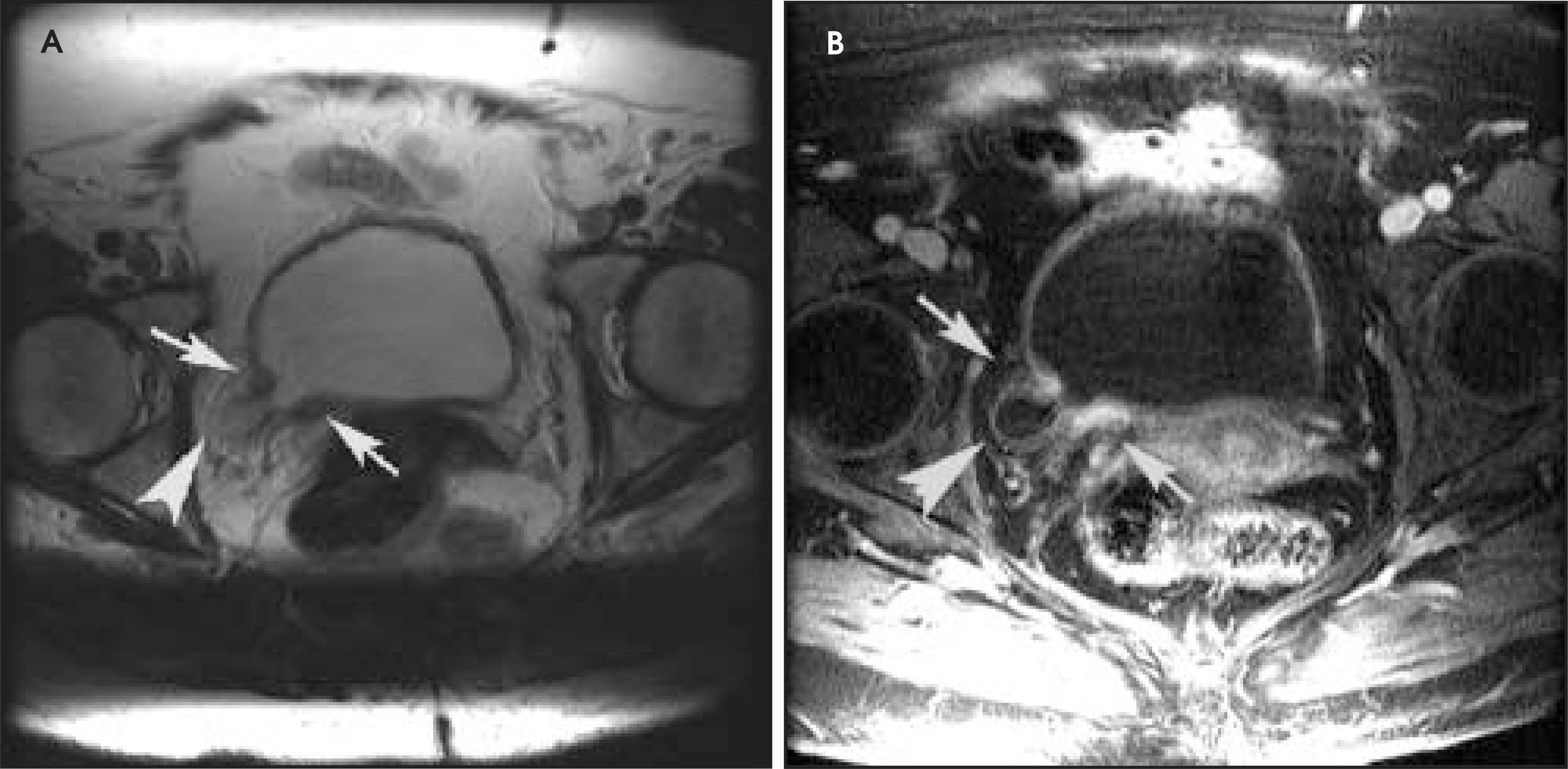
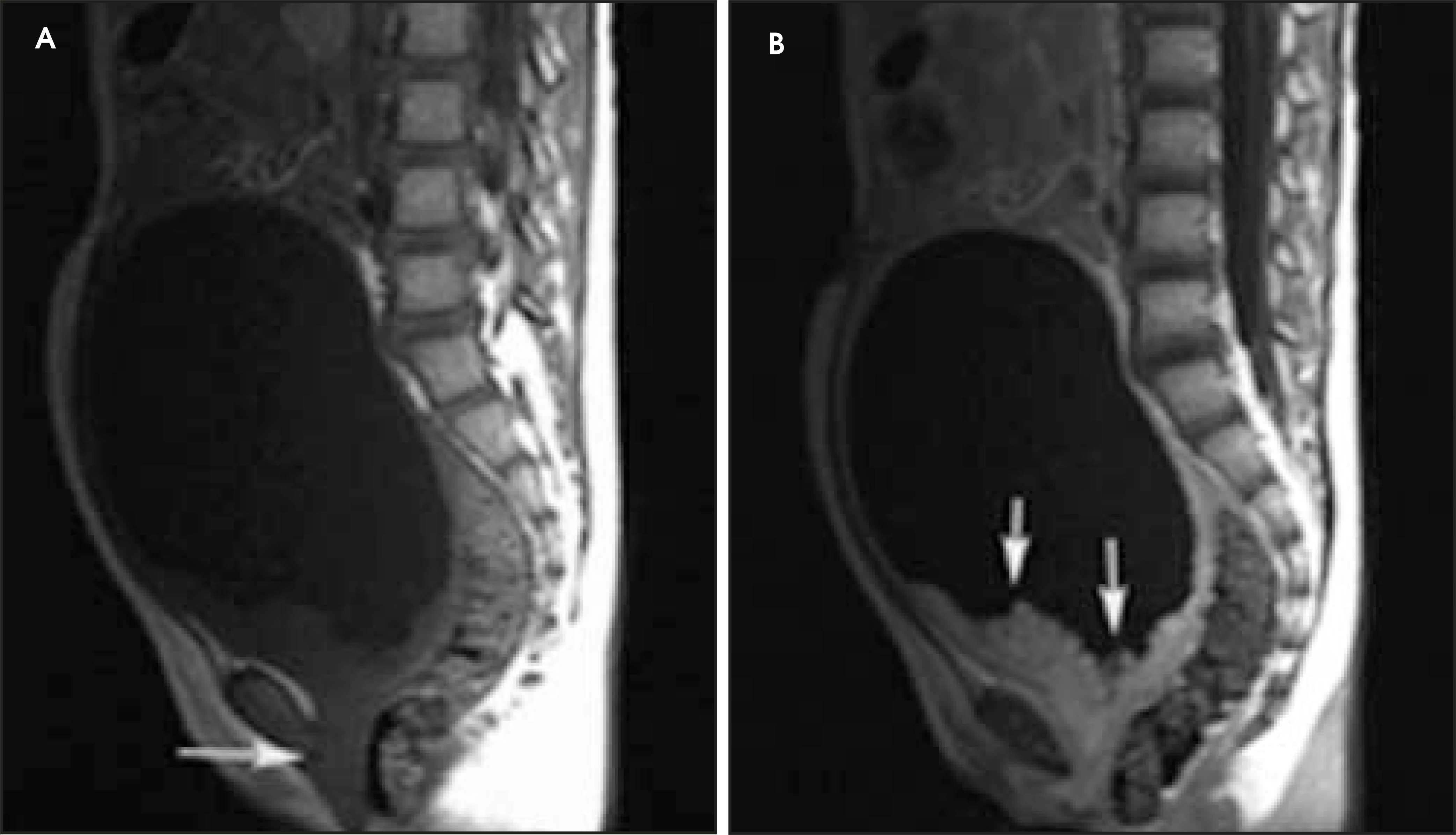
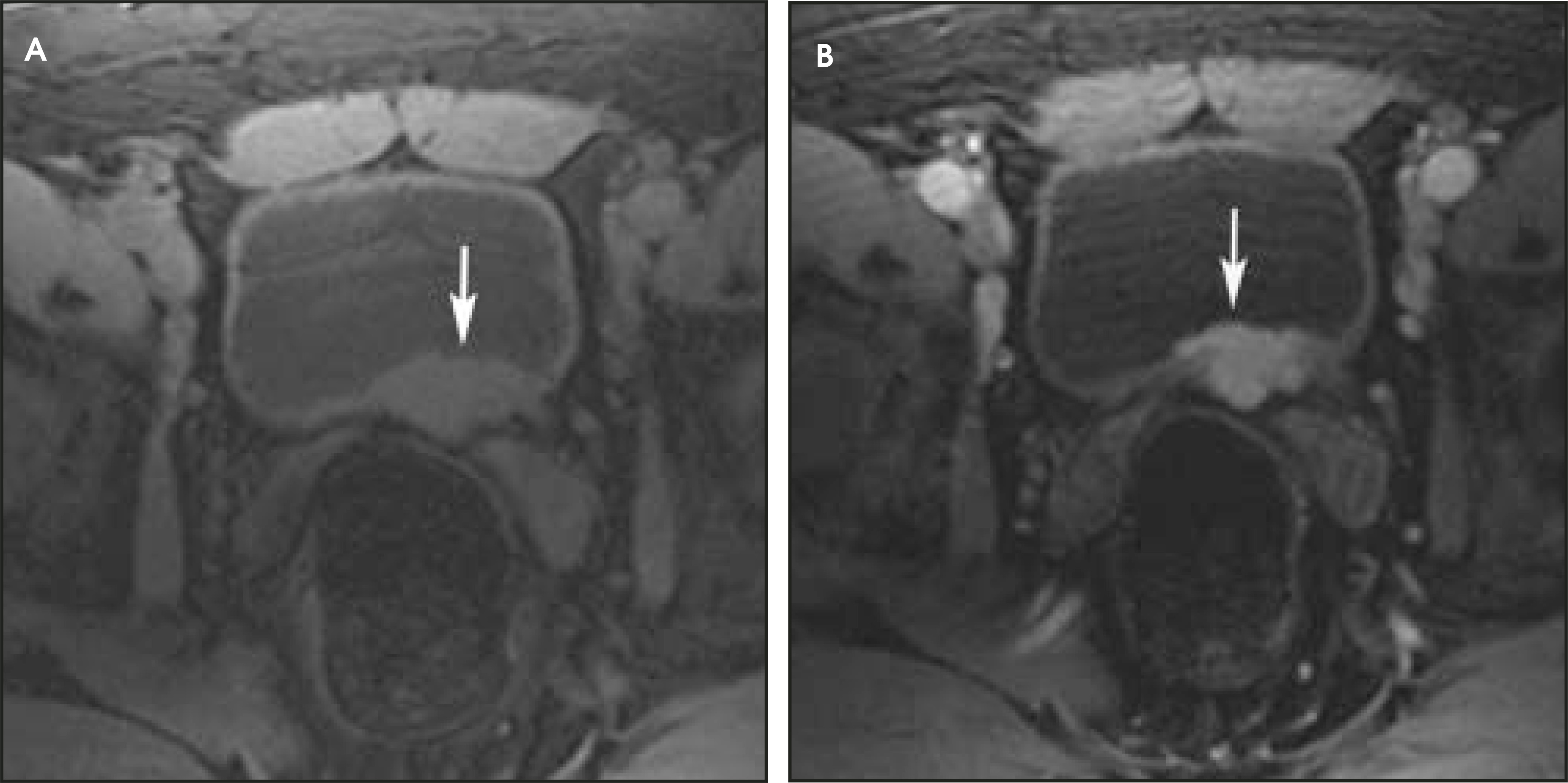
Urinary carcinomas have intermediate signal intensity, equal to that of muscle on the T1-weighted images. T1-weighted images are used to assess perivesical fat invasion and lymph node involvement. T2-weighted images aid in determining the depth of tumor infiltration into the bladder wall. On T2-weighted images, urine has high signal intensity, and the bladder wall appears hypointense. Bladder tumors have the same, or slightly higher, signal intensity as the bladder wall on T2-weighted images. An intact, low-signal-intensity muscle layer at the base of the tumor is classified as stage Ta or T1 (Figure 3); a disrupted low-signal-intensity muscle layer without infiltration of perivesical fat is classified as stage T2b (Figure 4).25
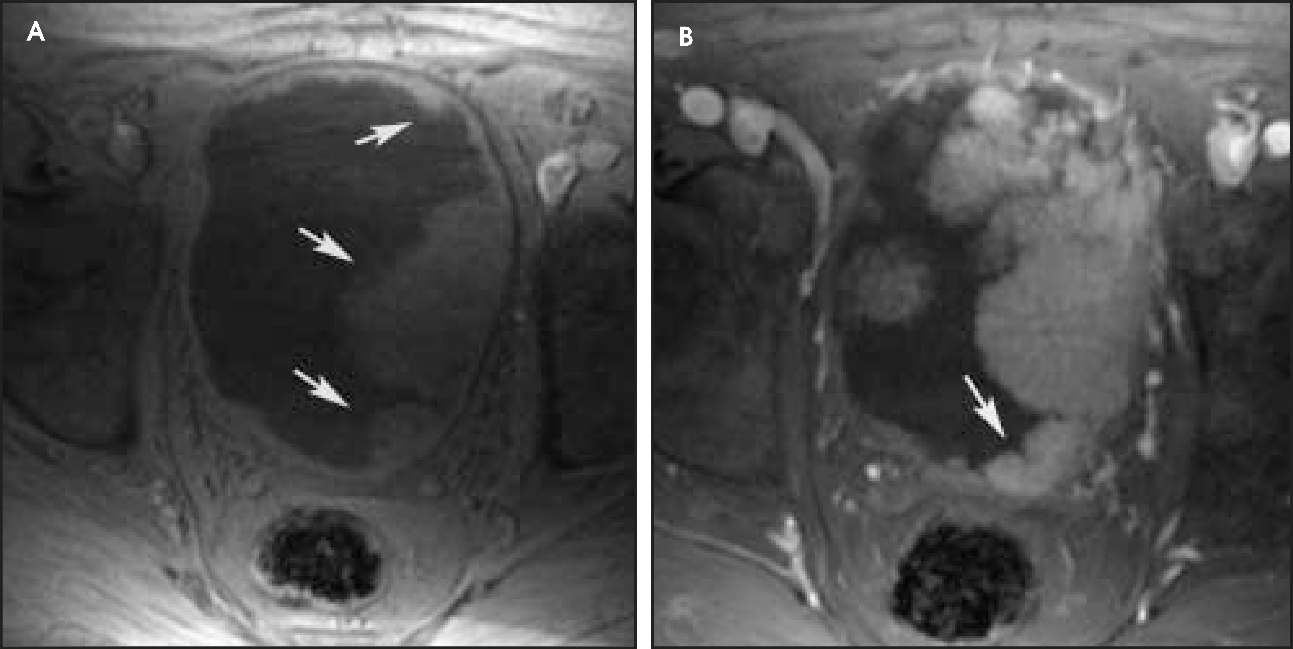
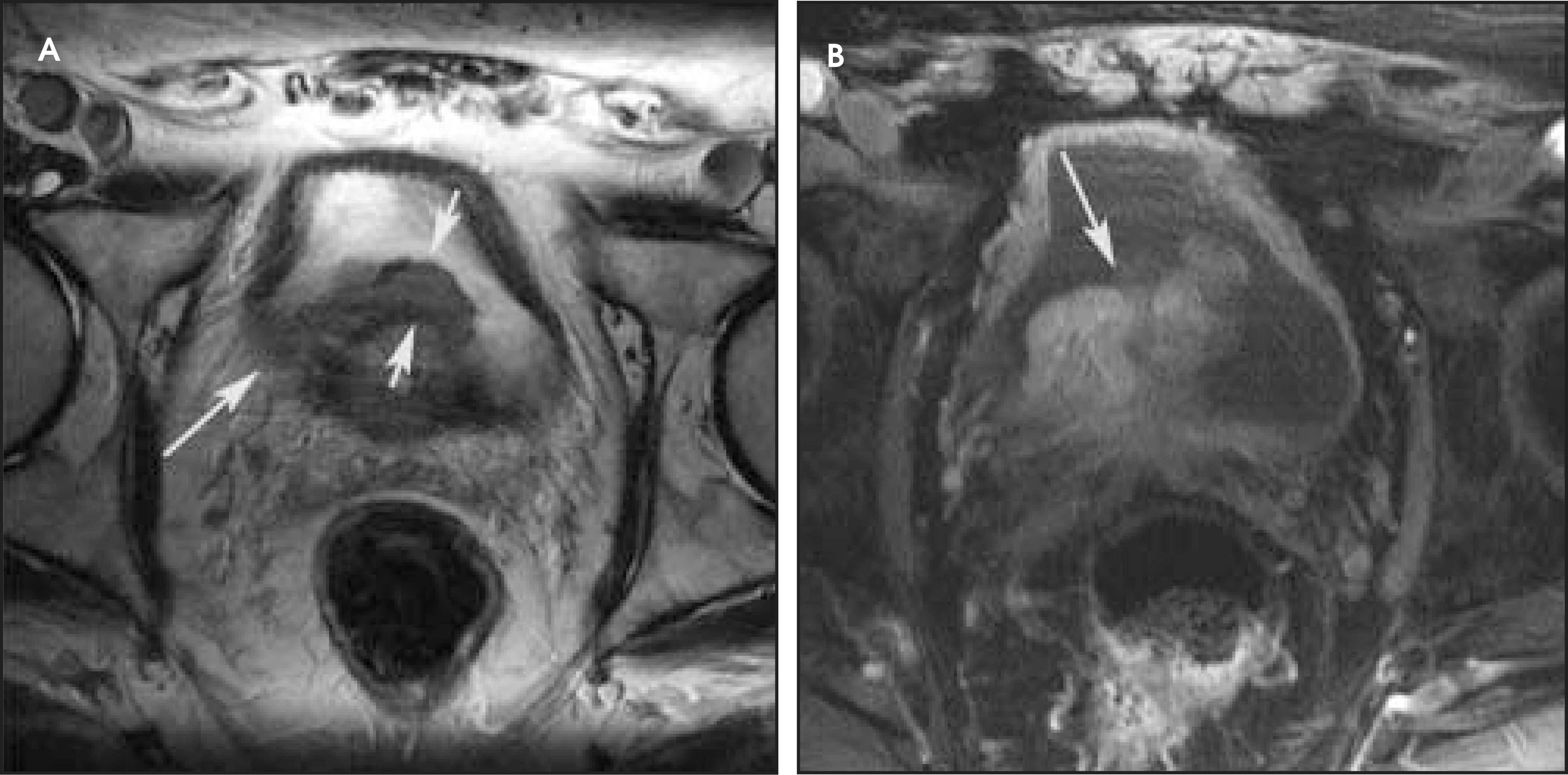
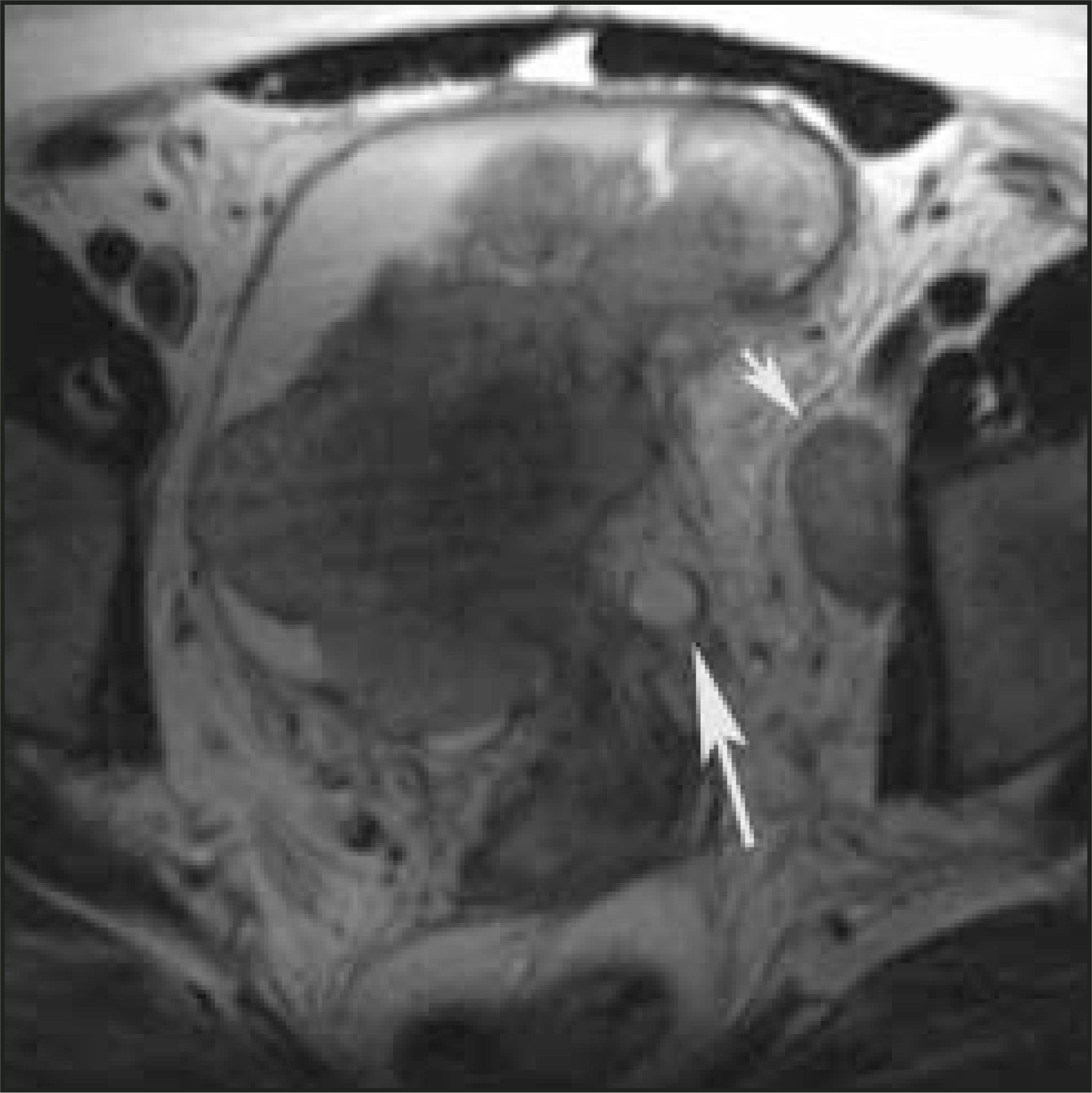
Urinary bladder carcinomas and their metastases develop neovascularization26; therefore, these tumors enhance earlier than the normal bladder wall.3 In the arterial phase of contrast enhancement, bladder tumors enhance more than the muscle of the bladder (Figure 5). Superficial tumors (those without muscle invasion, stage T1 and lower) may be differentiated from muscle-invasive tumors on contrast-enhanced images. On contrast-enhanced studies, a lesion with an irregular, shaggy outer border, and streaky areas of the signal intensity of the tumor in the perivesical fat is classified as stage T3b (Figure 6). A tumor extending into an adjacent organ or abdominal and pelvic sidewall with the same signal intensity as the primary tumor is classified as stage T4a or T4b, respectively (Figure 7).25 With the current imaging protocols using 1.5T MR scanners, MR imaging cannot differentiate stage Ta tumors from stage T1; and differentiation of stage T2a tumors (superficial muscle invasion) from stage T2b (deep muscle invasion) is problematic but sometimes can be better delineated on contrast-enhanced studies.2
Based on MR imaging features, it is not possible to differentiate between different histologic cell types of bladder tumors, but few clues may lead to the differentiation of transitional from non–transitional-cell carcinomas. Non–transitional-cell carcinomas are aggressive tumors that usually extend beyond the bladder wall at the time of initial diagnosis, whereas two thirds of transitional cell carcinomas are superficial. Non–transitional-cell carcinomas tend to cause marked bladder wall thickening and tend to be larger than transitional cell carcinomas.27 The non–transitional-cell carcinomas of the bladder include: squamous cell carcinoma, adenocarcinoma, small cell carcinoma, and carcinosarcoma.
Squamous cell carcinoma
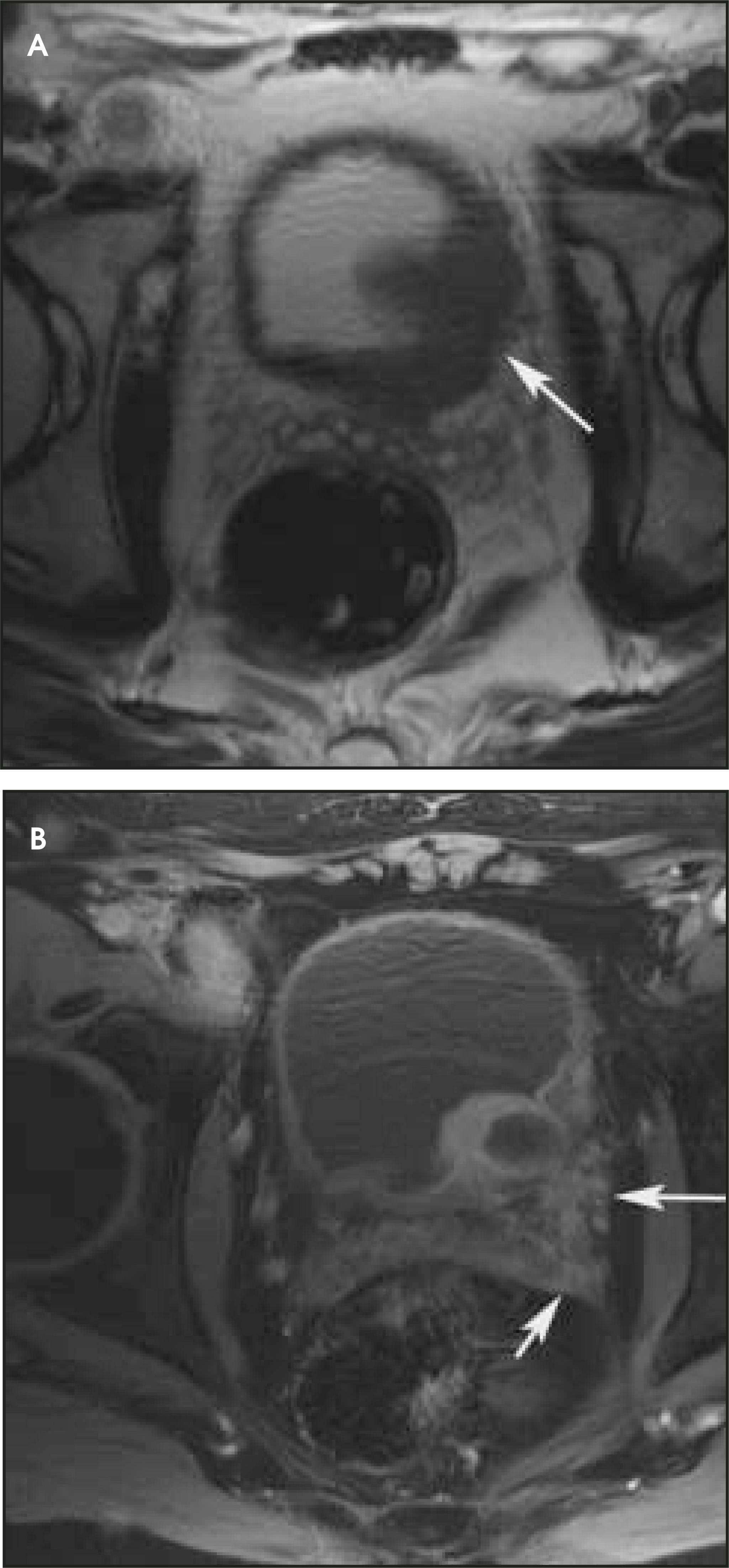
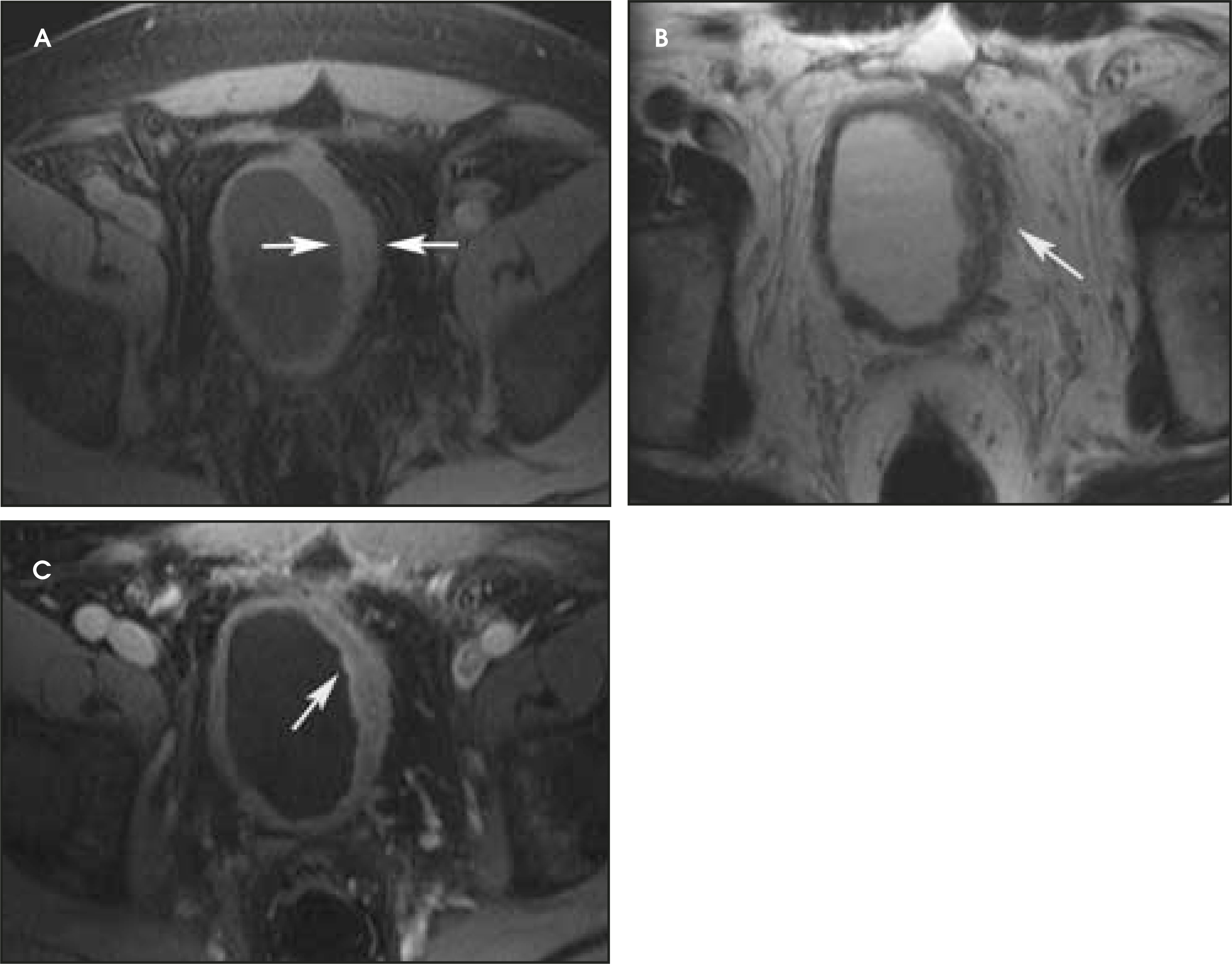
Squamous cell carcinoma is the most common non–transitional-cell bladder tumor, accounting for 3% to 7% of all bladder tumors in the United States. Approximately 80% of squamous cell carcinomas in Egypt are associated with chronic infection caused by schistosomiasis (bilharziasis).24 The disease is relatively more common in women, unlike transitional cell carcinoma, which is more common in men. The reported female-to-male ratio varies from 1.25:1 to 1.8:1.28 In contrast to transitional cell carcinomas, squamous cell carcinomas are often widespread and involve areas other than the base of the bladder. Most squamous cell carcinomas are solitary and large at the time of detection, with invasion of the muscular wall reported in <80% of patients (Figure 8). Metastases have been identified in at least 10% of patients at the time of diagnosis. Interestingly, metastases from squamous cell carcinomas of the urinary bladder often occur at sites other than the regional lymph nodes. Common metastatic sites include bone, lung, and bowel.29
Adenocarcinoma
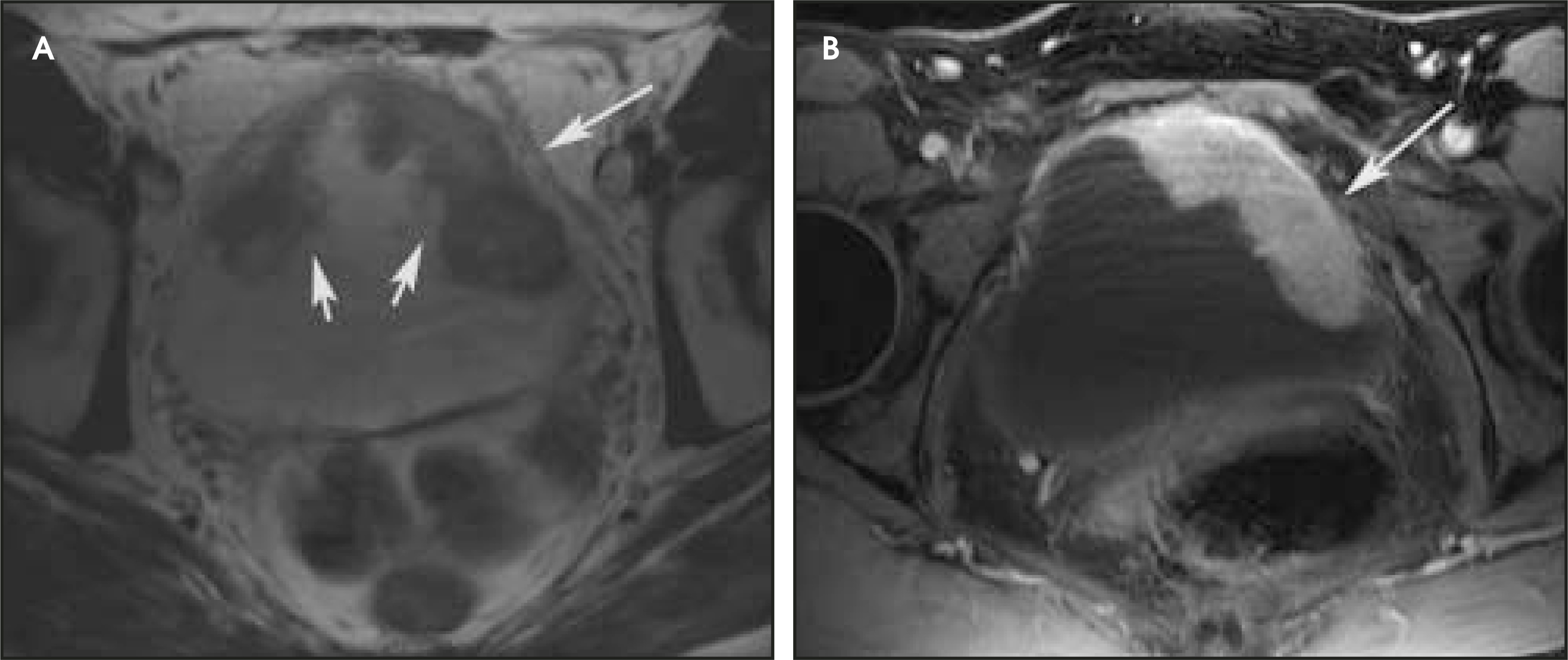
Adenocarcinoma of the urinary bladder is uncommon, accounting for 0.5% to 2% of all bladder malignancies.30 Adenocarcinomas are classified into 3 groups: primary, urachal, and metastatic.24 Like squa mous cell carcinomas, many adenocarcinomas probably occur in reaction to long-term mucosal irritation. Cystitis glandularis, bladder exstrophy, and urachal remnants are also associated with adenocarcinoma of the bladder. Cystitis glandularis has been observed in approximately 50% of adenocarcinoma cases located at the bladder base3,11,24,28,29; however, most au thors do not accept this entity as a predisposing factor for urothelial adenocarcinoma. Adenocarcinoma is observed in <10% of patients with bladder exstrophy, and <80% of urachal neoplasms are adenocarcinomas.29 Signet cell adenocarcinomas characteristically produce linitis plastica of the bladder (Figure 9).
Small cell carcinoma
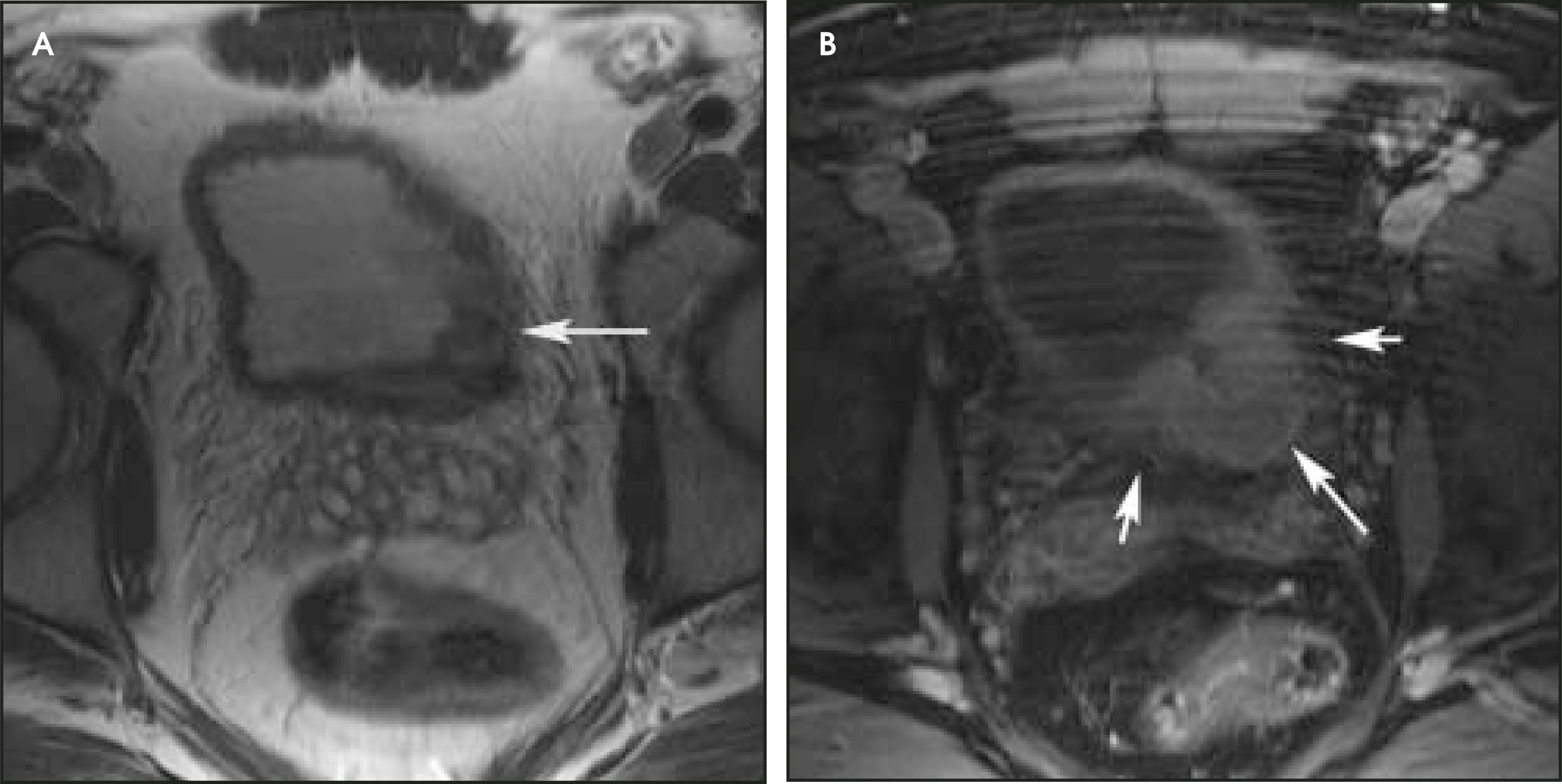
Small cell carcinoma of the bladder has also been called undifferentiated and poorly differentiated carcinoma. Most small cell carcinomas occur as a component of mixed carcinomas. Age, sex, and symptoms are comparable to those of transitional cell carcinomas. Cystoscopically, small cell carcinomas tend to be polypoid or nodular and often appear as ulcerated masses that cannot be distinguished from other high-grade bladder cancers (Figure 10). Metastatic spread occurs rapidly, and the most frequent sites are the regional lymph nodes, bones, and peritoneal cavity.29
Carcinosarcoma
Carcinosarcoma of the urinary bladder is a rare neoplasm; approximately 73 cases have been reported. They are most frequently observed in the female genital tract.31 Neither the etiology nor the pathogenesis of carcinosarcomas is currently known. Unlike transitional cell carcinomas, carcinosarcomas are highly aggressive tumors, with high recurrence rates and a poor prognosis. These tumors can present as large single tumors or multiple small- to medium-sized tumors. The tumors may be polypoid or sessile and may arise from anywhere in the bladder. Location, size, shape, and multiplicity do not help to differentiate carcinosarcomas from transitional cell carcinomas. Dynamic gadolinium-enhanced imaging in carcinosarcomas does not show early strong arterial enhancement, a feature differentiating this tumor from a transitional cell carcinoma. On T2-weighted imaging, the signal intensity of carcinosarcomas can be heterogeneous.32
Conclusion
MR imaging is a noninvasive examination that allows multiplanar visualization of the entire pelvis and offers superior soft-tissue contrast. It is a useful modality for the assessment of tumors of the urinary bladder. Imaging with gadolinium enhancement allows evaluation of the bladder tumor extent and adjacent organ involvement. MR imaging thus plays a critical role in improving staging accuracy, determining patient management, and assessing response to therapy.
References
- MacVicar A. Bladder cancer staging. BJU Int. 2000;86(1):111-122.
- Barentsz J, Witjes J, Ruijs J. What is new in bladder cancer imaging. Urol Clin North Am. 1997;24:583-602.
- Barentsz J, Jager G, Witjes J. MR imaging of the urinary bladder. Magn Reson Imaging Clin N Am. 2000;8:853-867.
- Barentsz J, Szh J. Genitourinary Radiology Diagnostic and Therapeutic. 1997:138-158.
- Chen M, Lipson S, Hricak H. MR imaging evaluation of benign mesenchymal tumors of the urinary bladder. AJR Am J Roentgenol. 1997;168:399-403.
- Silverberg E, Boring C, Squires T. Cancer statistics, 1990. CA Cancer J Clin. 1990;40:9-26.
- Including Cancer Trends: 1950-1985. 1987.
- Wynder E, Goldsmith R. The epidemiology of bladder cancer: A second look. Cancer. 1977;40:1246-1268.
- Clavel J, Cordier S, Boccon-Gibod L. Tobacco and bladder cancer in males: Increased risk for inhalers and smokers of black tobacco. Int J Cancer. 1989;44:605-610.
- Rosai J, Rosai J. Ackerman’s Surgical Pathology. 1996:1195-1196.
- Sobin L, Fleming I. TNM Classification of Malignant Tumors, fifth edition (1997). Union Internationale Contre le Cancer and the American Joint Committee on Cancer. Cancer. 1997;80:1803-1804.
- Barentsz J, Jager G, Mugler J. Staging urinary bladder cancer: Value of T1-weighted three-dimensional magnetization prepared-rapid gradient-echo and two-dimensional spin-echo sequences. AJR Am J Roentgenol. 1995;164:109-115.
- Stein J, Lieskovsky G, Cote R. Radical cystectomy in the treatment of invasive bladder cancer: Long-term results in 1,054 patients. J Clin Oncol. 2001;19:666-675.
- Fisher M, Hricak H, Tanagho E. Urinary bladder MR imaging. Part II. Neoplasm. Radiology. 1985;157:471-477.
- Kim B, Semelka R, Ascher S. Bladder tumor staging: Comparison of contrast-enhanced CT, T1- and T2-weighted MR imaging, dynamic gadolinium-enhanced imaging, and late gadolinium-enhanced imaging. Radiology. 1994;193:239-245.
- Tanimoto A, Yuasa Y, Imai Y. Bladder tumor staging: Comparison of conventional and gadolinium-enhanced dynamic MR imaging and CT. Radiology. 1992;185:741-747.
- Buy J, Moss A, Guinet C. MR staging of bladder carcinoma: Correlation with pathologic findings. Radiology. 1988;169:695-700.
- Amendola M, Glazer G, Grossman H. Staging of bladder carcinoma: MRI-CT surgical cor-relation. AJR Am J Roentgenol. 1986;146:1179-1183.
- Tachibana M, Baba S, Deguchi N. Efficacy of gadolinium-diethylenetriaminepentaacetic acid-enhanced magnetic resonance imaging for differentiation between superficial and muscle-invasive tumor of the bladder: A comparative study with computerized tomography and transurethral ultrasonography. J Urol. 1991;145:1169-1173.
- Husband J, Olliff J, Williams M. Bladder cancer: Staging with CT and MR imaging. Radiology. 1989;173:435-440.
- Narumi Y, Kadota T, Inoue E. Bladder tumors: Staging with gadolinium-enhanced oblique MR imaging. Radiology. 1993;187:145-150.
- Barentsz J, Jager G, van Vierzen P. Staging urinary bladder cancer after transurethral biopsy: Value of fast dynamic contrast-enhanced MR imaging. Radiology. 1996;201:185-193.
- Scattoni V, Da Pozzo L, Colombo R. Dynamic gadolinium-enhanced magnetic resonance imaging in staging of superficial bladder cancer. J Urol. 1996;155:1594-1599.
- Messing E, Catalona W, Walsh P. Chambell’s Urology. 1998:2340-2342.
- Tekes A, Kamel I, Imam K. MR imaging features of transitional cell carcinoma of the urinary bladder. AJR Am J Roentgenol. 2003;180:771-777.
- Dickinson A, Fox S, Persad R. Quantification of angiogenesis as an independent predictor of prognosis in invasive bladder carcinomas. Br J Urol. 1994;74:762-766.
- Tekes A, Kamel I, Chan T. MR imaging features of non-transitional cell carcinoma of the urinary bladder with pathologic correlation. AJR Am J Roentgenol. 2003;180:779-784.
- Ghoneim M, Krane R, Siroky M, Fitzpatrick J. Clinical Urology. 1994:680-681.
- Murphy W, Murphy W. Urological Pathology. 1997:98-111.
- Chan T, Epstein J. In situ adenocarcinoma of the bladder. Am J Surg Pathol. 2001;25:892-899.
- Perret L, Chaubert P, Hessler D. Primary heterologous carcinosarcoma (metaplastic carcinoma) of the urinary bladder: A clinicopathologic, immunohistochemical, and ultrastructural analysis of eight cases and a review of the literature. Cancer. 1998;82:1535-1549.
- Tekes A, Kamel I, Szarf G. Carcinosarcoma of the urinary bladder: Dynamic contrast-enhanced MR imaging with clinical and pathologic correlation. AJR Am J Roentgenol. 2003;181:139-142.
Citation
. MR imaging in the evaluation of bladder cancer. Applied Radiology. 2004;33(10):8-17. doi:10.37549/AR1287.