MR imaging of muscle injuries
Applied Radiology — Vol. 33 , Issue 4 , pp. 14 -26
DOI: 10.37549/AR1235
Published: April 1, 2004
Categories
Acute and chronic musculoskeletal injuries account for a substantial proportion of the problems for which patients seek medical attention.1 Muscle injuries also account for approximately half of all injuries that occur in sports-related activities.2 Even without a specific injury, sports-related muscle pain due to overuse is common in both the trained and untrained athlete. While muscle injuries are usually mild and self-limiting, the pain from these injuries can be severe and debilitating.3
Although clinical assessment is usually sufficient for deciding on clinical management of muscle injuries, imaging may be needed in some situations such as a complete muscle tear, which may need surgical repair, or injuries in high-caliber athletes during the competitive season of their sport.
This article will review muscle morphology, risk factors for injury, and the clinical spectrum of muscle injuries. The magnetic resonance (MR) imaging findings of muscle strain, contusion, hematoma, and herniation will also be discussed in detail.
Muscle morphology and risk factors
The major purpose of muscle function is to produce joint motion.4 Most muscles cross one joint, but many muscles span two joints. The most commonly strained muscles are those that cross two joints and perform eccentric, rather than concentric, contraction. Eccentric contraction occurs when a muscle fiber is forced to lengthen when the resisting force is greater than the force generated by muscle.4,5 Concentric contraction occurs if the resisting load is less than the force generated by the muscle, thus, the muscle shortens.4
Another factor in muscle injury is the proportion of the two types of muscle fibers within each muscle. Most muscles consist of both type 1 and type 2 fibers. Type 1 fibers have slower contraction, are more resistant to fatigue, and are used for extended activities. Type 2 fibers have a faster contraction time, are seen in muscles that cross two joints, and are optimal for short energy bursts.4,6 Muscles with a higher proportion of type 2 fibers are more prone to injury. Muscle strain injury is not only the result of contraction, but can also be the result of stretch on a muscle while the muscle is in contraction.7
There are many etiologies for muscle injuries that include: insufficient warm-up and stretching prior to exercise, lack of flexibility, inadequate muscle strength and endurance, abnormal muscle contraction during running, awkward running style, and return to activity before complete rehabilitation following injury.8,9 Athletic training reduces the severity and frequency of muscle injury.10 The clinical risk factors for muscle injury include: previous injury, increasing age, and a change to a higher competitive performance level.9
Muscle injury
The most commonly injured muscles are in the lower extremities. These include the rectus femoris, biceps femoris, semitendinosis, adductors, hamstrings, and medial head of the gastrocnemius.1,4,6,7,11-13 Some authors include the vastus medialis and soleus as other commonly injured mus-cles.4
There is a vast spectrum of injuries that can result from trauma to skeletal muscle. Muscle trauma includes both direct and indirect injuries. Contusion, laceration, and hematoma result from direct injury; indirect injuries include excessive muscle stretching producing muscle strain or tear.
Muscle strains are usually due to an indirect stretch-induced injury. These injuries can be quite painful and often occur in athletes participating in sports that require high-speed running, such as track, rugby, soccer, football, and basketball.12 These injuries can be debilitating and devastating to high-performance athletes and can result in time lost from work in the recreational athlete.12
These injuries are classified into the following grading system. Grade 1 strain consists of a minor degree of microscopic tearing with no permanent defect. Grade 2 strain represents an incomplete disruption of muscle fibers, which is also called a partial tear. Clinically, Grade 1 and Grade 2 strains can be inseparable, resulting in some loss of muscle function. Grade 3 strains are a complete rupture of muscle, with nearly complete or complete loss of muscle function.1-6,8,14-17 Grade 3 strains are usually associated with retraction, spasm, and shortening of muscle. The grade of injury is important in determining whether conservative or surgical intervention is needed.
Muscle strain almost uniformly occurs at the myotendinous junction, which is the weakest link within the muscle.1,3,5-8,11,12,14,16-19 Most tears are partial tears.12,20 Partial tears can be difficult to differentiate clinically from a complete tear when pain limits a complete physical examination of strength and range of motion. MR imaging can confirm the diagnosis of muscle strain and indicate the muscles involved; MR can also indicate the stages and degree of muscle involvement and can determine if the tear is incomplete or complete.
Muscle pain can also occur with excessive or unaccustomed exercise, especially following periods of inactivity. Pain or soreness that begins hours to days following a strenuous activity is termed delayed onset of muscle soreness (DOMS). The symptoms usually last only a few days.2,4,6,11,12
Imaging of muscle injury
As noted, muscle injuries are usually self-limited and do not require imaging. However, there are certain circumstances in which MR imaging may provide important diagnostic and prognostic information and may help guide the choice of treatment. MR can be used: in high-performance athletes to determine surgical versus conservative treatment; in patients with a soft-tissue mass with no history of trauma; to provide prompt diagnosis about the site and the severity of an injury before beginning therapy; to evaluate obscure muscle pain; to evaluate pain in difficult-to-detect, deeply injured muscles; to guide rehabilitation; and to evaluate cases in which there are complications from injury (ie, hematoma, seroma, herniation, and ossification).2,4,5,12,14,18
Precise definition of an injured muscle is important for optimal management in an athlete‘s performance or in laborers due to muscle weakness and where the injury may cause recurrent injuries.14 It is also important to localize a lesion and estimate the severity of muscle involvement, as well as to determine the presence or absence of associated abnormalities.14 In addition, MR imaging is often useful in evaluating patients who have had remote injury with persistent pain or a soft-tissue mass.
MR technique
The main objective in imaging an injured muscle is to determine the location and extent of involvement. Because of its multiplanar capability and high sensitivity to the hemorrhage and edema that result from soft-tissue injury, MRI is the technique of choice for imaging muscles, tendons, and ligaments.
Normal skeletal muscle is characterized by intermediate-to-low signal intensity on T1-weighted (short repetition time [TR]/short echo time [TE]) images and by low T2-weighted (long TR/long TE) signal intensity relative to other soft tissues.20,21 Muscle tears and other soft-tissue injuries are best depicted on T2-weighted images, which optimize contrast between edema and hemorrhage and the adjacent normal muscle.
Our protocol is to obtain T1-weighted spin-echo and T2-weighted fast spin-echo fat-suppressed images in the axial and at least one longitudinal plane, sagittal or coronal. A capsule is placed over the patient‘s point of maximal tenderness, or above and below the area of concern, to help localize the abnormality. For large body parts, such as the thigh, we use a large field-of-view for our axial images and image the opposite side for comparison. This allows evaluation of subtle changes in muscle bulk as well as determines the abnormal area at the injured site. For smaller, more distal body parts, a dedicated extremity coil is used with a small field-of-view to provide the necessary fine detail.
The axial plane is helpful to identify the specific muscle contours and determine the exact location of a lesion.4 Coronal and/or sagittal views are then obtained along the axis of the torn muscle to further evaluate the extent of injury. For example, coronal images are best for depicting injuries to the adductors, while sagittal images are best for evaluating the hamstring muscles.
T1-weighted images well-suited for anatomic detail and are less sensitive in depicting soft-tissue abnormalities, as most pathologic processes have long T1 relaxation times similar to muscle.20 T1-weighted images, however, are useful in evaluating subacute hematoma, fatty infiltration, and atrophy. There is good separation of the tendons and muscles from adjacent fat.
The T2-weighted images are best for identifying most muscle injuries, as the T2 relaxation time prolongation occurs with edema and hemorrhage. When uniform fat suppression is not possible on the T2-weighted images, short tau inversion recovery (STIR) sequences are an excellent alternative for assessing muscle edema and hemorrhage. Although this discrimination can be difficult on T1-weighted images, the edema-sensitive series helps distinguish a torn muscle from blood and edema. With a chronic injury, the T1-weighted images may be normal in small tears, but the T2-weighted images can have increased signal at the site of a symptomatic old tear.
MR Findings in muscle strains
MR imaging can confirm the diagnosis of a muscle strain as well as its severity and can indicate which muscle is involved. Imaging shows whether associated retraction or other confounding factors have occurred. Muscle strains are produced by indirect injury to muscle caused by excessive stretch or tension.6 As already noted, most strains occur at the myotendinous junction, which is the weakest link within the muscle. 1, 3,5-8,11,12,14,16-19
There are two major MR findings in an acute muscle tear: 1) deformity of the muscle, and 2) presence of high signal intensity on edema-sensitive series due to blood and edema. When muscle strain occurs, there is bleeding and edema at the site of the injury.4,18 In an incomplete tear, blood and edema often infiltrate between muscle bundles and produce a feathery appearance. The location of the injuries at the musculotendinous junction is defined by the “feather-like” pattern of edema. This pattern of edema is characteristic of a muscle strain.21 The blood and edema are limited by surrounding muscle fascia as they track between the myofascial planes. This tracking and confinement produces a “rim” of blood and edema.3,18 With acute tears, methemoglobin causes high signal areas on T1- and T2weighted images. As healing begins, muscle signal decreases, initially on the T1-weighted and then on the T2-weighted images.21 High signal on T1- and T2-weighted images can be seen long after injury; this signal is thought to be secondary to re-peated minimal hem-orrhage.21
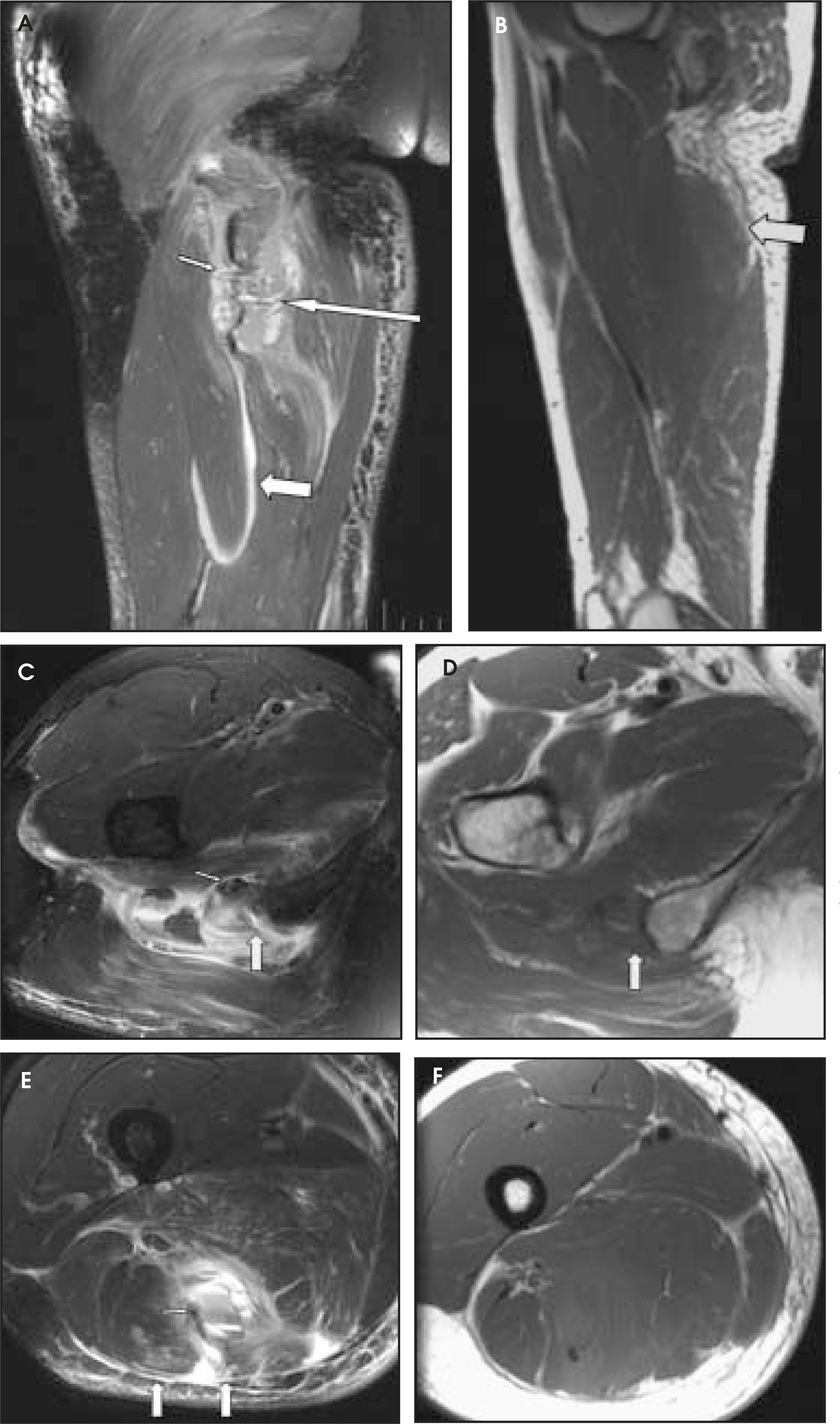
With Grade 1 muscle strain, intramuscular high signal is seen on T2-weighted images without disruption of muscle fibers (Figure 1) and perifascial fluid is seen to track along the intermuscular region. In Grade 2 muscle strains (partial tear), the myotendinous junction is partially torn. The tendon fibers are irregular and thinned with mild laxity. Associated muscle edema and hemorrhage occur with extension along the fascial planes between muscle groups (Figures 2 and 3). A hematoma at the myotendinous junction is pathognomonic for a Grade 2 strain.4 A Grade 3 strain is complete disruption of the myotendinous junction (Figure 4). Evaluation of the injured muscle can be limited because of extensive acute edema and hemorrhage. At times, it may be difficult to determine whether there is a partial or a complete tear. For a complete tear, the margins of the tendon are separated from each other and are irregular; the muscle can retract, and blood and edema can fill the defect between the torn muscle.
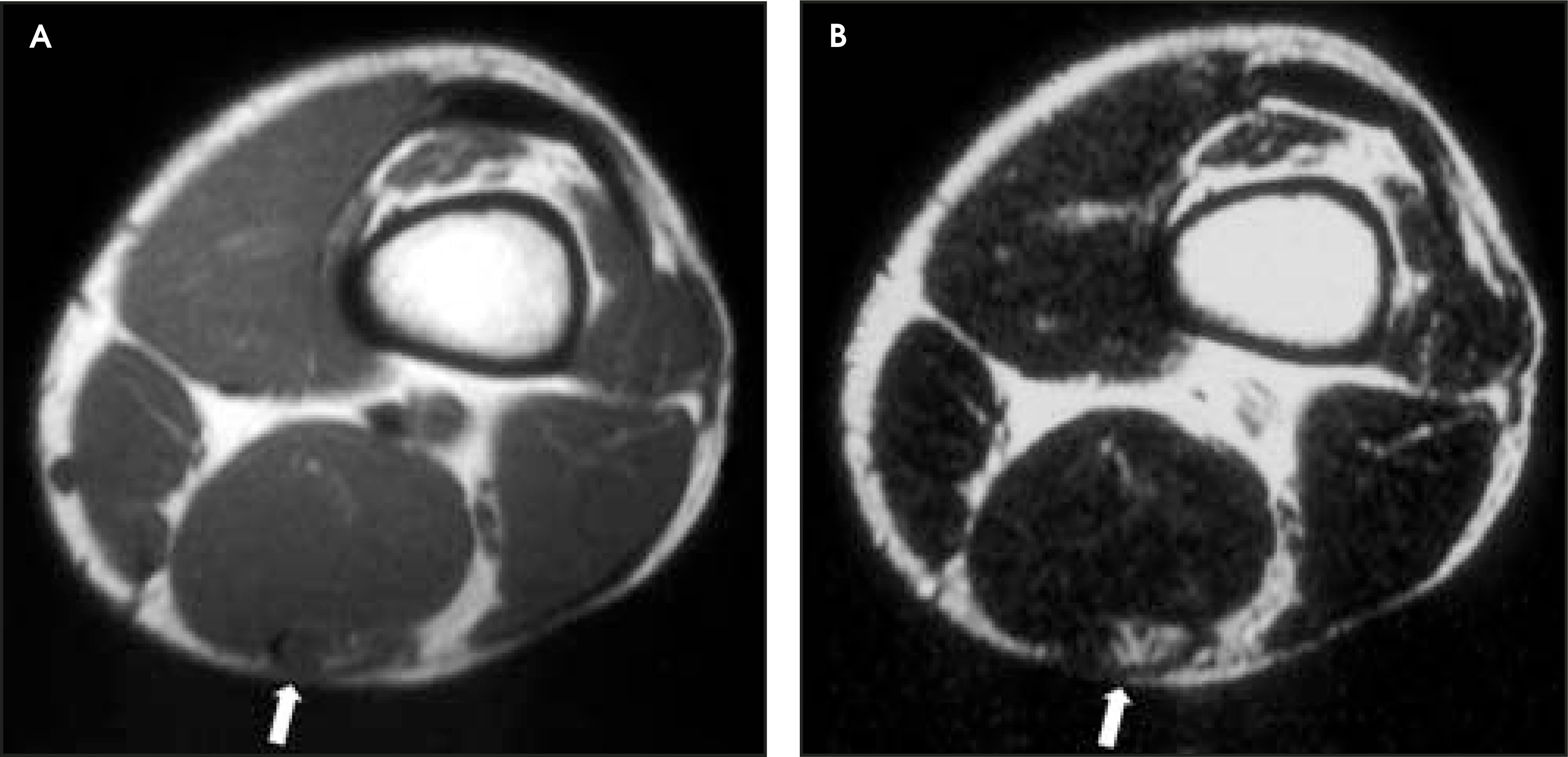
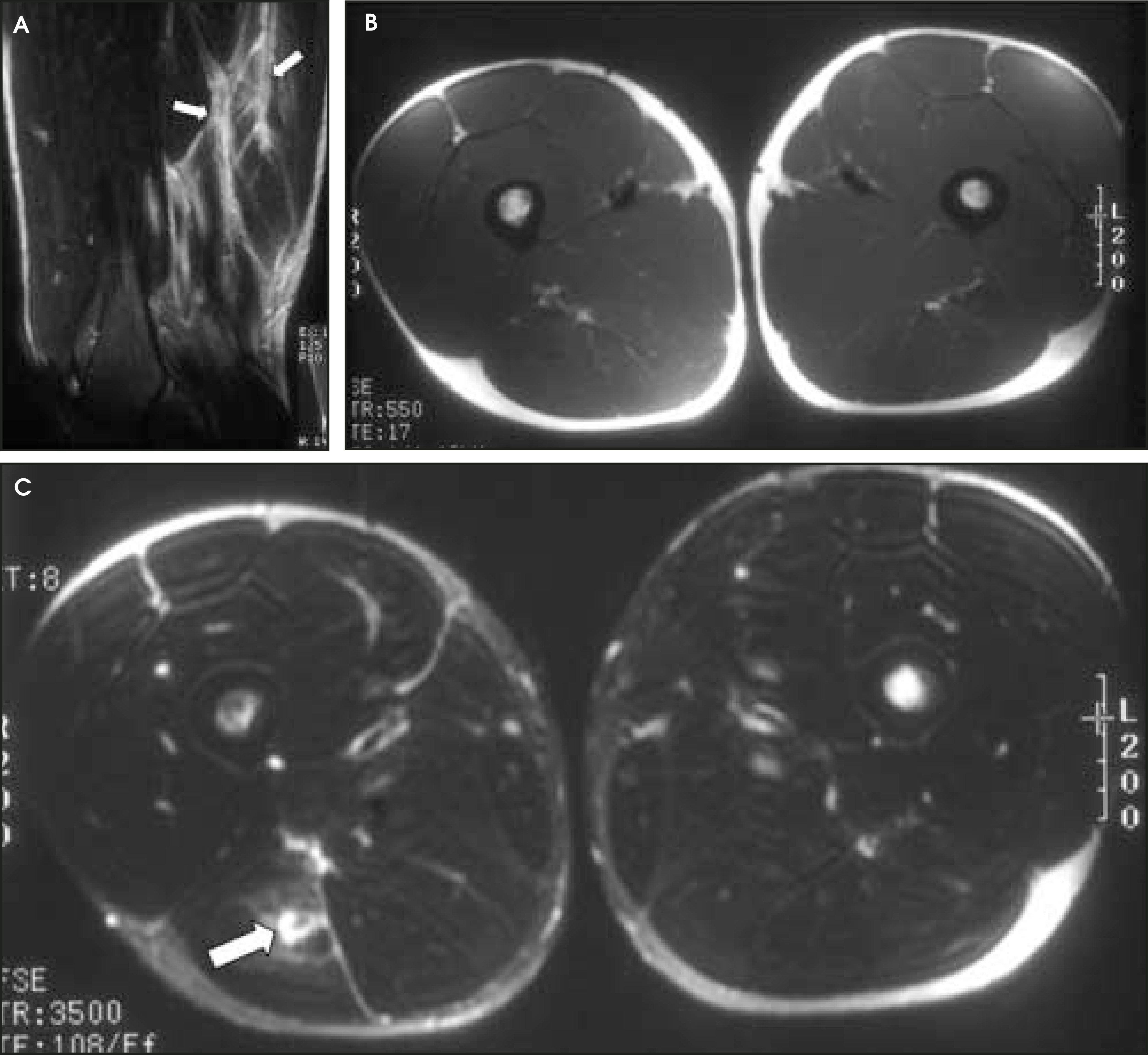
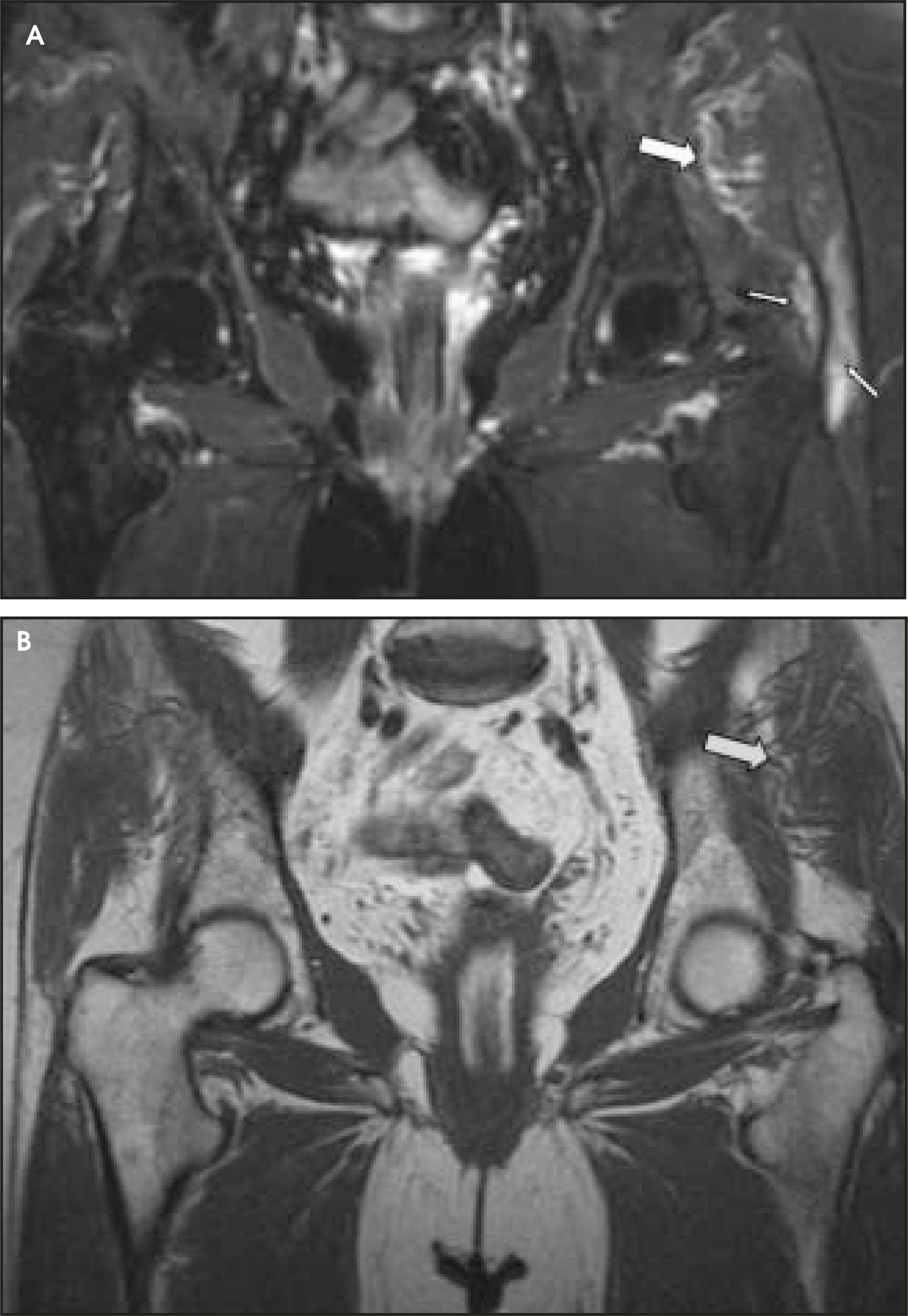
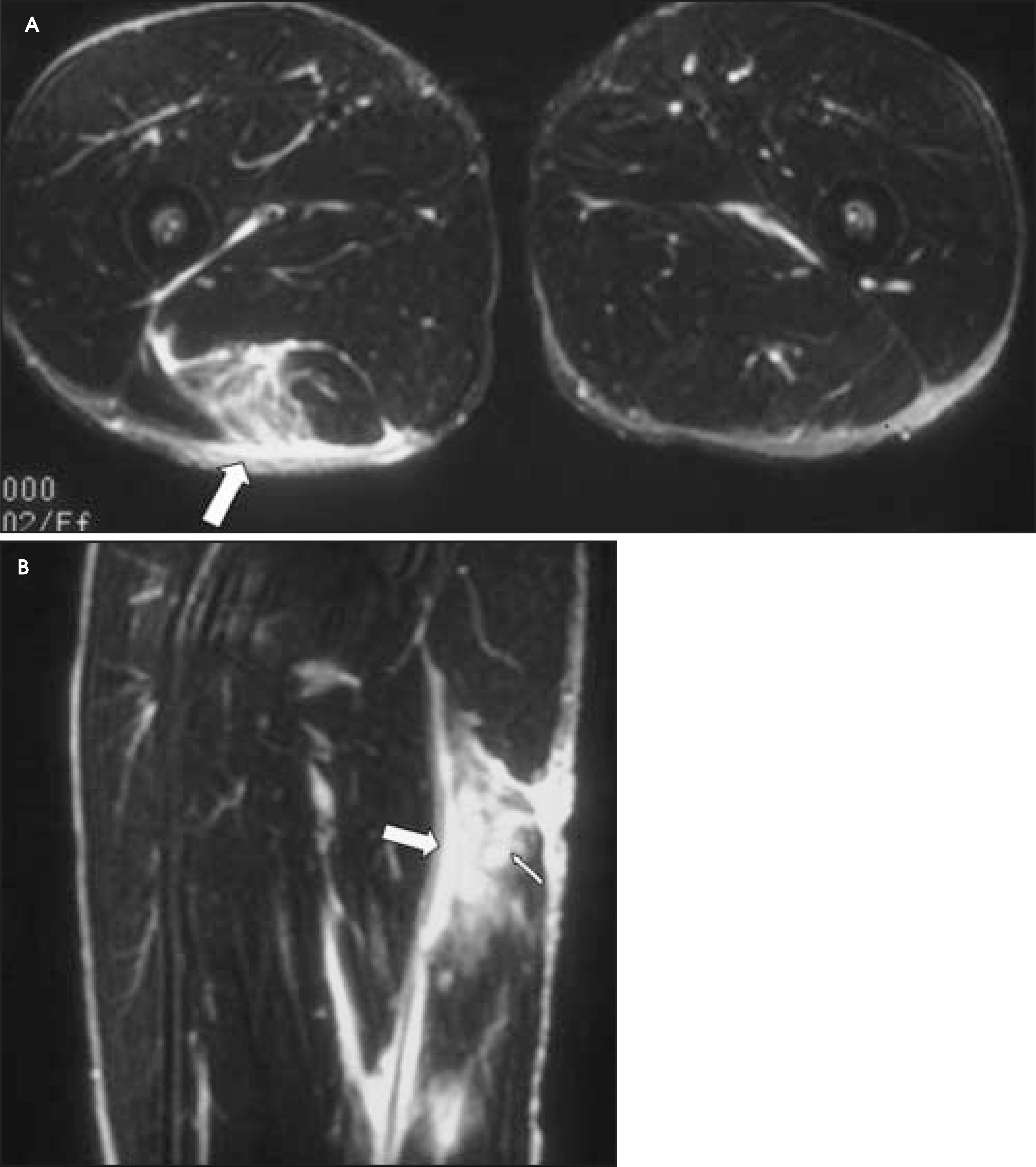
Typical findings of muscle strains are seen in injuries of the hamstrings and pectoralis major muscles. Strains of the hamstrings usually occur during a sudden, forceful accident with maximal contraction of the hamstring muscles while the hip is flexed and the knee is extended.22 In one study, hamstring strains accounted for 50% of the strain injuries in sprinters.8 Strain injuries most often occur at the proximal musculotendinous junction, with the biceps femoris the most commonly injured hamstring.16,18 One devastating common injury to the hamstrings is that often seen in the water-skier. Due to the massive force generated when the boat pulls the skier from the water, there can be avulsion of the hamstring muscle complex at or near the level of the proximal bone-tendon junction, with partial to complete tear of the muscle tendon unit22,23 (Figure 5).
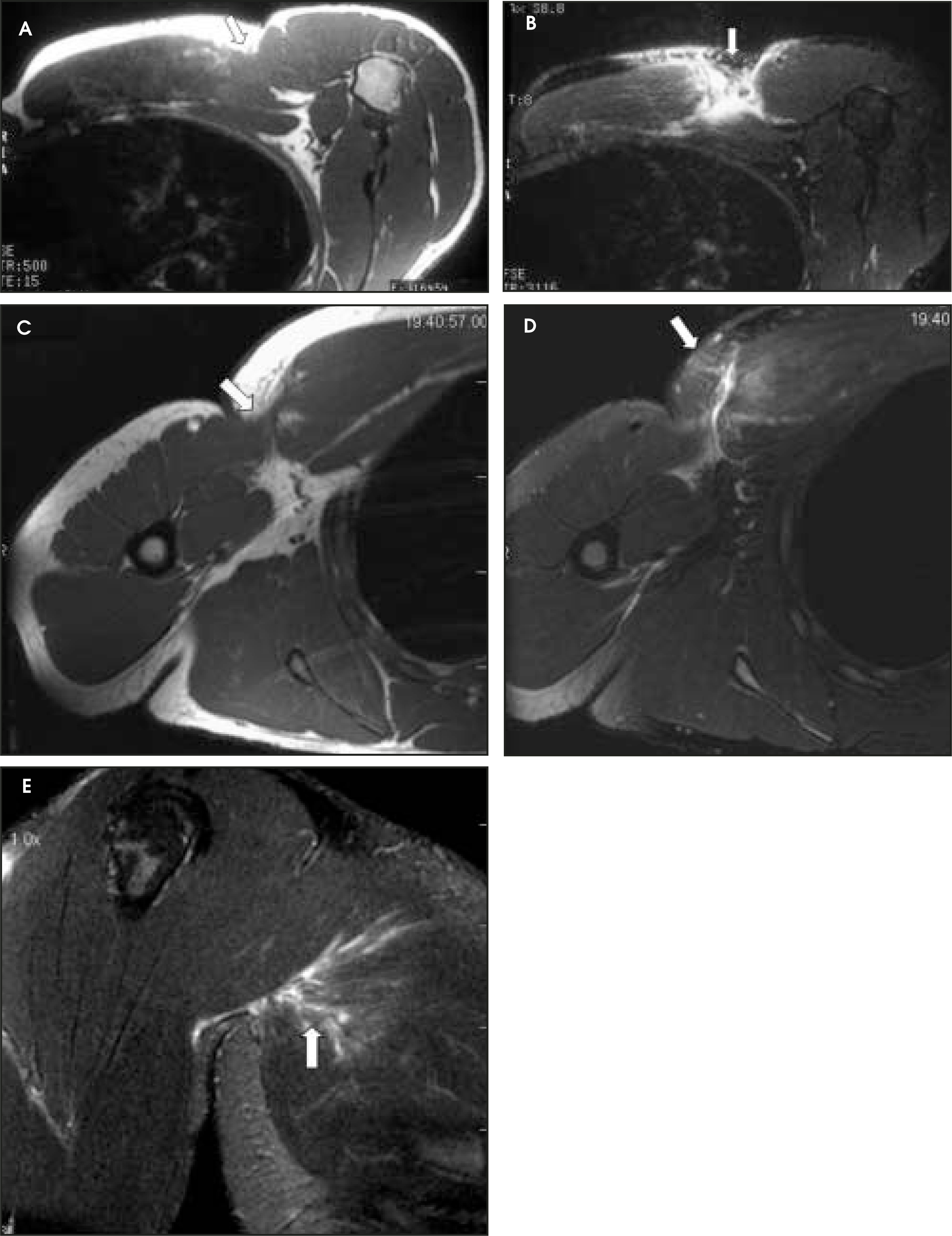
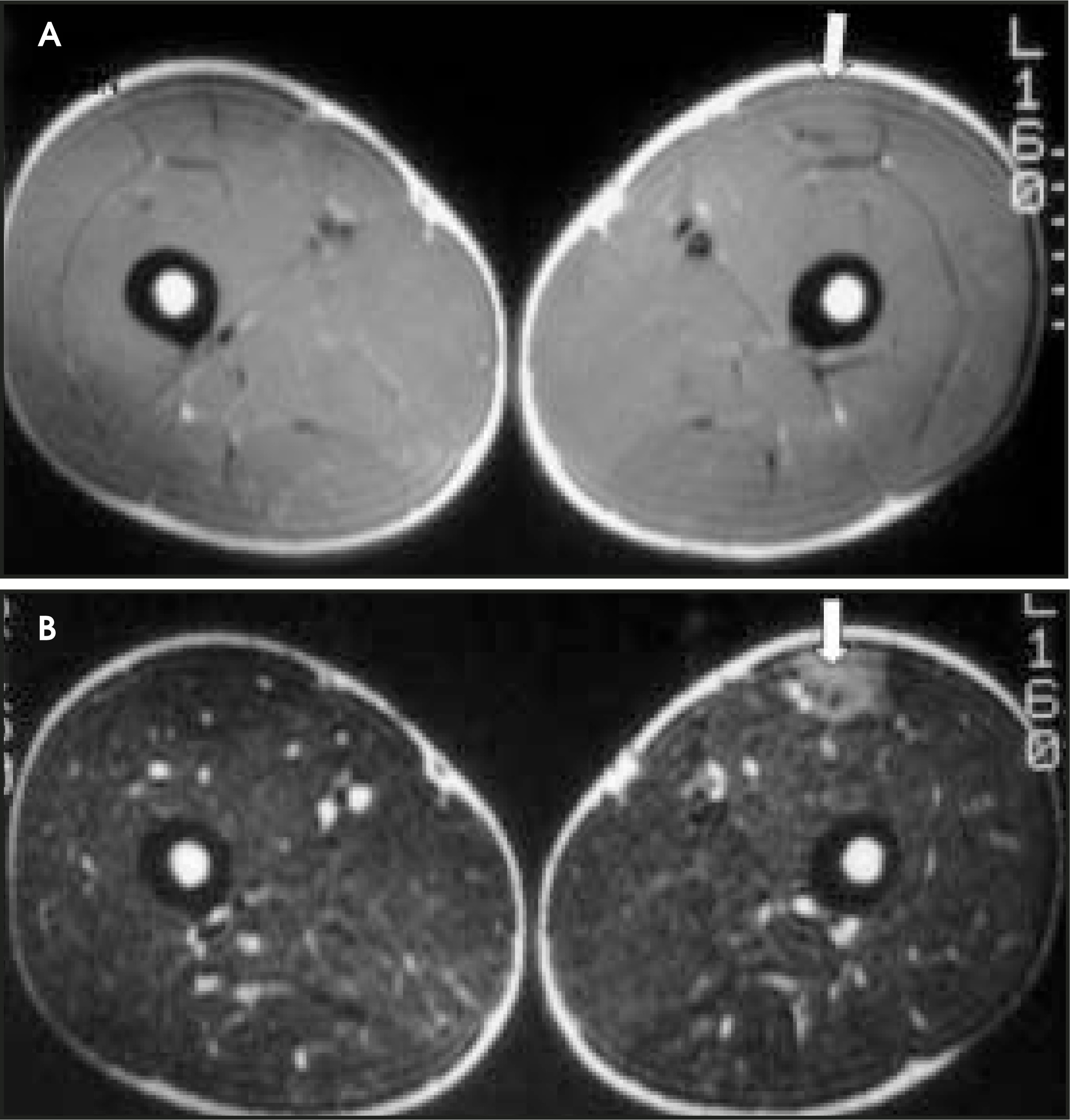
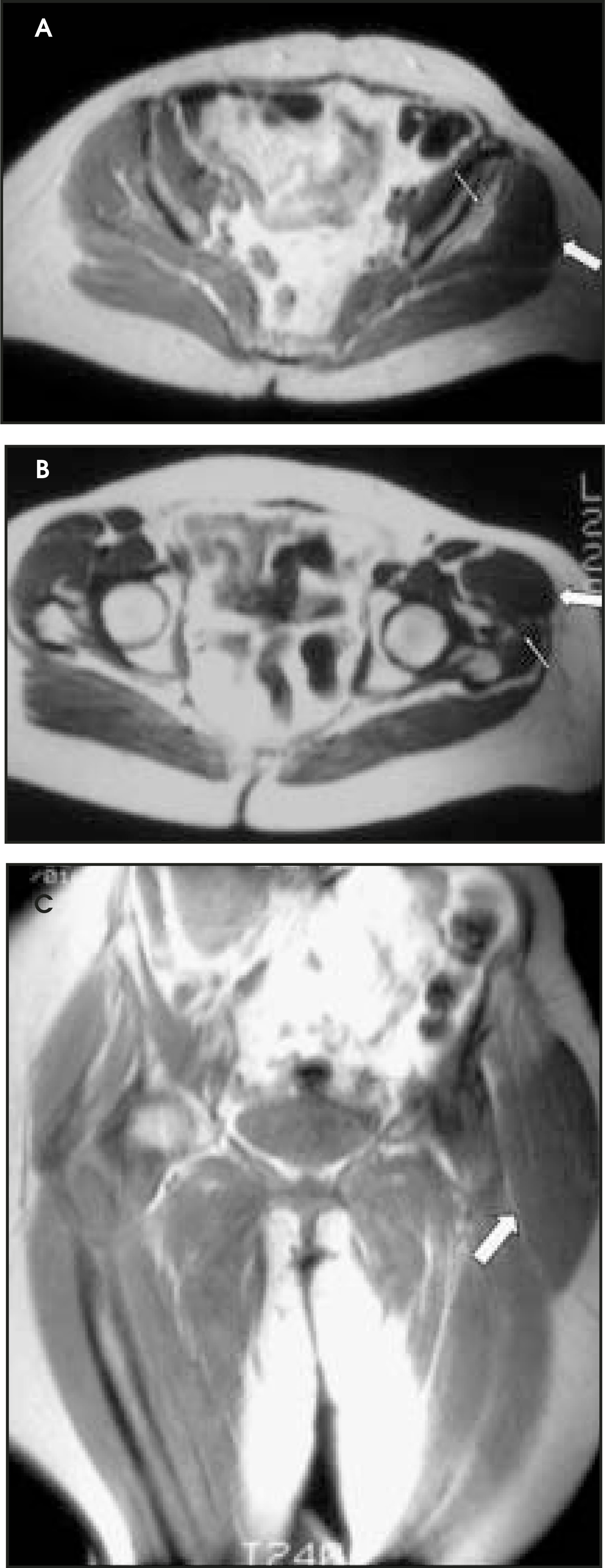
Rupture of the pectoralis major muscle is an uncommon sports-related injury, but it is increasing in frequency24,25 and is most often seen in weight lifters.26 The diagnosis is usually made clinically, although marked edema, pain, and decreased range of motion can make clinical assessment difficult.24 Tears usually occur at the musculotendinous junction or at the insertion onto the humerus (Figure 6). Partial tears at the musculotendinous junction are more common than complete tears, which tend to occur at the tendon-bone interface.25 Imaging in the axial and coronal planes is most useful in detecting pectoralis major injuries. A chronic tear can be detected as a defect at the site of a previous tear. There may be a clinically perceived mass that is isointense with normal muscle and can be detected because of retraction of the torn muscle (Figure 7). A strain typically involves the myotendinous junction, while chronic tears tend to involve the tendon.4 Old injuries can be associated with atrophy or compensatory hypertrophy of adjacent muscles. If the clinical question is to evaluate for a mass and there are typical characteristics seen on MR for an old injury in one muscle, the diagnosis is made without further work-up. Occasionally, MR imaging is ordered to evaluate for a mass following a remote injury. The study may reveal muscle hypertrophy (Figure 8).
Muscle contusion
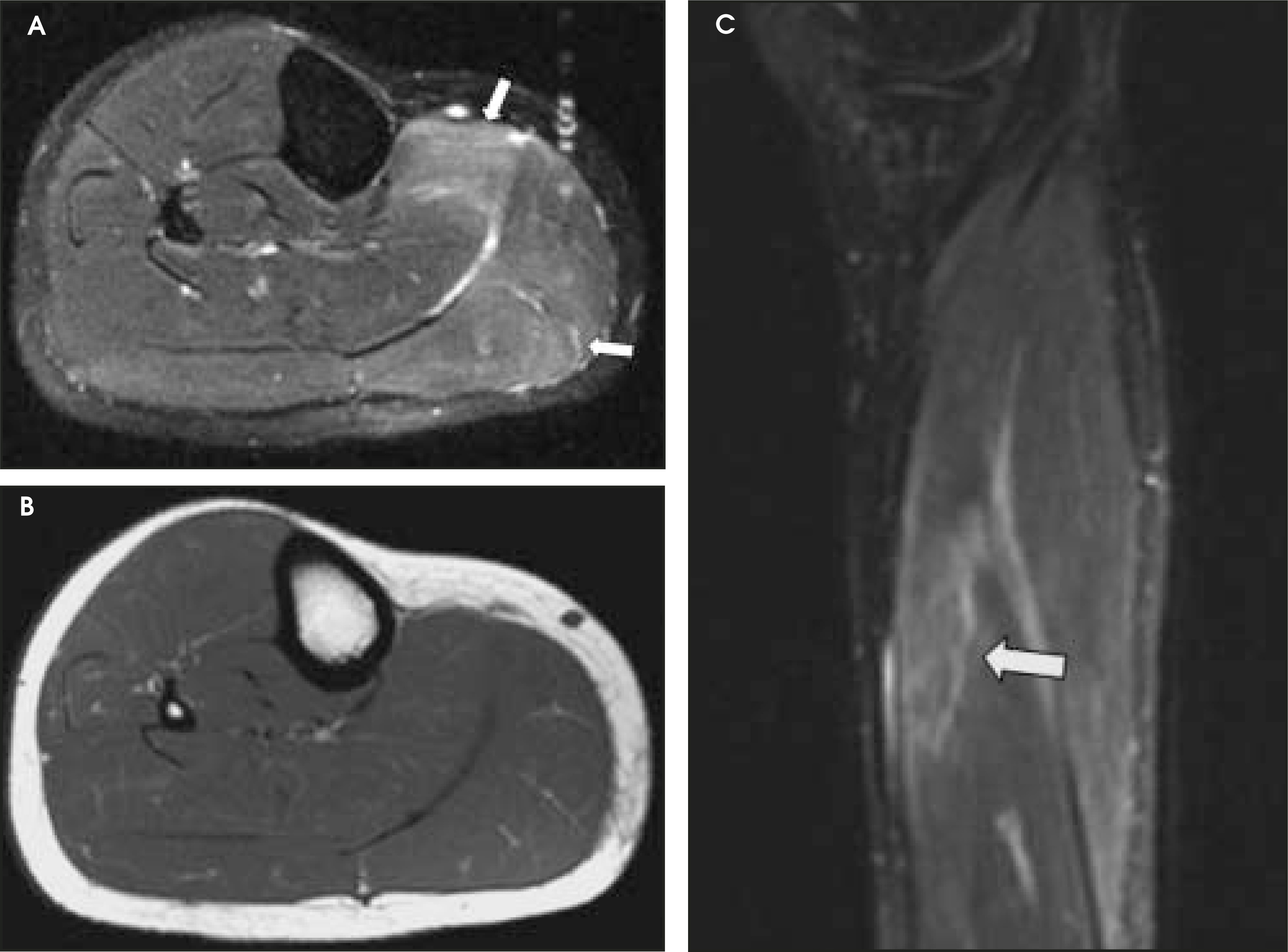
Contusions are common after direct trauma and are differentiated from muscle tears by the patient‘s ability to retain muscle strength. The MR appearance of a muscle contusion may resemble the appearance of a muscle strain (Figure 9). On MR, high T2 signal can be seen within the muscle, representing edema and hemorrhage. Increase in size of the muscle can be seen with the muscle remaining intact. This abnormal area can be isointense to muscle on T1-weighted images, depending on the age of the lesion. The high T2 signal is thought to represent inflammation, edema, and interstitial hemorrhage.3 The hemorrhage can have a feathery, stellate interstitial pattern of increased signal on T2-weighted imaging. These injuries may have the same appearance as Grade 1 and 2 strains. The typical appearance of intramuscular hemorrhage has high signal intensity on both T1- and T2-weighted images secondary to a combination of edema and methemoglobin. After the acute stage, there may be continued high signal on T1 images, which has been described as being secondary to recurrent hemorrhage.27
Muscle hematoma
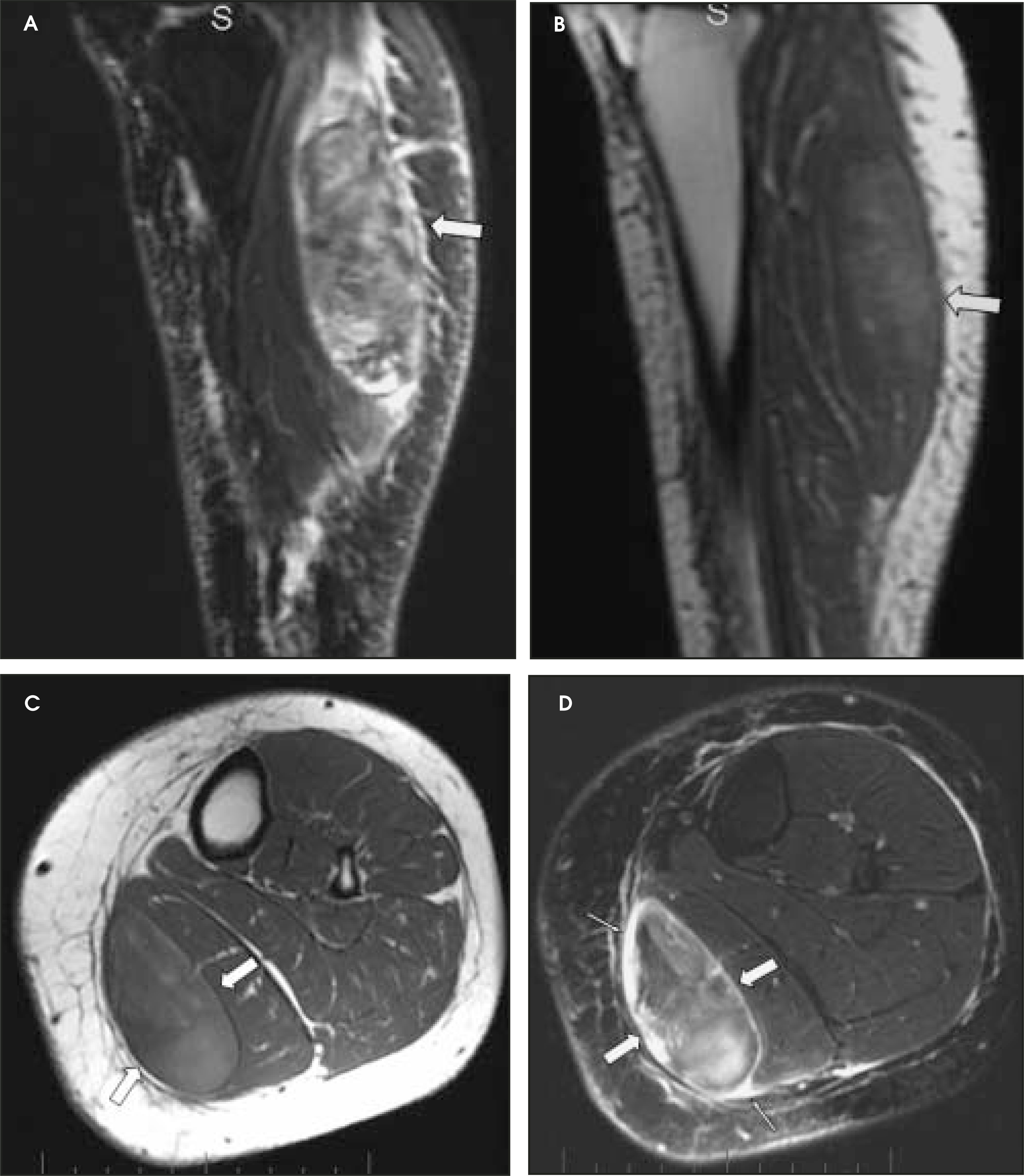
A hematoma, which is a collection of blood within a confined space, can develop within a muscle due to direct or indirect injury2,5,20 (Figure 10). Muscular hema-tomas are usually well-defined lobulated masses confined to a single muscle.6 Occasionally, surgical drainage may be needed because of severe pain from the soft-tissue compression caused by a large hematoma. The MR imaging findings of a hematoma are that of a well-defined increased mass within the muscle, often associated with an increase in muscle bulk, edema, or hemorrhage. The TR of hemorrhage is affected by the concentration of protein, methemoglobin, magnetic susceptibility at high field strength, and tissue clearance.6 Acute hematomas, <48 hours, will us ally be isointense on T1-weighted images. Subacute hematoma, <30 days, will have higher signal intensity than muscle on both T1- and T2-weight-ed images12,20 because of accumulation of methemoglobin. As the hematoma evolves, there is a wide range of MR signal intensity within the hematoma, depending on the age of the degradation products. In chronic hematomas, there is often a dark signal intensity rim secondary to hemosiderin. A seroma may develop if there is complete resorption of blood products.4,12
Muscle herniation
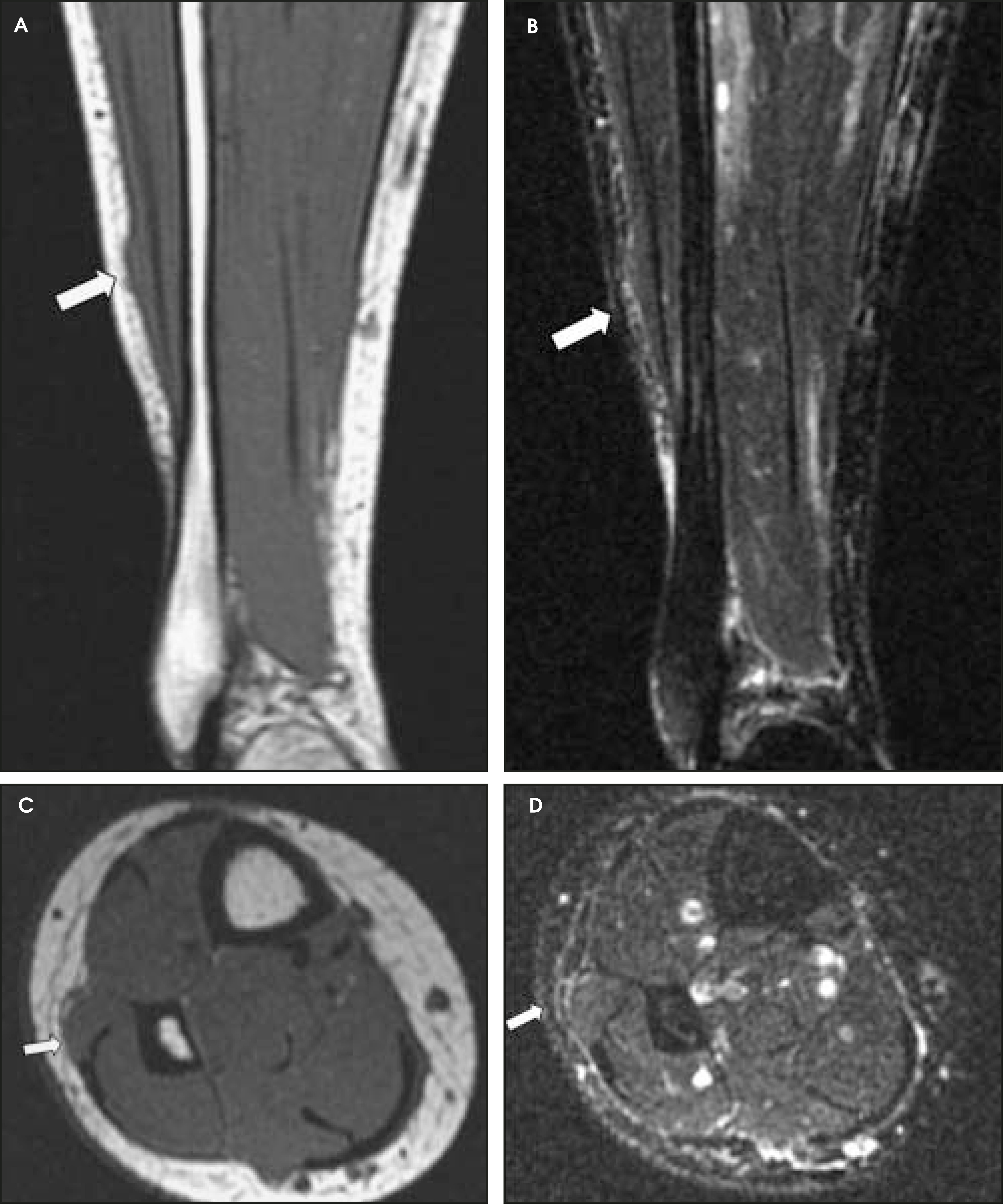
Muscle herniations are an uncommon complication of muscle injury. The lower extremity is most often involved in herniations and the tibialis anterior is the most frequently involved muscle.3,4,6,27-29 The most common cause of herniation is blunt trauma that produces a rent within the fascia through which the adjacent muscle can protrude.29 Another cause is muscle hypertrophy due to exercise, which can produce stretching of the vascular fenestrations of the fascia. The muscle can then herniate through these fenestrations.6,29 Usually this diagnosis is made clinically, but occasionally the findings on physical examination are equivocal and the patient is referred for imaging. A patient may complain of a mass and exertional pain at the site of injury weeks following an injury.3 If the herniation is small, pain may result only when the patient is actively exercising. MR imaging is superior to computed tomography for assessment of a muscle herniation because MR is able to distinguish the differing signal characteristics of the muscle and adjacent fascia. The fascial defect may not always be identified during MR imaging with the muscle relaxed. In this situation, dynamic imaging using short MR scans during muscle contraction may define the muscle herniation3,4,6,27-29 (Figure 11).
Conclusion
Muscle injuries are common and are usually diagnosed and treated without imaging. However, MR has a role in identifying muscle injuries, in determining the site and severity of injury, and in evaluating for complications, such as large hematomas or muscle herniation. The most important part of proper imaging of muscle injuries is having a careful clinical history and physical examination. The typical MR location and characteristic imaging features, in combination with the clinical history, should allow accurate diagnosis of a muscle injury.
References
- Ehman R, Berquist T. Magnetic resonance imaging of musculoskeletal trauma. Radiol Clin N Am.. 1986;24:291-319.
- Shellock F, Mink J, Deutsch A. MR imaging of muscle injuries. Appl Radiol. 1994;(2):11-16.
- Tuite M, De Smet A. MRI of selected sports injuries: Muscle tears, groin pain, and osteochondritis dissecans. Semin Ultrasound CT MR. 1994;15:318-340.
- El-Khoury G, Brandser E, Kathol M. Imaging of muscle injuries. Skeletal Radiol. 1996;25(1):3-11.
- Palmer W, Kuong S, Elmadbouh H. MR imaging of myotendinous strain. AJR Am J Roentgenol. 1999;173:703-709.
- Steinbach L, Fleckenstein J, Mink J, Weissman B. MR imaging of muscle injuries. Syllabus: ACategorical Course in Musculoskeletal Radiology. 1993:225-237.
- Garrett W. Muscle strain injuries. Am J Sports Med. 1996;24(6 suppl):S2-S8.
- Agre J. Hamstring injuries. Proposed aetiological factors, prevention, and treatment. Sports Med. 1985;2(1):21-33.
- Verrall G, Slavotinek J, Barnes P. Clinical risk factors for hamstring muscle strain injury: A prospective study with correlation of injury by magnetic resonance imaging. Br J Sports Med. 2001;35:435-439.
- Fleckenstein J, Weatherall P, Parkey R. Sports-related muscle injuries: Evaluation with MR imaging. Radiology. 1989;172:793-798.
- De Smet A, Best T. MR imaging of the distribution and location of acute hamstring injuries in athletes. AJR Am J Roentgenol. 2000;174:393-399.
- Nguyen B, Brandser E, Rubin D. Pains, strains and fasciculations. MRI Clin N Am. 2000;8:391-408.
- Varela J, Rodriguez E, Soler R. Complete rupture of the distal semimembranosus tendon with secondary hamstring muscles atrophy: MR findings in two cases. Skeletal Radiol. 2000;29:362-364.
- Fleckenstein J, Shellock F. Exertional muscle injuries: Magnetic resonance imaging evaluation. Top Magn Reson Imaging. 1991;3(4):50-70.
- Rubin S, Feldman F, Staron R. Magnetic resonance imaging of muscle injuries. Clin Imaging. 1995;19:263-269.
- Kujala U, Orava S, Jarvinen M. Hamstring injuries. Current trends in treatment and prevention. Sports Med. 1997;23:397-404.
- Speer K, Lohnes J, Garrett W. Radiographic imaging of muscle strain injury. Am J Sports Med. 1993;21:89-95.
- Brandser E, el-Khoury G, Kathol M. Hamstring injuries: Radiographic, conventional tomographic, CT, and MR imaging characteristics. Radiology. 1995;197:257-262.
- Garrett W, Rich F, Nikolaou P. Computed tomography of hamstring muscle strains. Med Sci Sports Exerc. 1989;21:506-514.
- Deutsch A, Mink J. Magnetic resonance imaging of musculoskeletal injuries. Radiol Clin North Am. 1989;27:983-1002.
- De Smet A, Fisher D, Heiner J. Magnetic resonance imaging of muscle tears. Skeletal Radiol. 1990;19:283-286.
- Orava S, Kujala U. Rupture of the ischial origin of the hamstring muscles. Am J Sports Med. 1995;23:702-705.
- Sallay P, Friedman R, Coogan P. Hamstring muscle injuries among water skiers. Functional outcome and prevention. Am J Sports Med. 1996;24:130-136.
- Lee J, Brookenthal K, Ramsey M. MR imaging assessment of the pectoralis major myotendinous unit: An MR imaging-anatomic correlative study with surgical correlation. AJR Am J Roentgenol. 2000;174:1371-1375.
- Connell D, Potter H, Sherman M. Injuries of the pectoralis major muscle: Evaluation with MR imaging. Radiology. 1999;210:785-791.
- Miller M, Johnson D, Fu F. Rupture of the pectoralis major muscle in a collegiate football player. Use of magnetic resonance imaging in early diagnosis. Am J Sports Med. 1993;21:475-477.
- De Smet A. Magnetic resonance findings in skeletal muscle tears. Skeletal Radiol. 1993;22:479-484.
- Mellado J, Palomar L. Muscle hernia of the lower leg: MRI findings. Skeletal Radiol. 1999;28:465-469.
- Zeiss J, Ebraheim N, Woldenberg L. Magnetic resonance imaging in the diagnosis of anterior tibialis muscle herniation. Clin Orthop. 1988;244:249-253.
Citation
. MR imaging of muscle injuries. Applied Radiology. 2004;33(4):14-26. doi:10.37549/AR1235.