MR imaging of the hip and pelvis: Current concepts
Applied Radiology — Vol. 32 , Issue 6 , pp. 14 -21
DOI: 10.37549/AR1189
Published: June 1, 2003
Categories
Traditionally, magnetic resonance (MR) imaging of the hip has been primarily used for evaluation of fractures and osteonecrosis; however, there is increasing appreciation of the utility of MR in diagnosing other musculoskeletal pathology. This article will review the more established indications for MR of the hip along with their pertinent findings, followed by a discussion of other abnormalities that we encounter with increasing frequency.
Vascular abnormalities
One of the well-known indications for MR imaging of the hip is for detection of vascular insults to the femoral head, the most common of which is avascular necrosis (AVN). In the early stages of this process, bone scans and radiographs may be normal.1 The MR appearance of AVN includes diffuse bone marrow edema, which becomes more focal in the femoral head with a central area of fatty marrow surrounded by a serpiginous line of low signal between the 10 o’clock and 2 o’clock positions (Figure 1).2 Less commonly, the “double-line sign” of a low signal area surrounding an inner line of high signal on T2-weighted images is seen. The stages of AVN visible on conventional radiography do not parallel those seen on MR imaging.3 MR is recommended when AVN is suspected clinically and the conventional radiographs are normal, and in patients with known AVN in one hip to assess for contralateral involvement, which is not an infrequent occurrence. Treatment includes core decompression, rotational osteotomy, and free vascularized fibular grafting.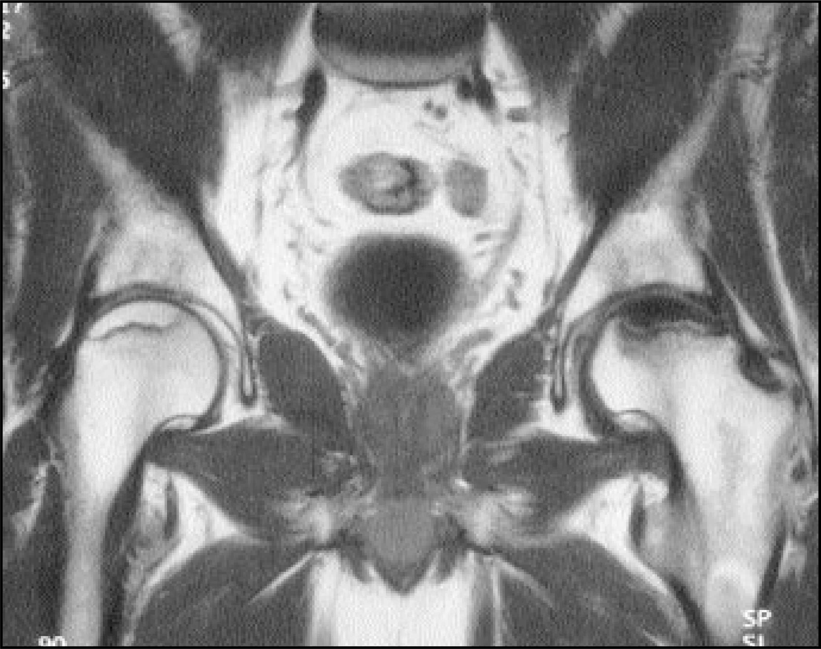
The current etiologic theory of idiopathic transient osteoporosis of the hip (ITOH) is that it is caused by a vascular insult.4 This abnormality has also been called transient painful bone marrow edema. Although more common in middle-aged men, it also can occur in pregnant women. Patients present with severe pain but no history of trauma.5 Conventional radiographs may show osteopenia, while MR imaging shows low T1 signal and high T2 signal in the femoral head extending to the inter-trochanteric region (Figure 2). There is often an associated moderate joint effusion. This pattern is nonspecific and can mimic early AVN and infection but resolves spontaneously, generally in 6 to 8 months, thus allowing distinction between AVN and ITOH. A follow-up MR is occasionally necessary to make the diagnosis.6-8 This process can also migrate to other joints. Treatment is protective weight-bearing.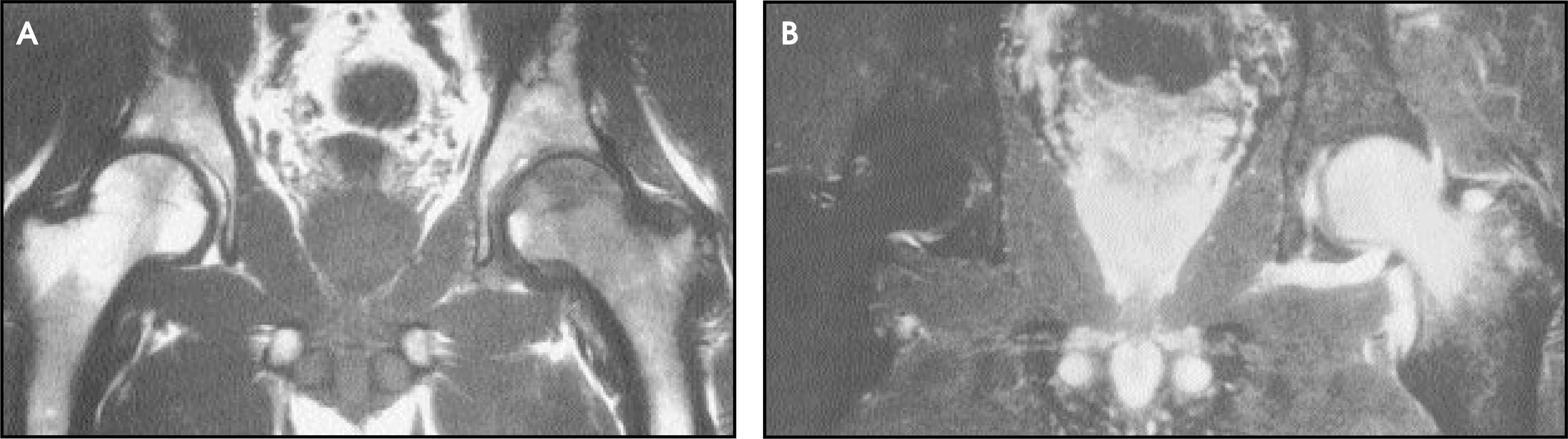
Fractures
Stress fractures and insufficiency fractures have been reported in all bones around the pelvis. Often, the initial radiographs are normal and, therefore, MR imaging is strongly indicated if there is suspicion of fracture, as it is sensitive and specific for identifying stress fractures. These fractures should be diagnosed in a timely fashion, especially when the femoral neck is involved, as patients should be protected to prevent progression to complete fracture, which can occur with continued unlimited weight-bearing. In such cases, MR imaging shows a linear low signal fracture line on T1- and T2-weighted images with associated surrounding edema (Figure 3).9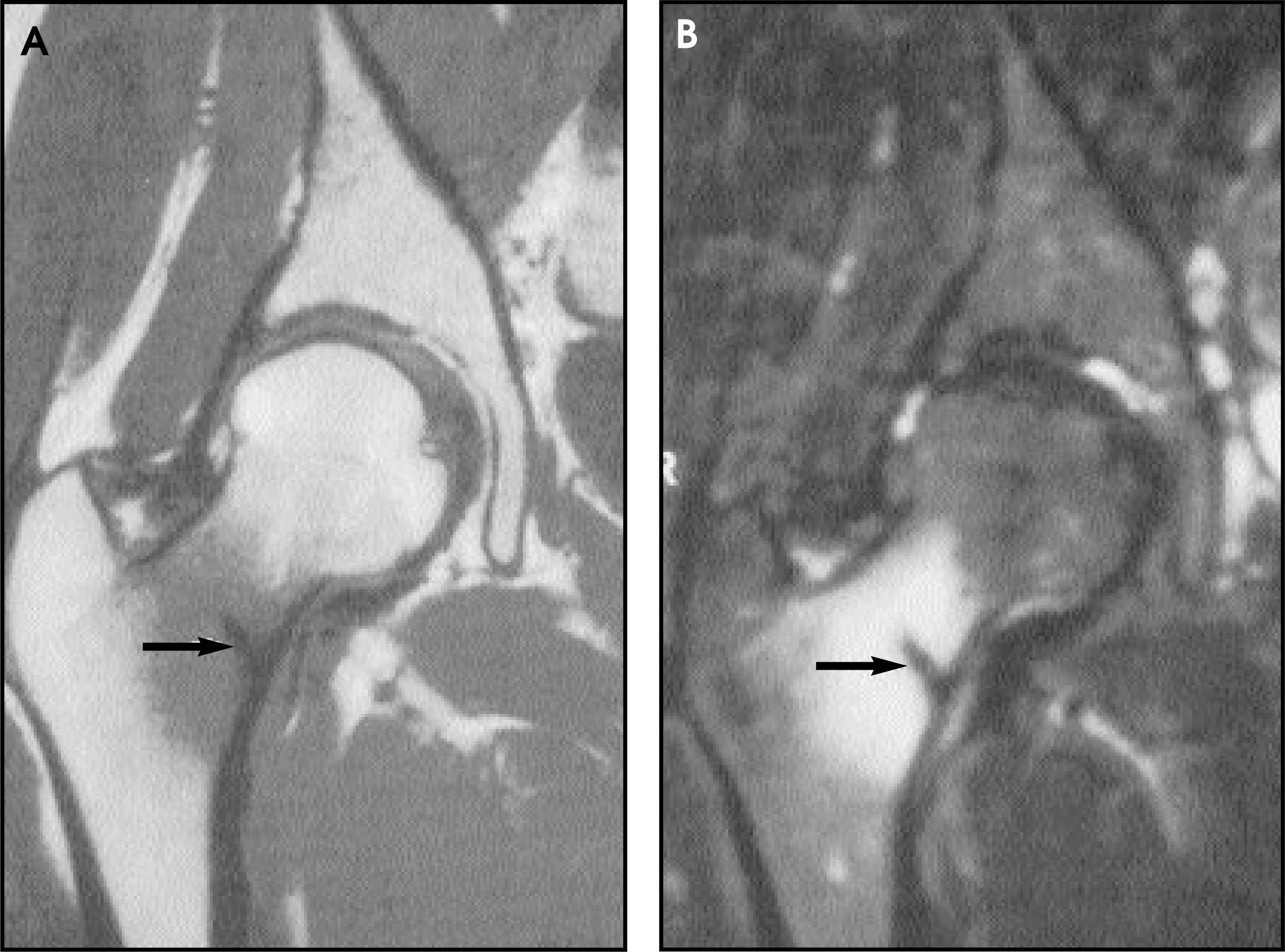
Sacral stress fractures have recently been described in long-distance runners. The patient’s symptoms mimic that of disk disease, for which they are often erroneously treated. Findings on MR examination show low signal on T1-weighted images and high T2 signal paralleling the sacroiliac joint (Figure 4). The treatment is rest with no further running until symptoms have resolved.10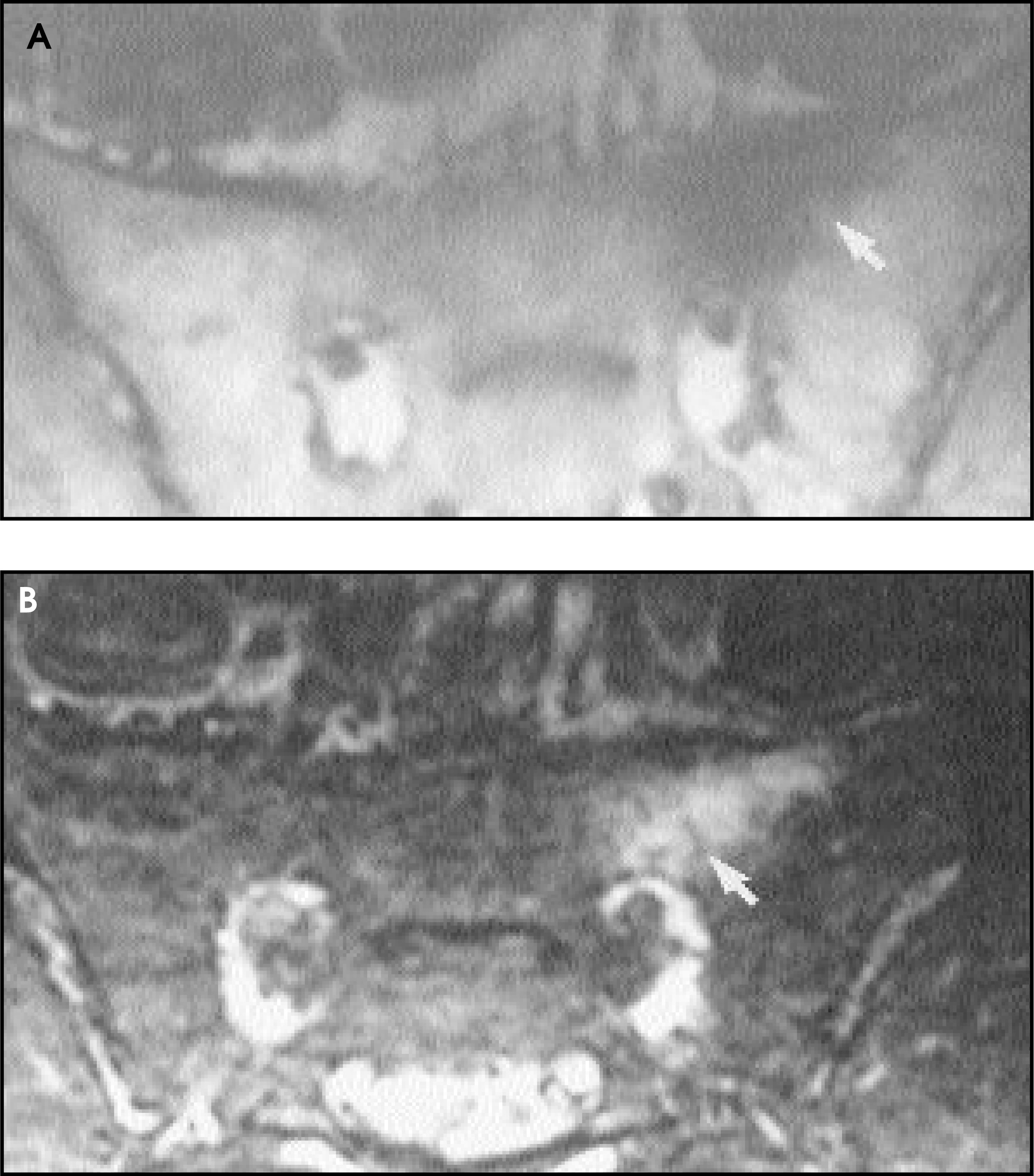
Insufficiency fractures are most common in patients with osteoporosis and those who have undergone radiation therapy. These fractures can be radiographically occult. Since the patient’s bones are osteopenic, even incidental trauma can result in a fracture. Therefore, lack of history of trauma should not dissuade physicians from considering fracture as a possible explanation for hip pain. Similarly, since weight-bearing is sometimes tolerated by the patient when the fracture is nondisplaced, a history of “weight-bearing” can be misleading. Bone scintigraphy was formerly the imaging modality of choice to diagnose radiographically occult insufficiency fractures, with delayed imaging often necessary in these osteopenic patients. With MR imaging, the fracture line is readily visible as linear low signal on T1- and T2-weighted images with surrounding edema (Figure 5).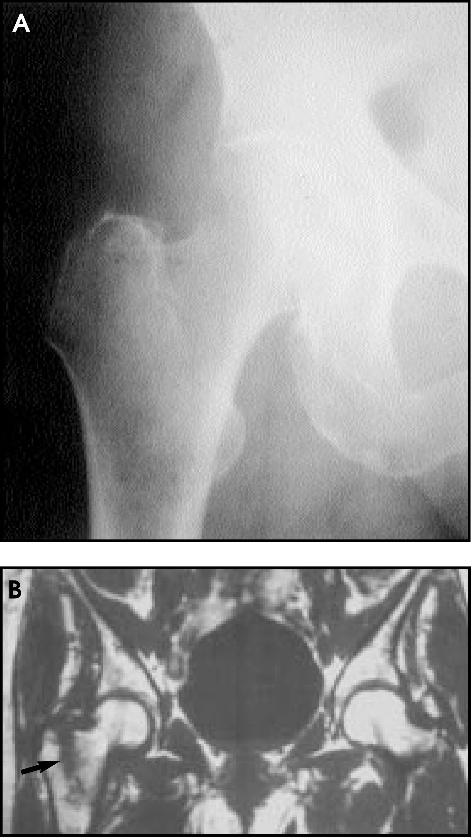
The most common locations for insufficiency fractures of the pelvis are subcapital, intertrochanteric, sacral, supra-acetabular, and pubic bones; often multiple fractures co-exist. Supra-acetabular insufficiency fractures are diagnosed by recognition of a curvilinear low signal fracture line that parallels the acetabular roof with adjacent edema, while sacral insufficiency fractures show the low signal fracture line paralleling the sacroiliac joint (Figures 6 and 7).11 Most commonly, the adjacent edema in sacral fractures does not cross the midline unless the fracture is bilateral. This can mimic metastatic disease when edema without a distinct fracture line is present. A search for surrounding soft-tissue extension and bone destruction supports a pathologic process.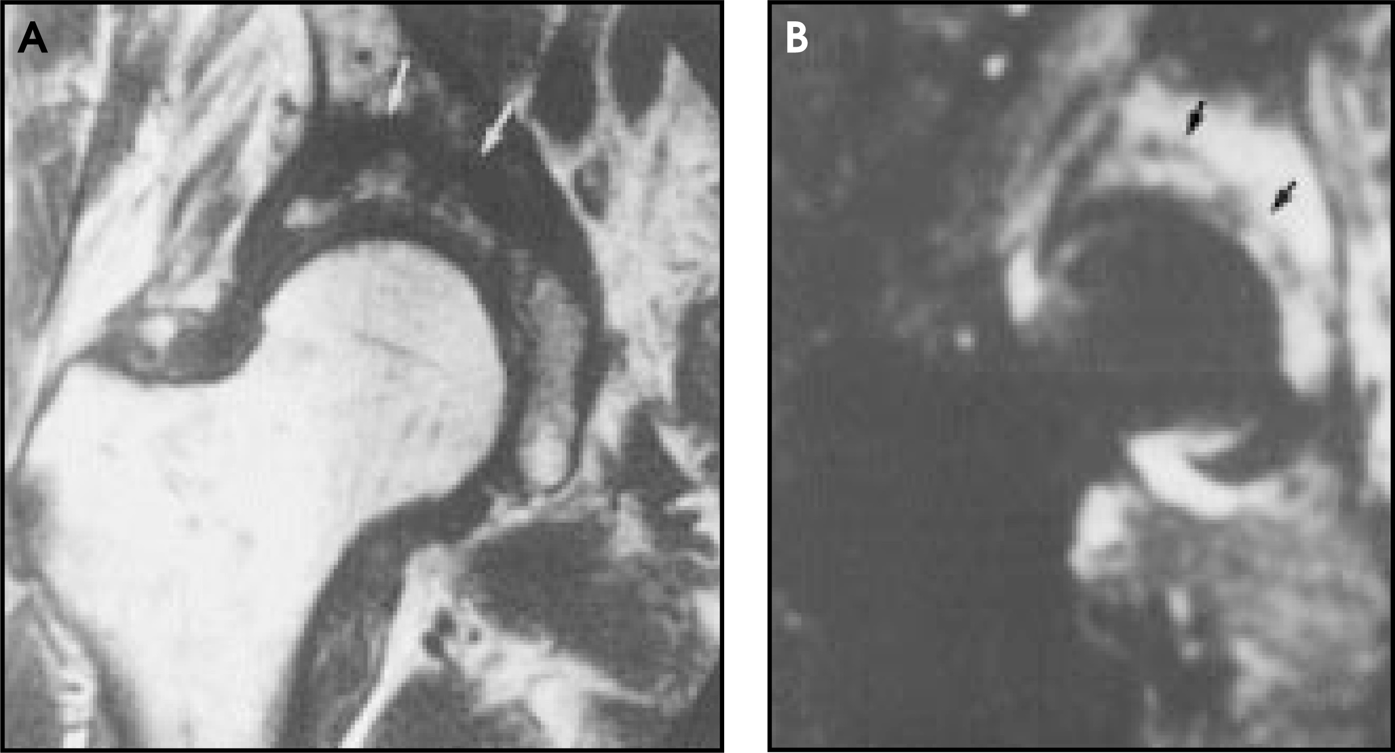
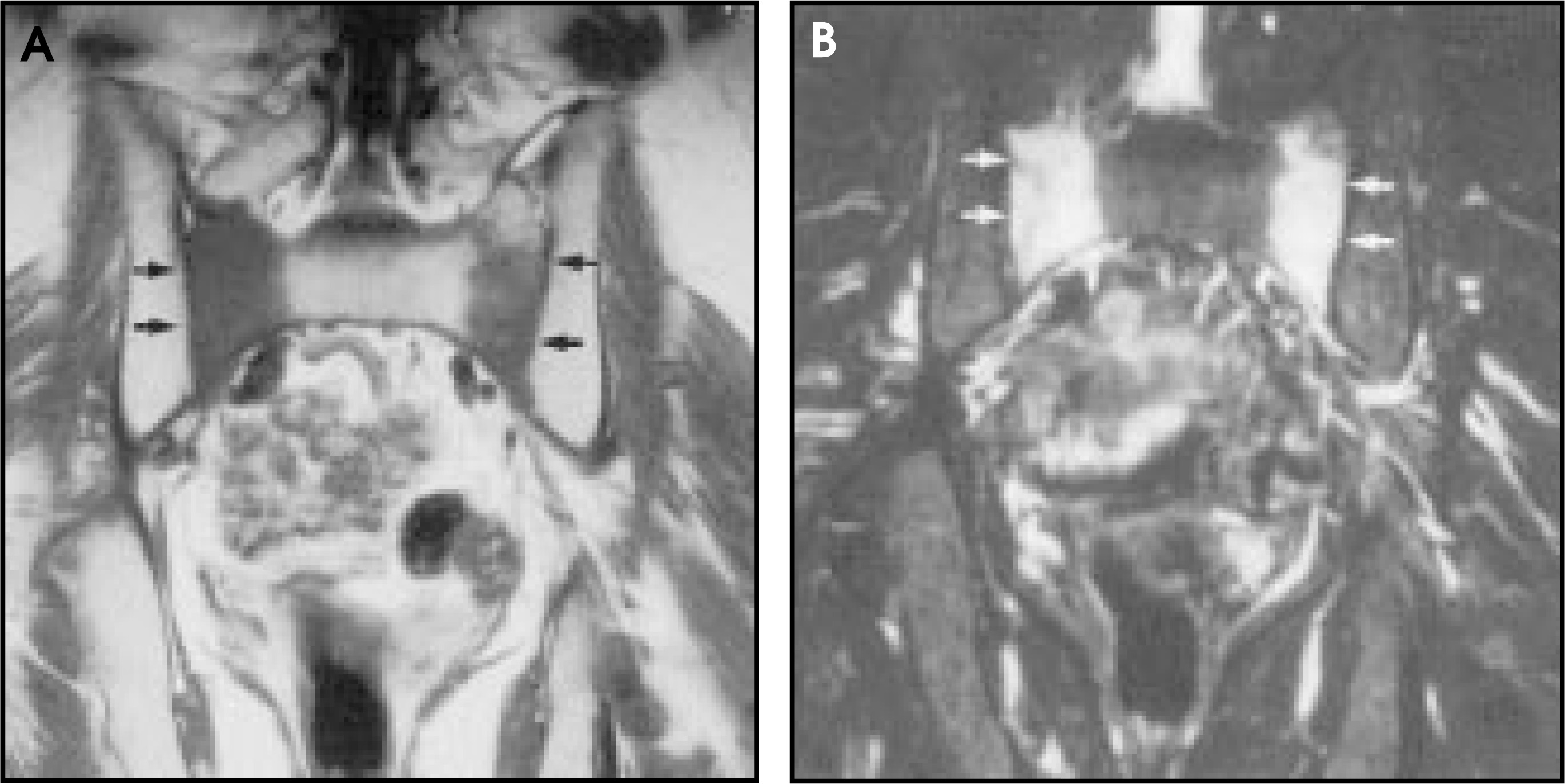
Salter-Harris fractures can occur at the hip and are most common in adolescence. Slipped capital femoral epiphysis (SCFE) affects males more often than females and has a higher incidence in overweight children. Radiographs may be equivocal and MR can be utilized to show the widened physis with increased signal on T2-weighted images as well as the medial and posterior displacement of the femoral head. Early AVN,which affects 15% of children with this process, can also be detected and can be a result of the injury to the physis during the Salter injury or as a result of the treatment.
Herniation pits
Herniation pit is a commonly seen aperture in the cortex of the femoral neck along its anterior-lateral surface. It is the result of ingrowth of fibrocartilaginous elements through a perforation in the cortex. Herniation pits can change in size due to mechanical forces and can be unilateral or bilateral, but are asymptomatic. MR shows a well-defined focus of low signal on T1-weighted images and increased signal on T2-weighted images in the typical anterolateral location.12
Soft tissues
Muscle/tendon abnormalities
MR imaging can clearly demonstrate occult fractures in the setting of pain, but can also reveal soft-tissue abnormalities related to the muscles and tendons of the pelvis when conventional radiography is normal. Muscle strains, hematomas, and tendon tears can account for pain when no fracture is seen.13
Muscle strains
The most common muscle strain at the pelvis involves the hamstrings, which originate from the ischial tuberosity. The quadriceps and adductor muscles can also be injured but less frequently. MRI findings in muscle strain show muscle enlargement on T1 with loss of normal intervening fat and possible hemorrhage, while T2-weighted images show a feathery interstitial increased signal from edema and hemorrhage with perifascial edema (Figure 8).14 Complete tears are readily diagnosed with MR imaging by noting the free edge of the tendon.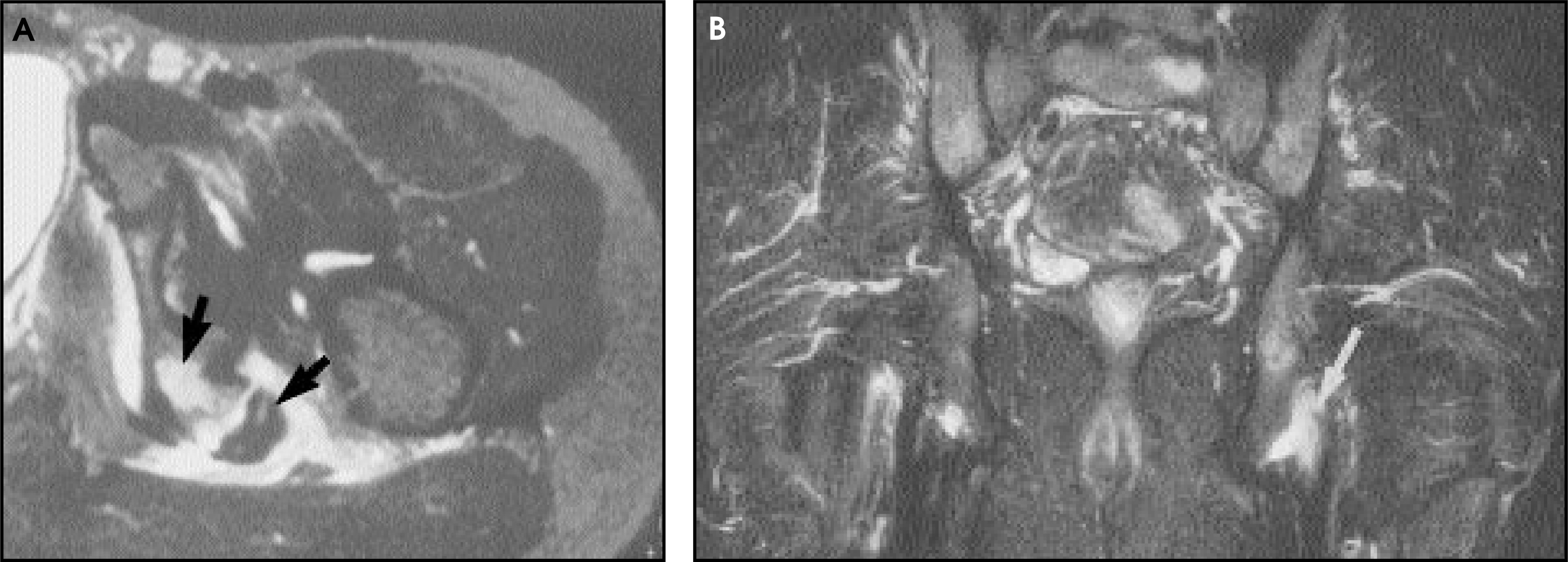
Gluteus medius tendon tears
Gluteus medius tendon tears have also been referred to as greater trochanter pain syndrome, as patients complain of chronic pain around the hip or groin, which can mimic intra-articular hip pathology. This entity is most often seen in older women, but can be seen in men also. This is associated with tears or tendinopathy of the gluteus medius or minimus and their associated muscles. MR imaging shows high signal in the muscles on T2-weighted images with increased signal in the tendons, which may be discontinuous or avulsed from the greater trochanter. Signal is isointense to musculature on T1-weighted images and is difficult to appreciate as abnormal.15,16
Piriformis syndrome
The sciatic nerve is intimately associated with the piriformis muscle and usually lies just anterior to the muscle. Variations do exist and the nerve can split through the muscle, or the nerve can split, with only a portion going through the muscle and a portion remaining superficial to the muscle belly. Because of these variations, compression, hypertrophy, or injury to the piriformis muscle can cause irritation of the sciatic nerve, mimicking radicular symptoms from disc pathology. MR can show asymmetry in muscle size, without abnormal signal in the muscle itself unless there has been trauma, in which case high signal on T2-weighted images may be seen due to edema and hematoma.
Bursae
The largest bursa in the body is the iliopsoas bursa, which is located anterior to the hip joint and adjacent to the femoral vessels and iliopsoas muscle. It is not seen unless it is distended by fluid, in which case it is identified as a well-defined area of low signal on T1-weighted images and increased on T2-weighted images immediately anterior to the iliopsoas muscle. After administration of gadolinium, the fluid does not enhance but the bursal lining does, thus differentiating this from a solid soft-tissue mass, which would enhance diffusely. If bursae are distended, they can cause pain, usually in patients with rheumatoid or osteoarthritis. This bursa can communicate with the hip joint in 15% of patients.17
Greater trochanter bursitis can also cause hip pain, usually laterally, and is secondary to repetitive hip flexion. It can mimic gluteus medius tendon tears clinically, which occur in a similar population. It is often associated with gluteus medius tendinopathy. MR imaging shows isointense fluid to muscle along the greater trochanter, which is increased in signal on T2-weighted images (Figure 9). Bursitis is treated with anti-inflammatory medications or direct steroid injection into the bursa.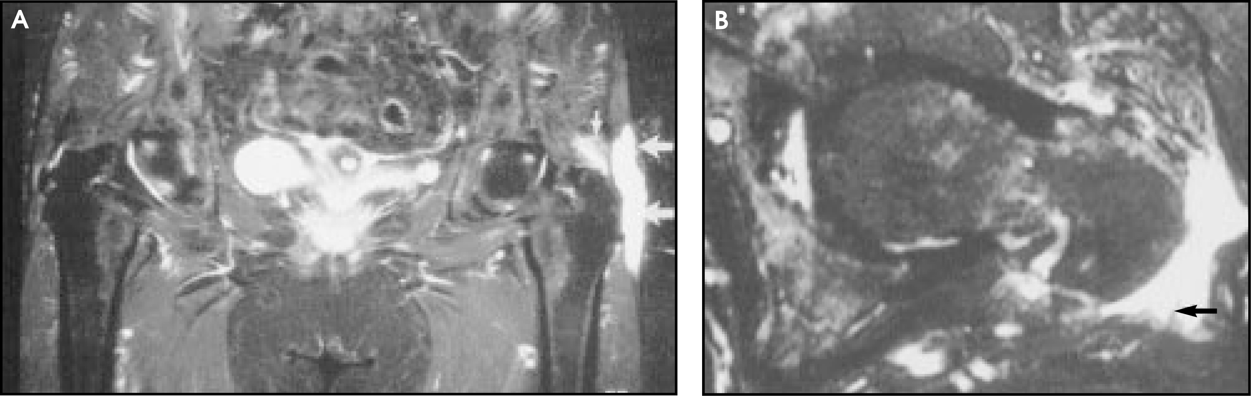
Labrum
The acetabular labrum is a rim of fibrocartilaginous tissue around the acetabulum that deepens the acetabular fossa and provides additional coverage of the femoral head. The labrum is normally triangular on axial and coronal MR images, is low in signal on all pulse sequences, and is thicker at the posterosuperior portion and thinner at the anteroinferior aspect.
Symptoms of labral tears include clicking, persistent pain, and decreased range of motion. Etiologies include a single traumatic event, chronic stress associated with athletic participation, or developmental dysplasia of the hip. Most tears occur along the anterior-superior labrum with chondral defects associated in 30% of labral tears.18
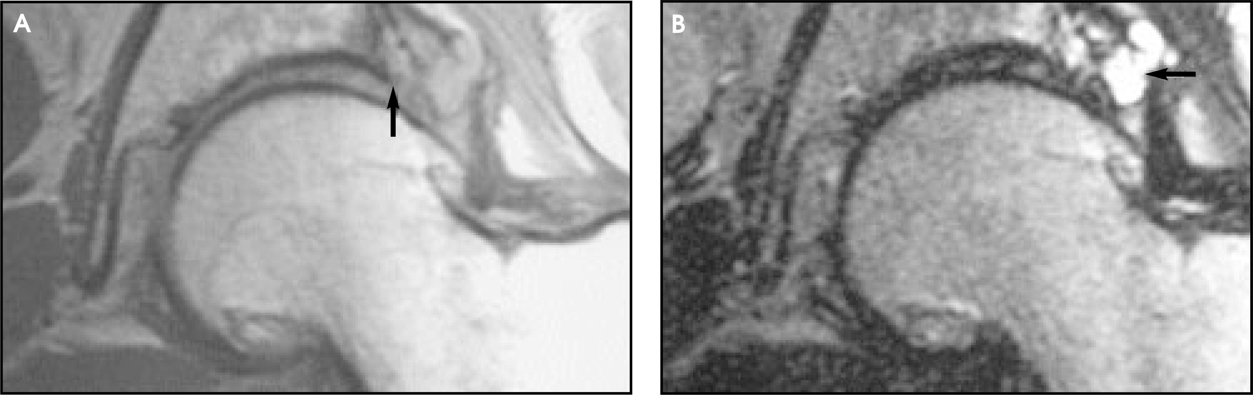
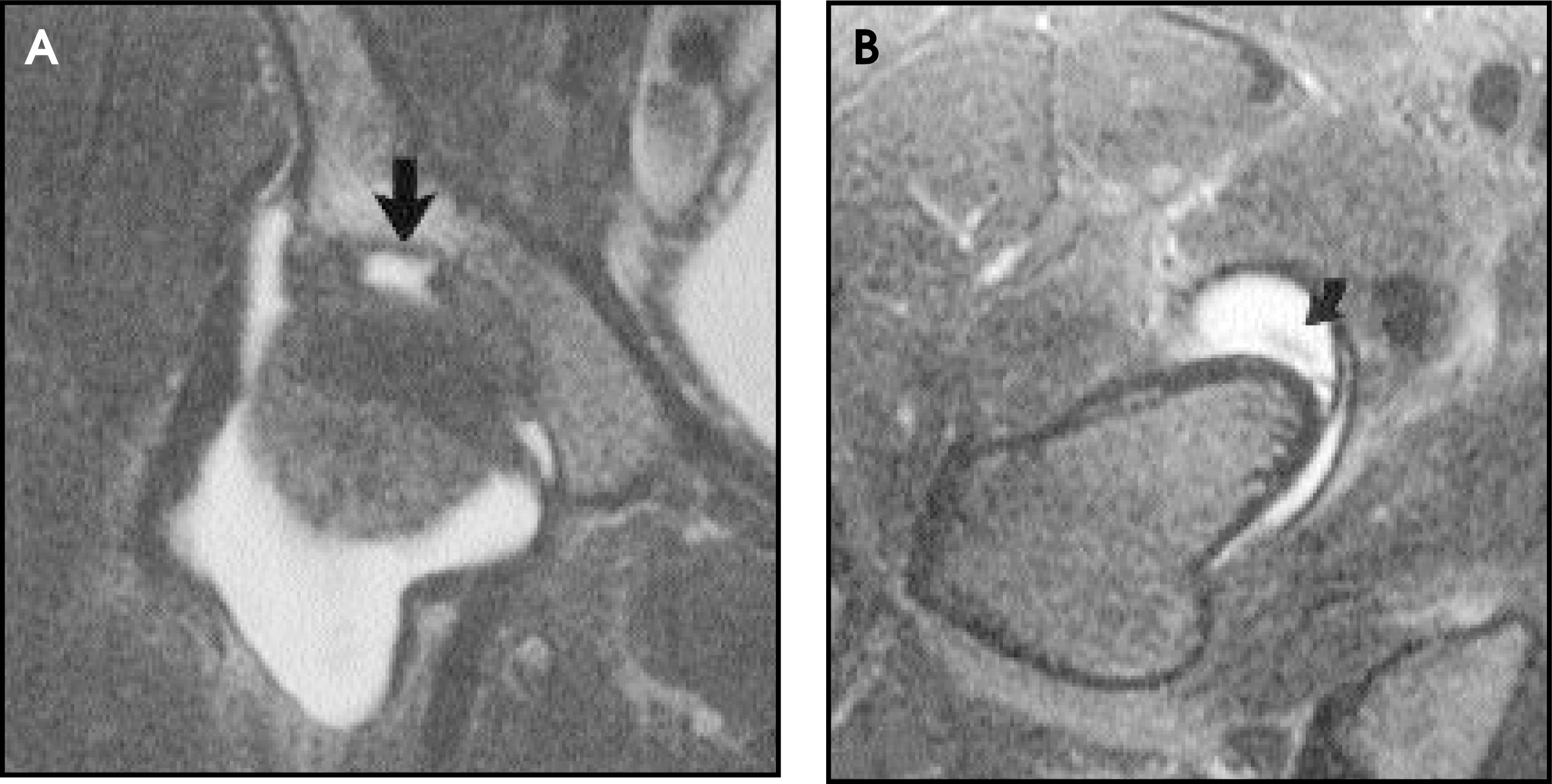
MR imaging with intra-articular administration of gadolinium and a smaller field of view to visualize only the hip in question, rather than the entire pelvis, is the most sensitive method of imaging labral tears. Findings include linear or diffuse high signal within the labrum, deformity of contour, or complete detachment (Figures 10 through 12). A paralabral cyst may also be visible and is a strong indicator of an underlying tear (Figure 13).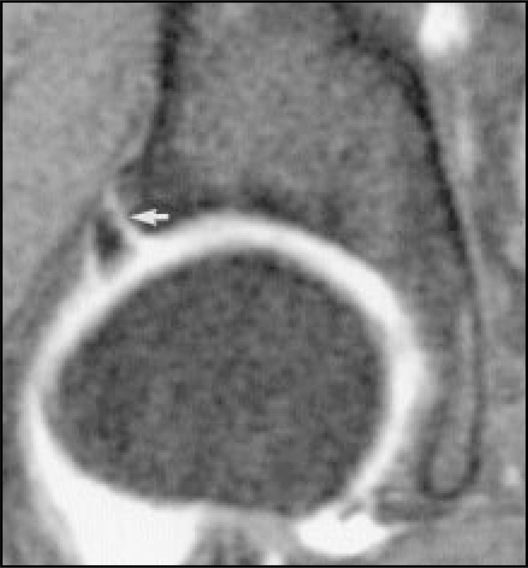
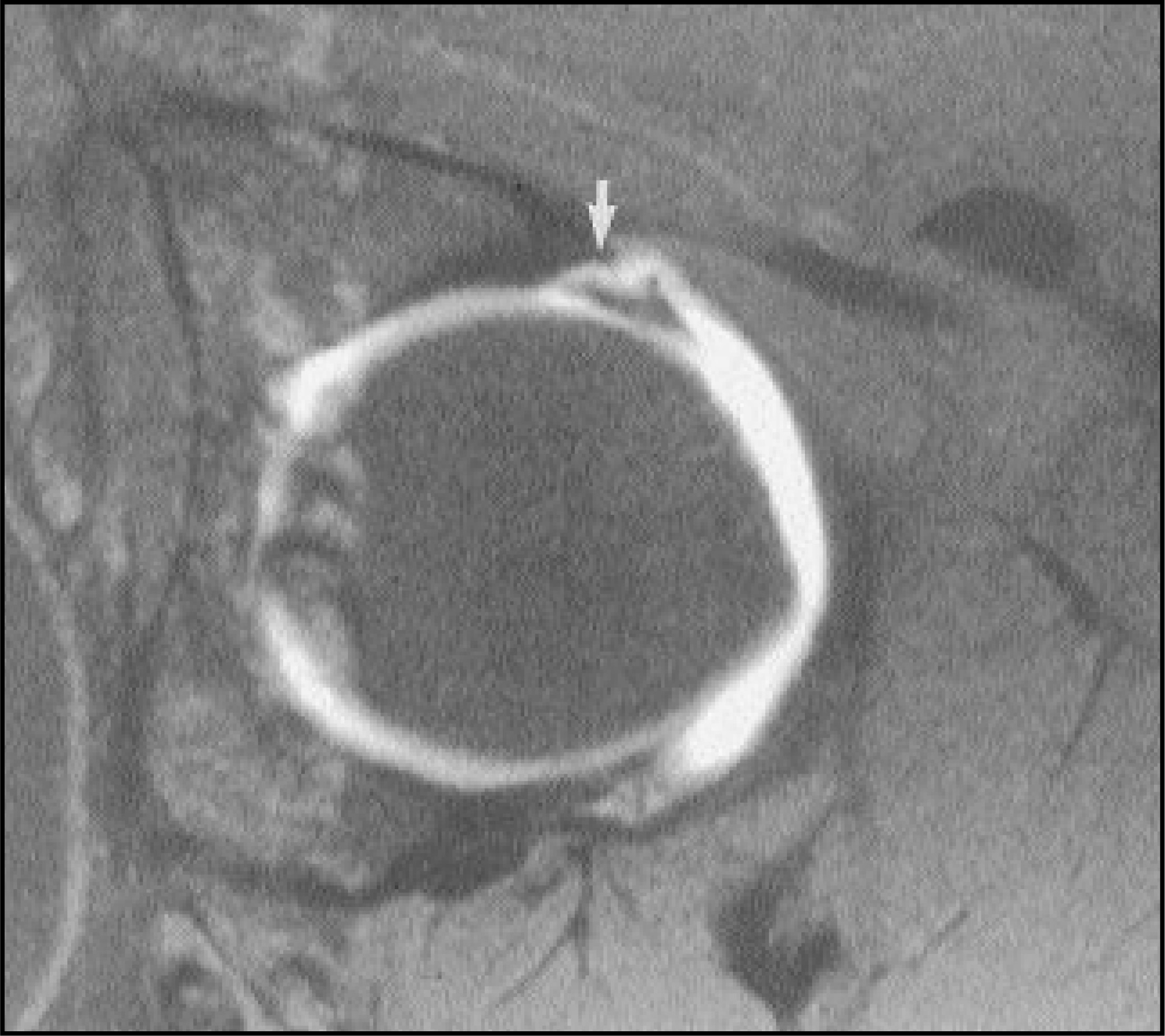
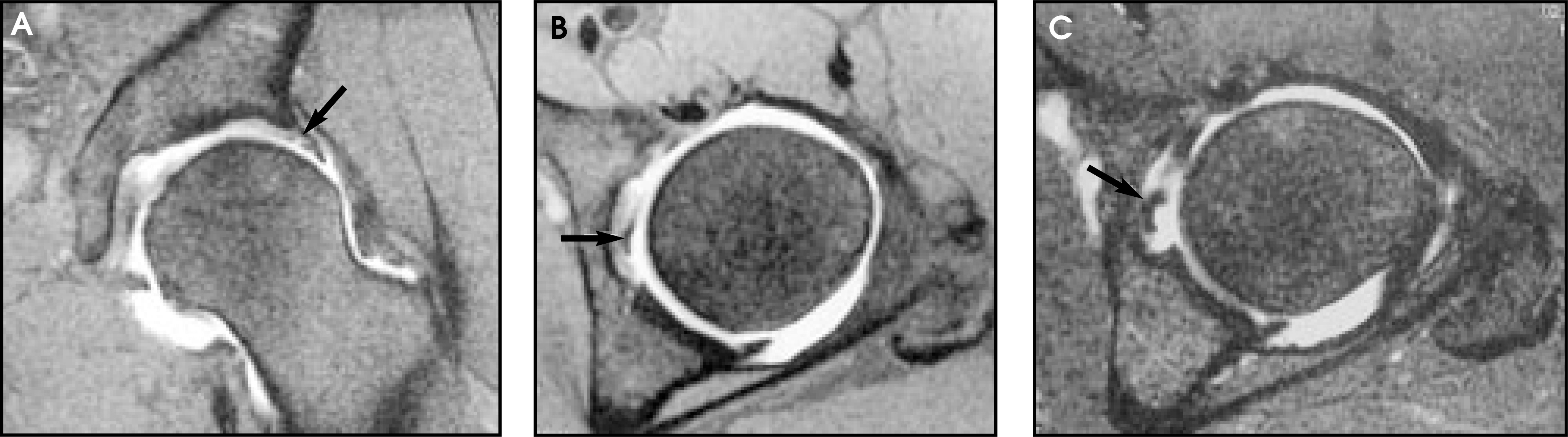
Pitfalls to be avoided in diagnosing labral tears include undercutting of the acetabular cartilage that extends medially deep to the labrum. In addition, the iliopsoas tendon courses anteriorly to the labrum and can mimic a tear (Figure 14).19 Surgery is generally indicated for symptomatic labral abnormalities.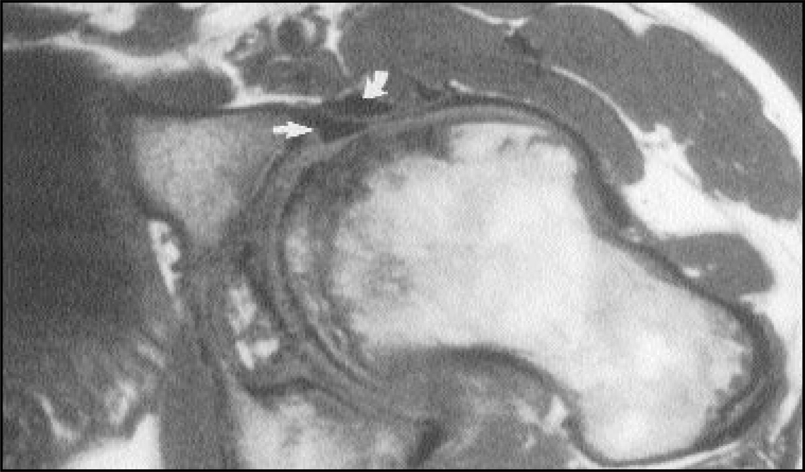
Osteochondral lesions
Osteochondral defects of the femoral head are yet another entity that can cause hip pain in the setting of normal plain radiographs. These are more common in athletes and there may or may not be a history of a specific traumatic event. MR imaging shows a wedge-shaped signal abnormality along the medial femoral head from the 10 o’clock to 11 o’clock positions (Figures 15 and 16). This area is smaller than that seen for AVN, and does not show serpiginous low signal as is seen with AVN. Several of these patients have also been shown to have delamination injuries at surgery.20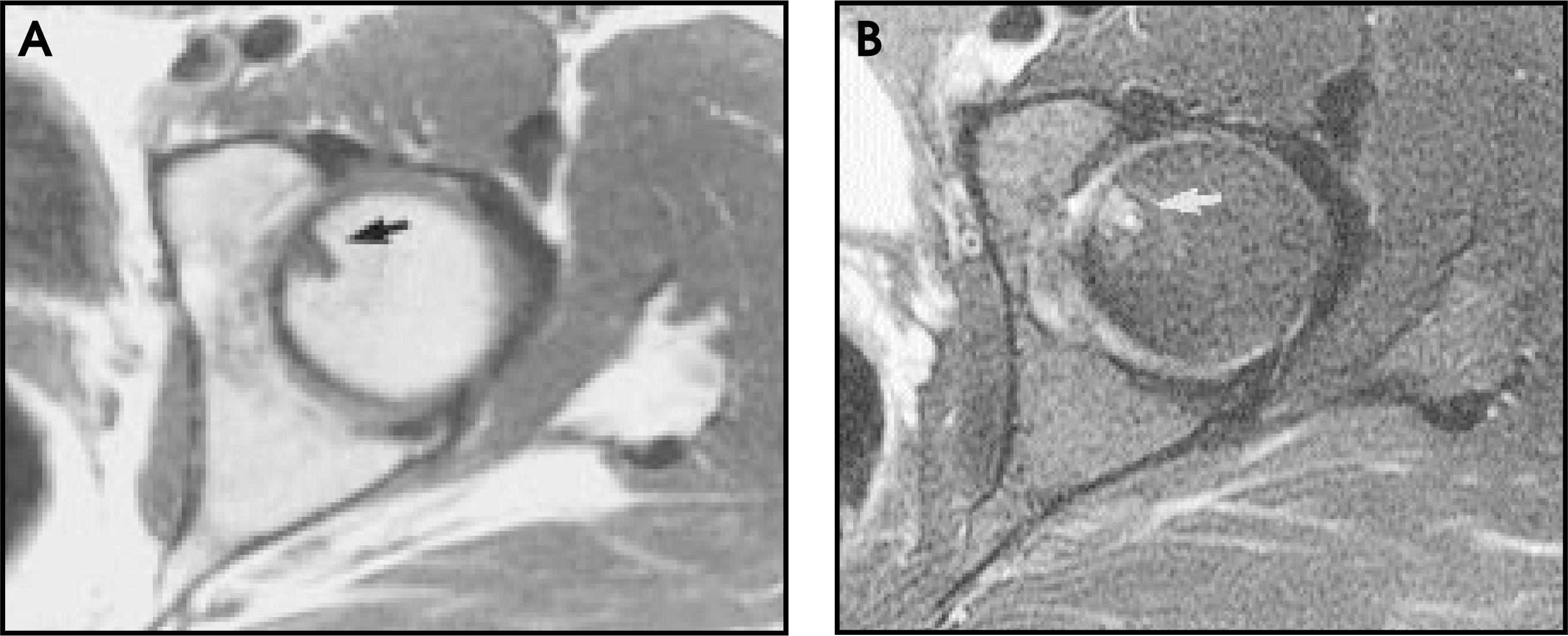
Conclusion
The more well-recognized etiologies of hip pain, such as vascular abnormalities and fractures, are often radiographically occult. Other potential sources of hip pain, such as labral pathology, muscle and tendon injuries, inflamed bursa, and osteochondral lesions, are diagnosed exclusively with MR imaging. Therefore, MR imaging is the modality of choice for evaluating pathology in and around the hip and pelvis. A patient with hip pain and negative plain films should undergo MR imaging to assess the source of pain, as the lesions presented are radiographically occult and management of the patient can be affected. AR
References
- Coleman B, Kressel H, Dalinka M. Radiographically negative avascular necrosis: Detection with MR imaging.. Radiology.. 1988;168:525-528.
- Beltran J, Herman L, Burk J. Femoral head avascular necrosis: MR imaging with clinical-pathological and radionuclide correlations.. Radiology.. 1988;166:215-220.
- Mitchell D, Rao V, Dalinka M. Femoral head avascular necrosis: Correlation of MR imaging, radiographic staging, radionuclide imaging, and clinical findings.. Radiology.. 1987;162:709-715.
- Major N, Helms C. Idiopathic transient osteoporosis of the hip.. Arthritis Rheum.. 1997;40:1178-1179.
- Curtiss P, Kincaid W. Transitory demineralization of the hip in pregnancy: A report of three cases.. J Bone Joint Surg [Am].. 1959;41:1327-1333.
- Guerra J, Steinberg M. Distinguishing transient osteoporosis from avascular necrosis of the hip.. J Bone Joint Surg [Am].. 1995;77:616-624.
- Yamamoto T, Kubo T, Hirasawa Y. A clinicopathologic study of transient osteoporosis of the hip.. Skeletal Radiol.. 1999;28:621-627.
- VandeBerg B, Malghem J, Lecourt F. Idiopathic bone marrow edema lesions of the femoral head: Predictive value of MR imaging findings.. Radiology.. 1999;212:527-535.
- Stafford S, Rosenthal D, Gebhardt M. MRI in stress fracture.. AJR Am J Roentgenol.. 1986;147:553-556.
- Major N, Helms C. Sacral stress fractures in long distance runners.. AJR Am J Roentgenol.. 2000;174:727-729.
- Otte M, Helms C, Fritz R. MR imaging of supra-acetabular insufficiency fractures.. Skeletal Radiol.. 1997;26:279-283.
- Nokes S, Vogler J, Spritzer C. Herniation pits of the femoral neck: Appearance at MR imaging.. Radiology.. 1989;172:231-234.
- Bogost G, Lizerbram E, Crues J. MR imaging in evaluation of suspected hip fracture: Frequency of unsuspected bone and soft-tissue injury.. Radiology.. 1995;197:263-267.
- Ishikawa K, Kai K, Mizuta H. Avulsion of the hamstring muscles from the ischial tuberosity: A report of two cases.. Clin Orthop.. 1988;232:153-155.
- Chung C, Robertson J, Cho G. Gluteus medius tendon teres and avulsive injuries in elderly women: imaging findings in six patients.. AJR Am J Roentgenol.. 1999;173:351-353.
- Kingzett-Taylor A, Tirman P, Feller J. Tendinosis and tears of gluteus medium and minimus muscles as a cause of hip pain: MR imaging findings.. AJR Am J Roentgenol.. 1999;173:1123-1126.
- Pritchard R, Shah H, Nelson C, FitzRandolph R. MR and CT appearance of iliopsoas bursal distention secondary to diseased hips.. J Comput Assist Tomogr.. 1990;14:797-800.
- Fitzgerald R. Acetabular labrum tears: Diagnosis and treatment.. Clin Orthop. 1995;311:60-68.
- Czerny C, Hoffman S, Urban M. MR arthrography of the adult acetabular capsular-labral complex: Correlation with surgery and anatomy.. AJR Am J Roentgenol.. 1999;173:345-349.
- Weaver C, Major N, Garrett W, Urbaniak J. Femoral head osteochondral lesions in painful hips of athletes: MR imaging findings.. AJR Am J Roentgenol.. 2002;178:973-977.
Citation
. MR imaging of the hip and pelvis: Current concepts. Applied Radiology. 2003;32(6):14-21. doi:10.37549/AR1189.