MRI of cervical spine neoplasms
Applied Radiology — Vol. 32 , Issue 12 , pp. 26 -38
DOI: 10.37549/AR1207
Published: December 1, 2003
Categories
Tumors of the cervical spine are relatively rare, but are of great importance due to their potentially devastating clinical effects and their challenging radiographic appearance. This article will address the clinical, pathologic, and radiographic characteristics of a variety of cervical spine tumors. These tumors are subdivided according to their point of origin: intramedullary, intradural-extramedullary, and extradural. Although this classification occasionally may be inexact owing to the propensity for some spinal tumors to traverse compartments,1 it provides a convenient and meaningful framework by which to approach the majority of cervical spine tumors.
Intramedullary tumors
Intramedullary tumors include gliomas (ependymomas, astrocytomas, and gangliogliomas), and nonglial tumors (such as hemangioblastomas, lymphoma, and metastases). Gliomas account for the majority (90% to 95%) of intramedullary spinal cord tumors.2
Ependymomas
Ependymomas are the most common intramedullary neoplasm in adults and usually occur in the cervical region.2 These lesions arise from ependymal cells that line the central canal and therefore tend to be central in location with respect to the spinal cord. Ependymomas tend to produce symmetric spinal cord expansion and usually have solid and cystic components. Cysts can be intratumoral, located within the solid tumor, or peritumoral (“polar”), occurring at the cranial or caudal aspects of the tumor. These cysts are not specific for ependymomas and can be seen with astrocytomas and gangliogliomas. Intratumoral cysts should be resected with the solid tumor, as they may contain tumor cells within them.3 However, polar cysts do not contain malignant cells3 and, therefore, need not be resected; although they may be drained surgically, they usually decompress spontaneously upon removal of the solid component.2
Ependymomas are characterized by slow growth, and compress rather than infiltrate adjacent spinal cord tissue, generally yielding a cleavage plane that aids in surgical resection.2 Almost all spinal cord ependymomas are low grade, classified as either grade I or grade II by the World Health Organization (WHO); malignant ependymomas are quite rare.2 On magnetic resonance imaging (MRI), ependymomas are generally iso- to hypointense on T1-weighted images and hyperintense on T2-weighted images (Figure 1). The solid components of ependymomas usually enhance avidly, although the degree of enhancement may vary considerably.3 In addition, ependymomas can hemorrhage, resulting in the “cap sign,” a hypointense rim at the periphery of the tumor on T2-weighted imaging that is related to hemosiderin deposition from prior hemorrhage.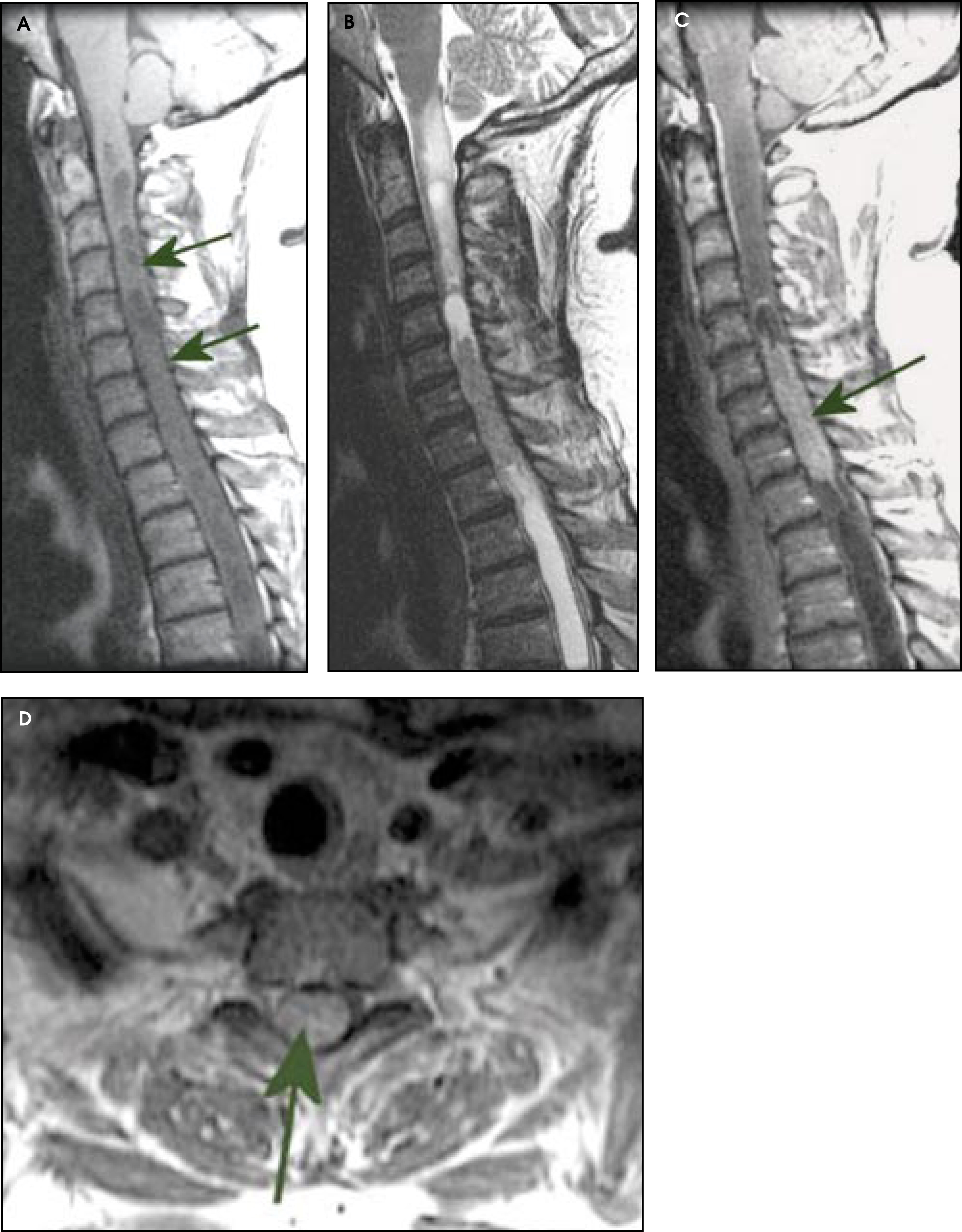
Astrocytomas
Astrocytomas are the second most common intramedullary neoplasm in adults, although they are the most common in children. Astrocytomas result from neoplastic transformation of astrocytes. In contradistinction to ependymomas, astrocytomas are located eccentrically within the spinal cord. The vast majority of spinal cord astrocytomas in adults are of low malignancy, classified either as pilocytic (grade I) or low-grade astrocytomas (grade II), although anaplastic astrocytomas and glioblastomas rarely occur.1,2 However, spinal cord astrocytomas tend to infiltrate the cord and are, therefore, difficult to resect completely.2 Astrocytomas are typically iso- to hypointense on T1-weighted imaging and hyperintense on T2-weighted imaging. They generally demonstrate patchy, irregular enhancement and have poorly defined margins. Like ependymomas, they can have intratumoral or polar cysts but do not tend to hemorrhage and, therefore, do not usually display a cap sign.2
Rarely, astrocytomas arise following radiation therapy, either for a primary central nervous system lesion, or for a lesion occurring outside the spine (Figure 2). There are no specific imaging findings for radiation-induced astrocytomas, and the main differential for these lesions is tumor recurrence (if there was a spinal cord tumor present initially) or radiation necrosis.4 The literature suggests that radiation-induced astrocytomas tend to be of a higher grade than idiopathic astrocytomas.5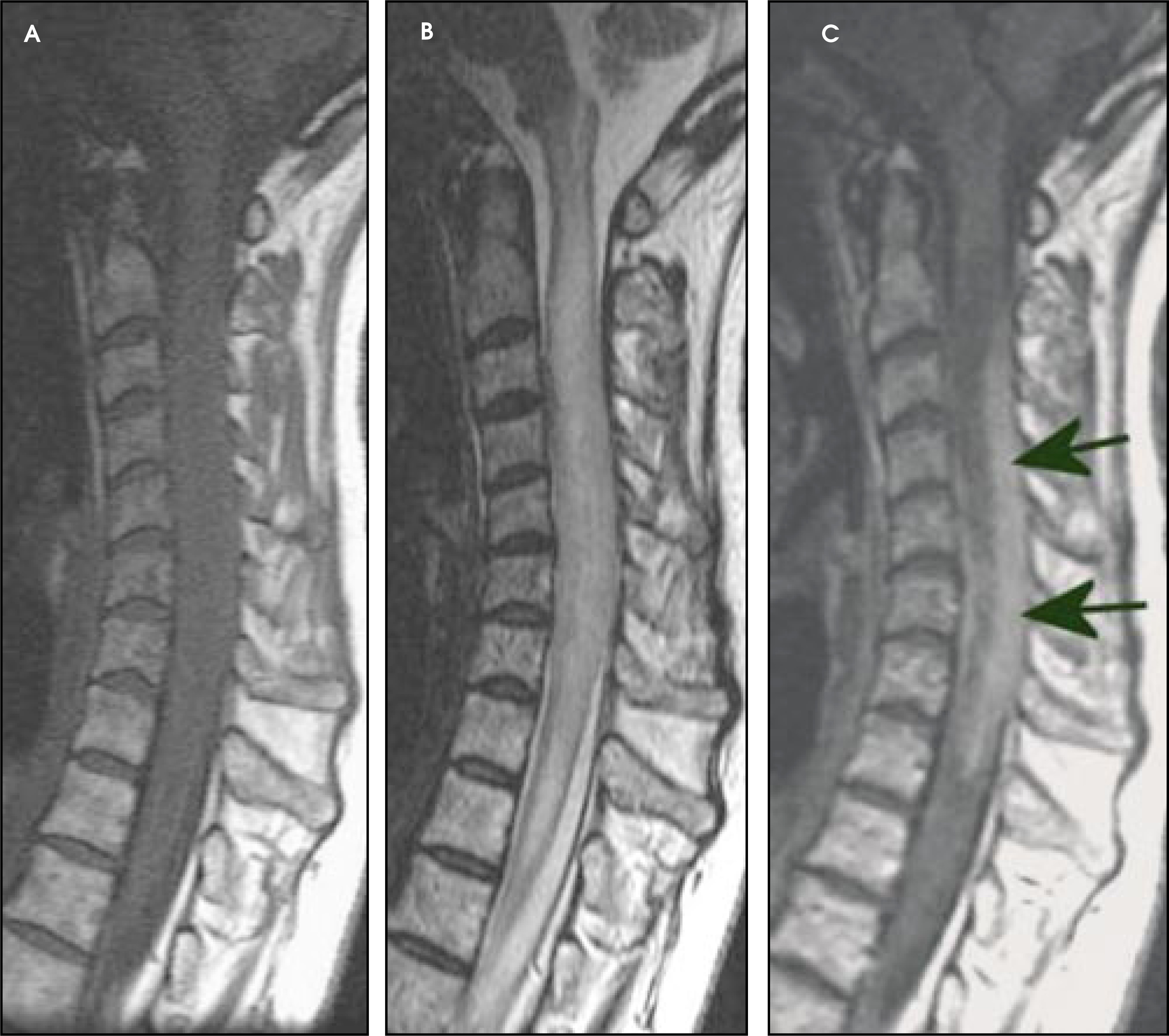
Gangliogliomas
Gangliogliomas are composed of a mixture of ganglion cells and neoplastic glial elements; the majority of neoplastic glial cell types are of the astrocytic subtype.2,6 These tumors tend to have a low malignant potential, slow growth, and a relatively good prognosis.2,6 Gangliogliomas are most common in children and young adults, with a mean age of 12 years at presentation.6 Gangliogliomas tend to be extensive on presentation, occupying an average length of 8 vertebral segments, compared with ependymomas and astrocytomas, which average 4 vertebral segments in length.6 Like astrocytomas, gangliogliomas tend to be eccentrically located within the spinal cord.6 Tumoral cysts are more common in gangliogliomas than in either astrocytomas or ependymomas.2 Chronic bony changes, including scoliosis and erosions, are often seen with gangliogliomas due to their relatively slow growth; these are rarely seen with ependymomas or astrocytomas.6 T1 signal characteristics of gangliogliomas are most often mixed, possibly secondary to the fact that gangliogliomas have a dual cell population composed of ganglion cells and glial elements.6 T2 signal characteristics of gangliogliomas are generally hyperintense, although surrounding edema is not as commonly seen as with ependymomas or astrocytomas.2,6 The majority of gangliogliomas show patchy enhancement.2,6 Other tumors of the glioma series such as oligodendrogliomas, pleomorphic xanthoastrocytomas, and dysembryoplastic neuroepithelial tumors are extremely uncommon in the adult spinal cord.
Hemangioblastomas
Hemangioblastomas are nonglial, highly vascular neoplasms of unknown cell origin that occur in the posterior fossa and spinal cord.2 In the spine, they are most commonly located in the subpial aspect of the dorsal cervical spine.7 They may extend exophytically into the subarachnoid space and have been reported in the extradural space.2 Most spinal hemangioblastomas occur sporadically, but approximately one-third of cases occur in association with von Hippel-Lindau’s disease7; in patients with this syndrome, hemangioblastomas are often multiple, and this necessitates screening of the entire spine with T1-weighted sagittal images (Figure 3). Hemangioblastomas are hypointense to isointense on T1-weighted imaging and hyperintense on T2-imaging and enhance homogeneously following gadolinium.7 Due to the vascularity of the lesion, prominent flow voids may be seen with MRI. Spinal hemangioblastomas may be associated with syringes that are usually more extensive than those seen with ependymomas or astrocytomas.7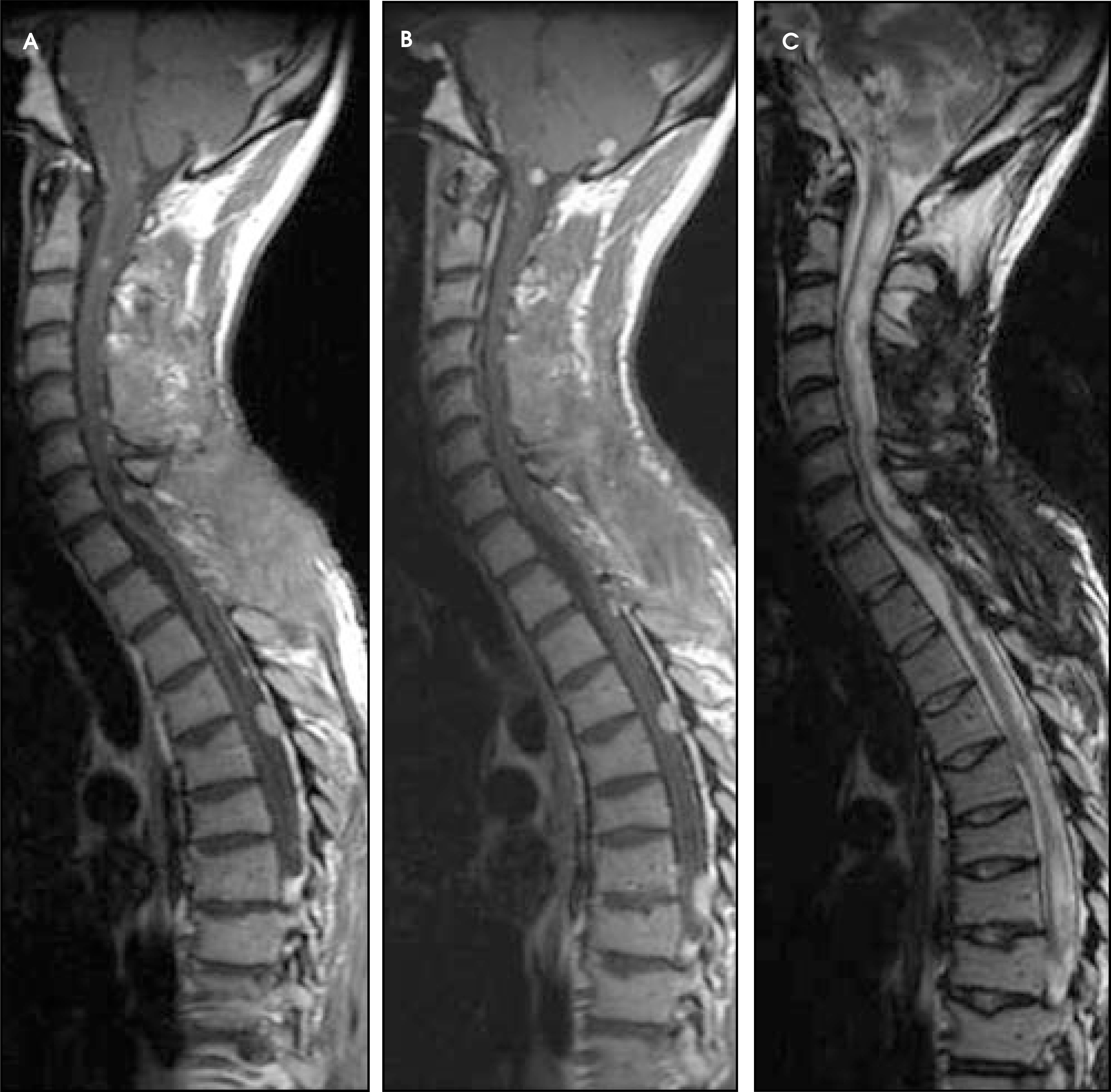
Intramedullary lymphoma
Primary intramedullary spinal lymphomas are extremely rare, with only 15 cases reported in the literature.2 These tumors are of the non-Hodgkin variety and can occur in both the immonocompromised8 and immunocompetent patients.9 The majority of these tumors occur in the cervical or thoracic regions of the spinal cord.8,9 They are solid tumors without necrosis that, on MR imaging, demonstrate marked T2 prolongation and enhance following gadolinium administration.10 Clinically, these patients initially respond to steroid treatment for a short time,10 but like primary lymphomas of the brain, the tumors usually recur after treatment.
Intramedullary metastases
Intramedullary spinal metastases are rare and represent approximately 2% of all metastases to the central nervous system; these usually involve the cervical cord.2 The most common primary tumors that metastasize to the spinal cord include lung, breast, colon, lymphoma, and kidney11; rarely, intramedullary spinal cord metastasis may be the initial presentation of the malignancy.12 The prognosis for patients with intramedullary spinal cord metastasis is poor; two-thirds of patients die within 6 months.2 On MRI, intramedullary spinal cord metastases are T1-hypointense, T2-hyperintense, and demonstrate homogeneous enhancement2 (Figure 4). As with metastases to the brain, the amount of surrounding edema is out of proportion to the size of the lesion.2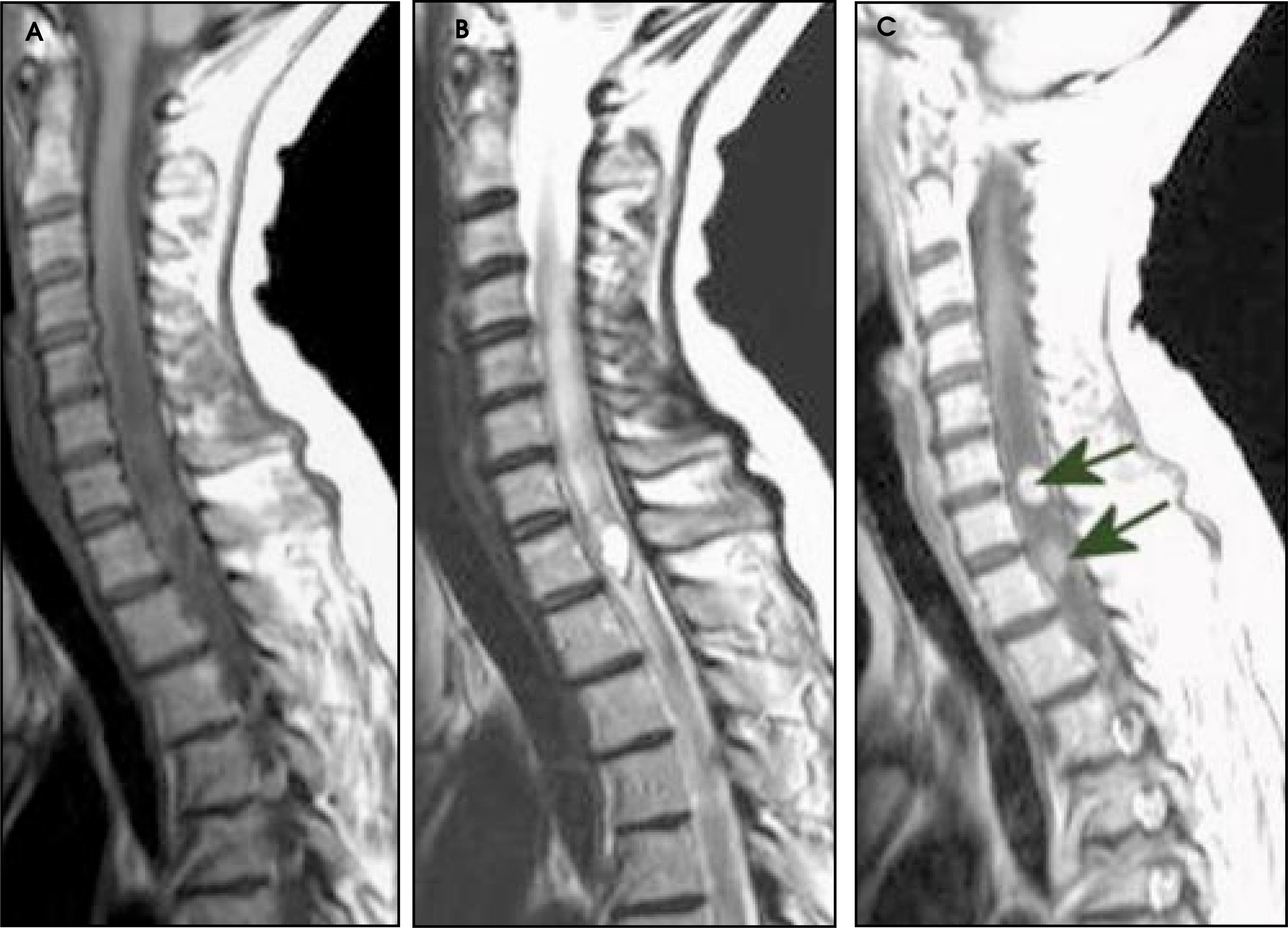
Intradural-extramedullary tumors
Since the arachnoid is essentially continuous with the dura in the spine, intradural lesions are located in the subarachnoid space. Intradural-extramedullary tumors include benign lesions (such as meningiomas and schwannomas) and metastatic lesions.
Meningiomas
Spinal meningiomas have similar characteristics to intracranial meningiomas. They have a strong female predominance with a peak occurrence in the fifth and sixth decades.13 The cervical spine is the second most common intraspinal location for meningiomas; these more often arise in the thoracic region.13 Typically, these lesions demonstrate T1 and T2 signal that is isointense with the spinal cord and display intense homogeneous enhancement following gadolinium administration13 (Figure 5). A dural tail may be seen, reflecting tumor spread or reactive changes in the dura adjacent to the tumor. These lesions are generally pathologically benign, and although they may exert considerable mass effect upon the spinal cord, symptoms may be relatively mild due to their slow growth.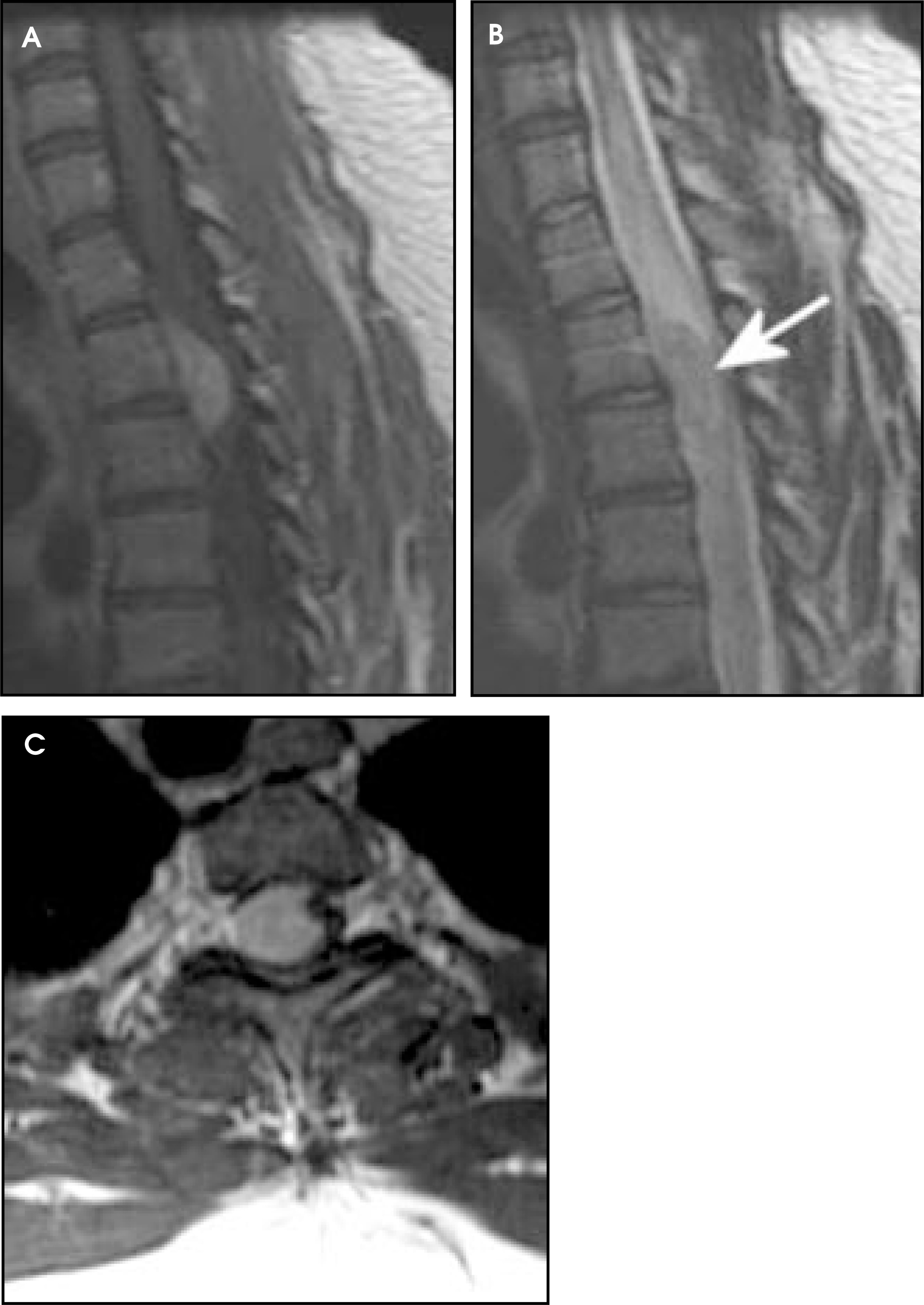
Schwannomas
Schwannomas are eccentric, exophytic nerve sheath tumors that are most common in the lower thoracic spine and lumbar spine.1 Schwannomas are usually moderately hyperintense on T2-weighted imaging, but may contain foci of T2 shortening centrally, likely relating to dense areas of collagen and Schwann cells.1 They are slightly hyperintense on noncontrast T1-weighted images1 and generally show homogeneous enhancement. Schwannomas rarely undergo malignant transformation; zones of fluidic signal within these tumors usually represent cystic degeneration, not necrosis.
Neurofibromas
Neurofibromas generally occur in association with neurofibromatosis (especially NF-1) and can occur at any level of the spine. They are usually dumbbell-shaped lesions that may expand the neural foramina and demonstrate homogeneous enhancement. They are generally isointense on T1 and slightly hyperintense on T2-weighted images. Like schwannomas, these masses follow the course of the nerve from which they arise and, therefore, usually have intradural and extradural components. However, unlike schwannomas, neurofibromas are tumors of the nerves themselves and cannot be removed without destroying the function of the parent nerve root.
Intradural metastases
Metastasis to the dura may arise from a variety of primary malignancies, most commonly breast cancer, lung cancer, and melanoma. Tumors of the central nervous system (glioblastoma multiforme and posterior fossa ependymomas) may produce “drop metastases.” These metastatic lesions usually appear as small, round, multifocal lesions that enhance and stud the surface of the cord. Lymphomatous metastases, on the other hand, tend to produce a more diffuse pial enhancement, as may breast and prostate metastases. In a patient with metastatic disease, these radiographic appearances generally indicate extramedullary-intradural disease. It should be noted, however, that multiple cerebral spinal fluid analyses are far more sensitive than imaging studies in the determination of this process.
Extradural tumors
Extradural lesions may arise from bone, fat, vessels, lymph nodes, or the surrounding extramedullary neural elements within the epidural space. Benign osseous lesions include osteochondroma and aneurysmal bone cyst. Malignant osseous lesions include osteosarcoma, chordoma, non-Hodgkin’s lymphoma, multiple mye-loma, and metastatic disease.
Osteochondromas
Osteochondromas are benign osseous lesions composed of cancellous bone surrounded by cortical bone and contain marrow within them.1 They are developmental lesions, rather than true neoplasms.14 They most commonly affect the appendicular skeleton, but can affect the spine in 2% of cases, most commonly involving the cervical region.14,15 Osteochondromas are usually exophytic pedunculated lesions but can be sessile, and they almost always arise from the posterior elements of the vertebral bodies.15 Malignant transformation occurs in 1% of solitary osteochondromas and in 3% to 5% of patients with hereditary multiple exostoses.14 If an osteochondroma undergoes continued growth in the adult patient, becomes painful, or recurs after resection, the possibility of malignant degeneration into an osteosarcoma or chondrosarcoma should be considered.14,15 Enlargement of the cartilagenous cap after skeletal maturity is a relatively sensitive indicator of malignant transformation. On MRI, osteochondromas are heterogeneous due to various degrees of maturity and calcification in the areas of enchondral ossification, and the cartilagenous cap demonstrates T1 hypointensity and T2 hyperintensity, reflecting its high water content.14
Aneurysmal bone cysts
Aneurysmal bone cysts are pathologically benign lesions that are expansile, lytic lesions filled with chronic blood products. They have a predilection to occur within the posterior elements of the spine but may also occur in the vertebral bodies.1 They classically appear multiloculated with heterogeneous fluid-fluid levels within the loculations, reflecting hemorrhage with sedimentation.16 These lesions may enhance heterogeneously. The most concerning aspect of aneurysmal bone cysts is their propensity to undergo pathologic fracture; even trivial trauma to these lesions may result in fracture with spinal cord compression.
Chordomas
Chordomas are malignant tumors that arise from remnants of the notochord that travels through the central vertebral bodies. These lesions have a 2:1 male predominance1 with a mean age of 50 years at presentation.16 They most commonly involve the sacrum but may also involve the clivus and upper cervical spine.16,17 In the cervical spine, chordomas tend to be midline in location, sparing the posterior elements.17 They are destructive, locally aggressive lesions that may be associated with prominent exophytic soft-tissue masses.17 As they enlarge, they tend to involve adjacent vertebral bodies and extend into the adjacent paraspinal tissues and epidural space; they may even grow into and expand neural foramina, potentially mimicking nerve sheath tumors.17,18 Uncommonly, they can arise solely in the musculature of the perivertebral space, presumably from extraosseous notochordal nests.17 Chordomas are low to intermediate in signal on T1 and classically demonstrate significant T2 prolongation19 due to the presence of their signature physaliferous cells (clear cells with intracytoplasmic vacuoles and abundant mucin).16 These lesions enhance heterogeneously following gadolinium administration19 and have amorphous calcifications in 50% to 75% of cases1 (Figure 6). Chordomas generally have a poor prognosis due to local recurrence following resection.18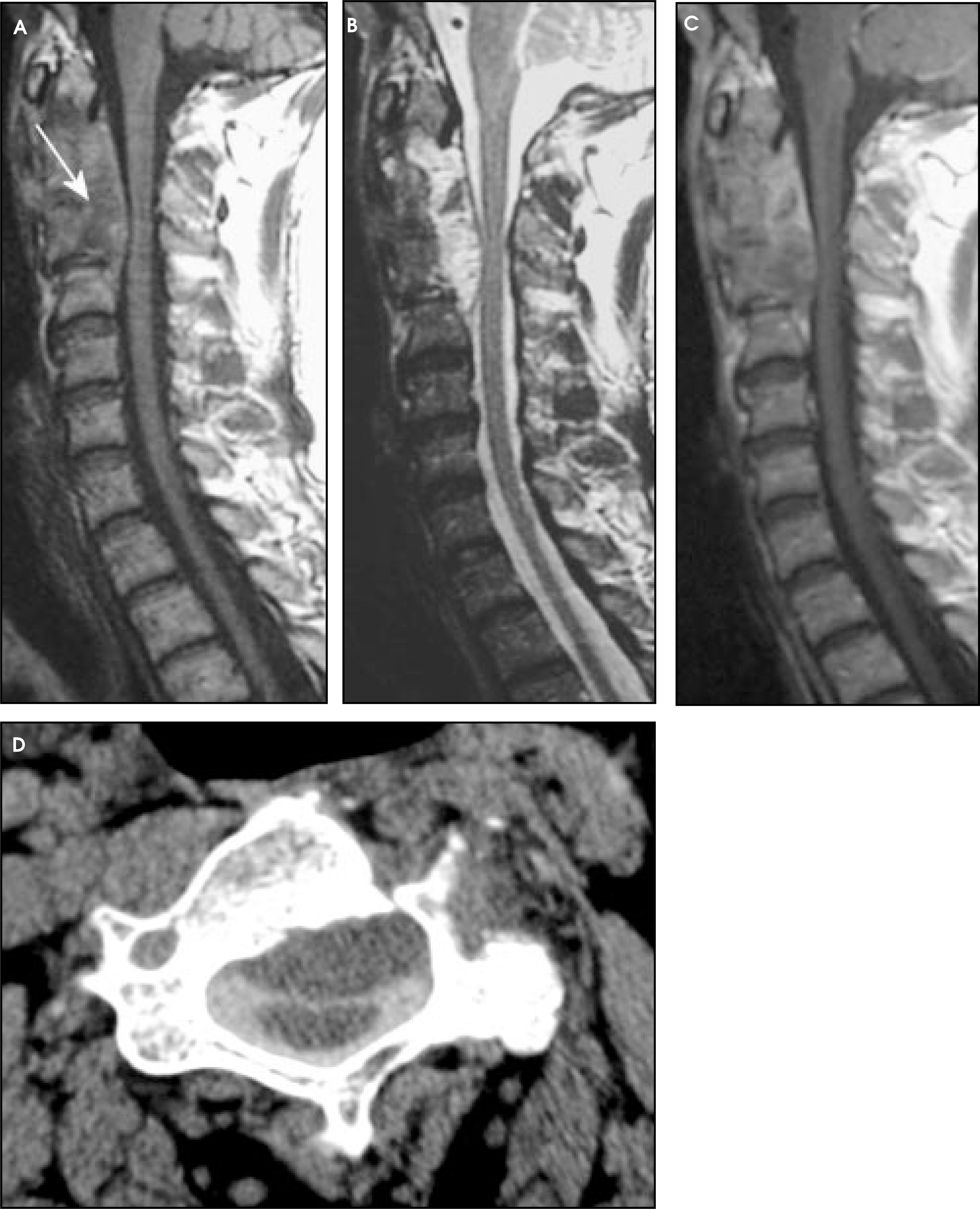
Multiple myeloma
Multiple myeloma is a malignancy characterized by monoclonal proliferation of malignant plasma cells.16 Multiple myeloma may involve any level of the spine. On plain film or CT, they usually appear as focal lytic lesions, but often the disease may present innocuously, appearing only as diffuse osteopenia. On MRI, myeloma deposits are multifocal, well-defined lesions that demonstrate T1 hypointensity and T2 hyperintensity with respect to normal marrow.16 Plasmacytomas are large, focal myeloma deposits that may show gadolinium enhancement. These lesions are expansile lytic masses that may extend into the epidural space; as with other tumors of the spine, they may undergo pathologic fracture (Figure 7).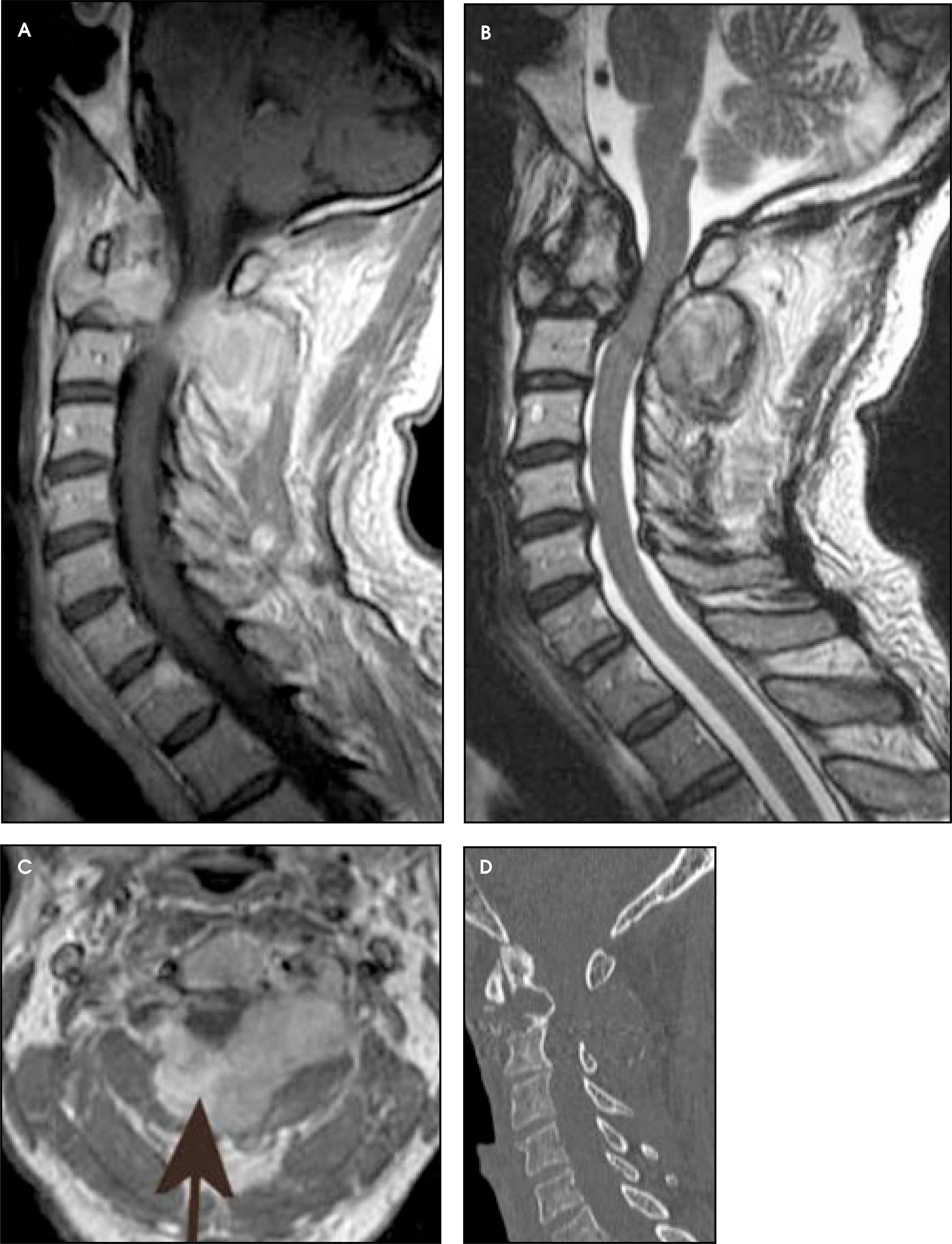
Lymphoma
Lymphoma of the spine most commonly involves the extradural compartment2 (Figure 8). It generally presents as a solid lesion that demonstrates T1 isointensity and T2 hypointensity. The T2 signal characteristics are related to the fact that lymphomas have a high nuclear-to-cytoplasmic ratio. Moreover, some authors have shown that because lymphomas are highly cellular tumors with a reduced extracellular matrix, there is more restricted diffusion on diffusion-weighted images than is seen with other tumors, such as high-grade astrocytomas.20 Lymphomas demonstrate robust enhancement and may narrow the spinal canal, resulting in compression of the spinal cord. Accurate diagnosis is of particular importance with lymphomas, since radiation therapy can result in rapid reduction in the size and compressive effects of these lesions.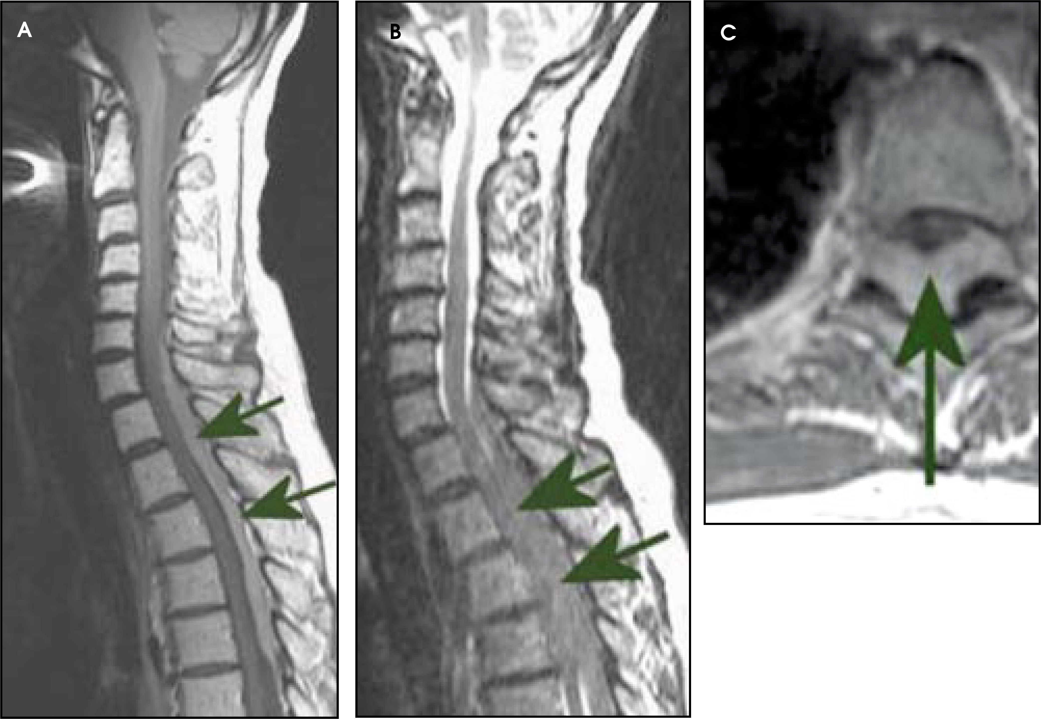
Metastases to bone
Metastases to bone generally present as T1-hypointense and T2-hyperintense lesions that replace normal marrow. Pathologic fractures can be difficult to differentiate from benign osteoporotic fractures. However, osteoporotic fractures more commonly cause anterior wedging and the abnormal signal tends to spare the pedicles and posterior processes, in contradistinction to metastatic lesions. In addition, benign compression fractures may be associated with a well-defined linear signal abnormality across the vertebral body1 and tend to reveal sparing of the marrow interspersed within the affected vertebrae. The most common metastases to bone include those from cancers of the breast, prostate, lung, as well as renal cell carcinoma and melanoma.
Conclusion
Tumors of the cervical spine can be classified according to their location: intramedullary, intradural-extramedullary, and extradural. Consideration of the location of cervical spine tumors, as well as their signal characteristics, growth patterns, and clinical associations, can lead the radiologist to arrive at a reasonable differential diagnosis for tumors occurring in this sensitive region.
References
- Sze G, Atlas S. Magnetic Resonance Imaging of the Brain and Spine. 2002:1715-1767.
- Koeller K, Rosenblum R, Morrison A. Neoplasms of the spinal cord and filum terminale: Radiologic-pathologic correlation. RadioGraphics. 2000;20:1721-1749.
- Kahan H, Sklar E, Post M, Bruce J. MR characteristics of histopathologic subtypes of spinal ependymoma. AJNR Am J Neuroradiol. 1996;17:143-150.
- Phuphanich S, Jacobs M, Murtagh F, Gonzalvo A. MRI of spinal cord radiation necrosis simulating recurrent cervical cord astrocytoma and syringomyelia. Surg Neurol. 1996;45:362-365.
- Grabb P, Kelly D, Fulmer B, Palmer C. Radiation-induced glioma of the spinal cord. Pediatr Neurosurg. 1996;25:214-219.
- Patel U, Pinto R, Miller D. MR of spinal cord ganglioglioma. AJNR Am J Neuroradiol. 1998;19:879-887.
- Baker K, Moran C, Wippold F. MR imaging of spinal hemangioblastoma. AJR Am J Roentgenol. 2000;174:377-382.
- Bekar A, Cordan T, Evrensel T, Tolunay S. A case of primary spinal intramedullary lymphoma. Surg Neurol. 2001;55:261-264.
- Caruso P, Patel M, Joseph J, Rachlin J. Primary intramedullary lymphoma of the spinal cord mimicking cervical spondylotic myelopathy. AJR Am J Roentgenol. 1998;171:526-527.
- Herrlinger U, Weller M, Kuker W. Primary CNS lymphoma in the spinal cord: Clinical manifestations may precede MRI detectability. Neuroradiology. 2002;44:239-244.
- Gasser T, Pospiech J, Stolke D, Schwechheimer K. Spinal intramedullary metastases. Report of two cases and review of the literature. Neurosurg Rev. 2001;24:88-92.
- Schiff D, O’Neill B. Intramedullary spinal cord metastases: Clinical features and treatment outcome. Neurology. 1996;47:906-912.
- Yoshiura T, Shrier D, Pilcher W, Rubio A. Cervical spinal meningioma with unusual MR contrast enhancement. AJNR Am J Neuroradiol. 1998;19:1040-1042.
- Murphey M, Choi J, Kransdorfv M. Imaging of osteochondroma: Variants and complications with radiologic-pathologic correlation. RadioGraphics. 2000;20:1407-1434.
- Quirini G, Meyer J, Herman M, Russell E. Osteochondroma of the thoracic spine: An unusual cause of spinal cord compression. AJNR Am J Neuroradiol. 1996;17:961-964.
- Llauger J, Palmer J, Amores S. Primary tumors of the sacrum: Diagnostic imaging. Am J Roentgenol. 2000;174:417-424.
- Wippold F, Koeller K, Smirniotopoulos J. Clinical and imaging features of cervical chordoma. AJR Am J Roentgenol. 1999;172:1423-1426.
- Zacay G, Eyal A, Shacked I. Chordoma of the cervical spine. Ann Otol Rhinol Laryngol. 2000;109:438-440.
- Meyers S, Hirsch W, Curtin H. Chordomas of the skull base: MR features. AJNR Am J Neuroradiol. 1992;13:1627-1636.
- Guo A, Cummings T, Dash R, Provenzale J. Lymphomas and high-grade astrocytomas: Comparison of water diffusibility and histologic characteristics. Radiology. 2002;224:177-183.
Citation
. MRI of cervical spine neoplasms. Applied Radiology. 2003;32(12):26-38. doi:10.37549/AR1207.