MRI of the hip
Applied Radiology — Vol. 31 , Issue 11 , pp. 33 -39
DOI: 10.37549/AR1139
Published: November 1, 2002
Categories
Recent technologic advances in gradient strength, pulse sequence, and surface coil design allow for unprecedented soft-tissue resolution with magnetic resonance imaging (MRI). The musculoskeletal system is only one of the areas for which MRI has become the modality of choice for evaluation of bone and soft-tissue pathology. The hip is one of the areas most frequently imaged with MR in symptomatic patients whose conditions cannot be resolved with plain films. Common indications for hip MRI include: trauma with suspected occult fracture, stress and insufficiency fractures, labral tears, osteochondritis dissecans, tendon tears, septic arthritis, osteomyelitis, osteonecrosis, transient osteoporosis or bone marrow edema, and other conditions common to all joints, such as bursitis, synovial chondromatosis, synovial osteochondromatosis, inflammatory conditions, and bone and soft-tissue tumors. In this article, the imaging technique and MRI manifestations of these entities in the adult hip are summarized.
Technical notes
The first decision to make with hip MRI is whether to image both hips simultaneously or only the symptomatic hip. It is an important decision since it will influence other decisions such as coil and pulse sequence selection, as well as whether or not to inject contrast material, intra-articularly or intravenously. This decision is compounded by the common situation in which no clinical information is provided to the radiologist. As a general guideline, imaging of both hips simultaneously may be appropriate if one is looking for osteonecrosis (given the frequency of bilateral involvement) or metastasis. In most other clinical situations, single hip imaging provides better spatial and contrast resolution, since a dedicated surface coil can be used.
When bilateral hip imaging is chosen, the body coil, preferably phase array, is used.1 The following set of pulse sequences is recommended: T1-weighted coronal and fast-spin echo (FSE) T2-weighted or short tau inversion recovery (STIR) axial. If there are positive findings, one may choose at this time to stop or to switch to single hip imaging. This is done by using a dedicated surface coil, such as a flexible coil, for better anatomical resolution of small structures such as the acetabular labrum, or for better evaluation of the articular surfaces or subchondral area of the femoral head. When single hip imaging is indicated, the following pulse sequences are recommended: Coronal T1-weighted high resolution with 512 matrix, axial T2-weighted FSE or STIR and sagittal two-dimensional (2D) or three-dimensional (3D) gradient-recalled echo (GRE) (fast low-angle shot [FLASH], spoiled gradient-recalled acquisition in a steady state [SPGR]) or T2-weighted or STIR sequences.
Intravenous contrast material may be useful to assess tissue viability in osteonecrosis and in cases of suspected infection or bone or soft-tissue tumors. T1-weighted high-resolution images with fat saturation in the axial sagittal and coronal planes are recommended when using intravenous contrast material. The most recent innovation is the so-called “indirect” MR arthrogram (IMRA), replacing the intra-articular or “direct” MR arthrogram (DMRA). Indirect MR arthrogram is performed by injecting, intravenously, a standard dose of a gadolinium chelate (1 mmol/kg). This is then followed by 10 to 15 minutes of gentle exercise before T1-weighted fat-saturated imaging with high resolution in all three orthogonal planes, in addition to a T2-weighted sequence, preferably in the axial plane. This technique provides enough diffusion of contrast material through the synovium to outline the articular surfaces and produce some distension of the capsule and its recesses. An added advantage of IMRA is that it produces enhancement of hyperemic tissues. Furthermore, if a pre-existing joint effusion is present, the injected contrast material mixes with the joint fluid, giving an excellent arthrographic effect with high resolution provided by the use of the T1-weighted fat-saturated images with 512 matrix.
The DMRA technique entails the placement of a needle into the hip joint, generally using fluoroscopic guidance, and injecting 10 to 15 mL of dilution of a gadolinium chelate (0.1 mL) and saline (20 mL). The patient is then taken immediately into the MRI suite, and the same pulse sequences described for IMRA are used. The main advantage of DMRA over IMRA is the full distension of the joint capsule and its recesses. The disadvantages, however, are significant: DMRA requires additional scheduling for fluoroscopy and is an invasive procedure with added morbidity.
The indications for IMRA or DMRA include assessment of the articular surfaces when studying osteochondritis dissecans and assessment of the acetabular labrum.
Normal anatomy
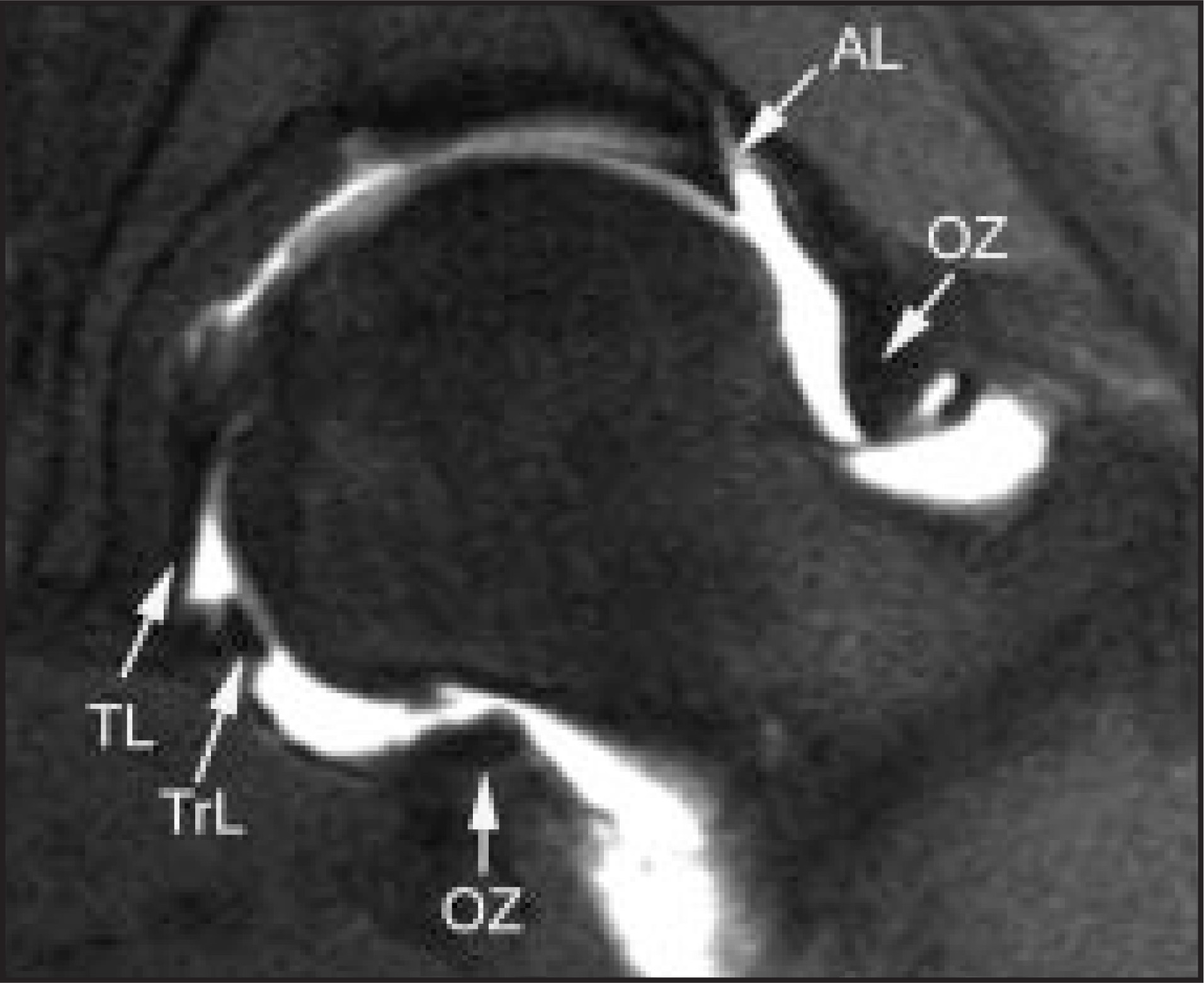
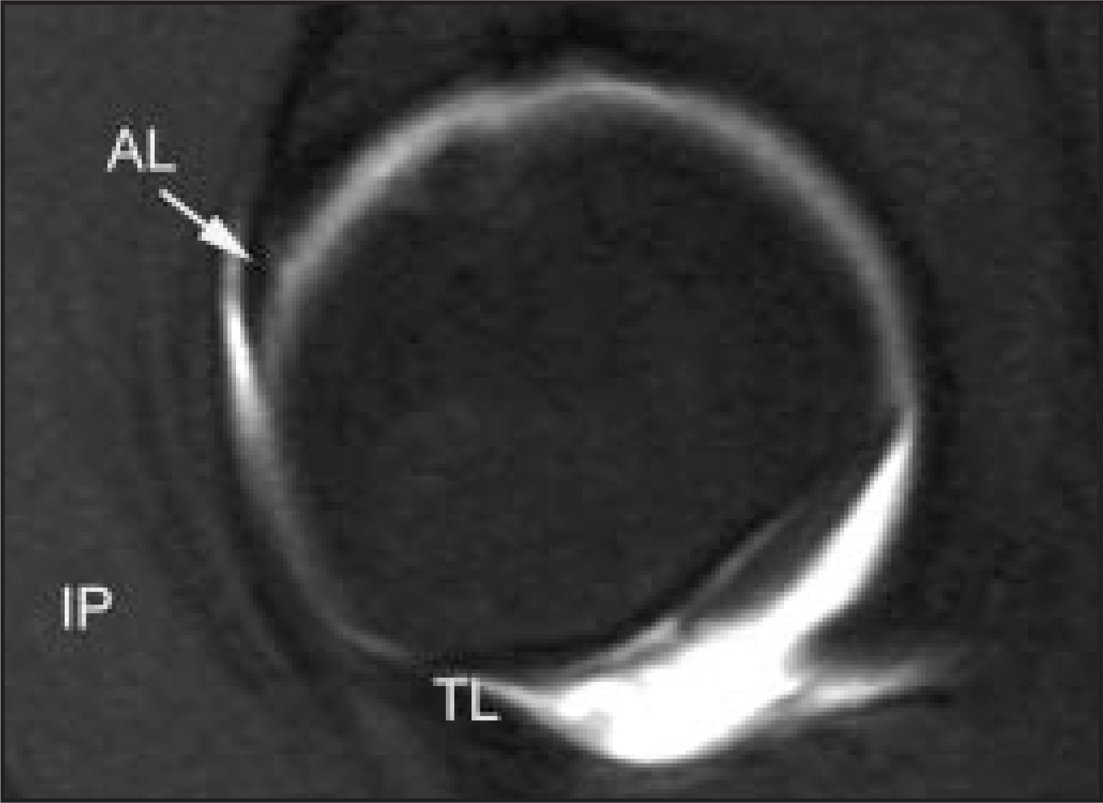
The hip is a ball-and-socket joint composed of the acetabulum and the femoral head. The acetabulum is partially covered by articular cartilage, a horseshoe shape opened inferiorly. The central portion or acetabular fossa is filled by fibrofatty tissue. The teres ligament originates in this fossa and inserts into the fovea capitis of the femoral head. A rim of fibrous tissue called the “acetabular labrum” surrounds the acetabulum. The joint capsule extends from the acetabular margin to the femoral neck, and it is reinforced anteriorly and posteriorly by a series of capsular ligaments. Figures 1, 2, and 3 depict the normal MRI anatomy of the hip in the three orthogonal planes.
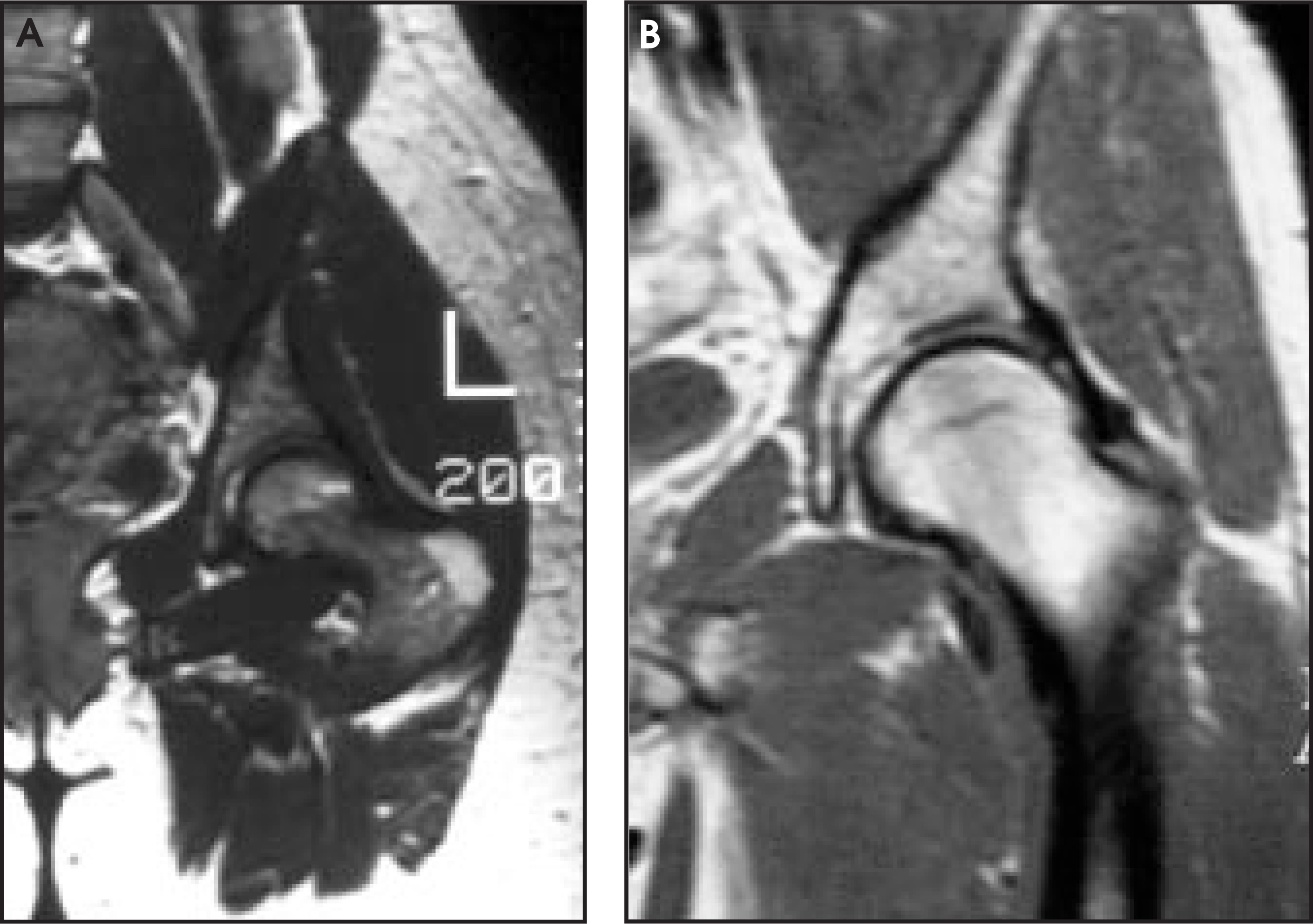
The normal patterns of the trabecular structure and bone marrow of the proximal femur vary with the age of the patient. As the patient becomes older, the trabecular pattern, formed by the primary and secondary trabeculae, becomes thinner and tends to be less conspicuous on MRI, due to osteoporosis. Similarly, the red marrow, which in young patients is very prominent in the femoral neck and intertrochanteric region, diminishes in the elderly (Figure 4).2,3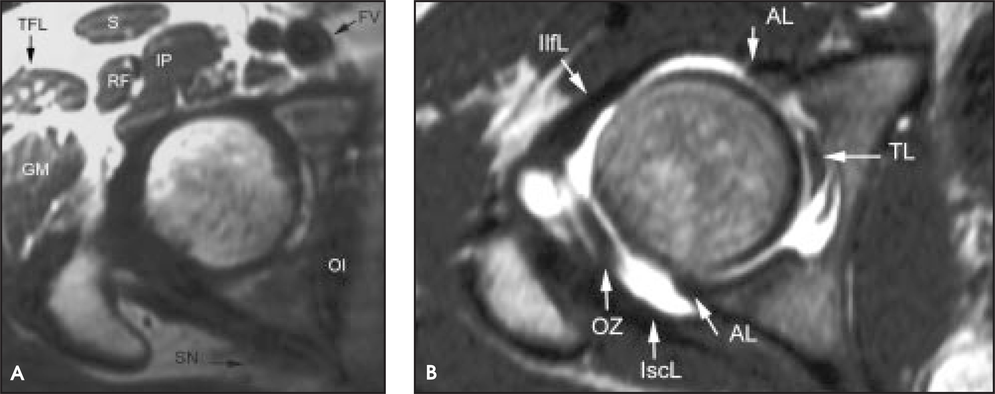
Trauma
Occult fractures in the elderly are one of the most frequent indications for MRI of the hip (Figure 5). In these generally osteoporotic patients, plain films may not demonstrate the fracture line, and bone scintigraphy may demonstrate increased uptake only several days after the trauma. MRI is highly sensitive for detecting the fracture and the surrounding edema immediately after the traumatic event.4-6 Subchondral fractures of the femoral head are now a well-recognized entity that may lead to extensive bone-marrow edema and the eventual collapse of the femoral head with subsequent bone resorption and rapid development of osteoarthritis. Subchondral fractures are mostly occult on plain films and may be related to stress, rather than direct trauma. This entity may be misdiagnosed as osteonecrosis or transient osteoporosis of the hip (TOH) because of the extensive bone-marrow edema that is seen during the first few months. In fact, TOH and subchondral fracture may be the same condition. Another disease that may be linked to subchondral fracture of the femoral head is rapidly destructive osteoarthritis or Pastel disease. MRI demonstrates bone-marrow edema, joint effusion, and a faintly seen subchondral hypointense line that represents the fracture. This line is better seen once the bone-marrow edema subsides (Figure 6).7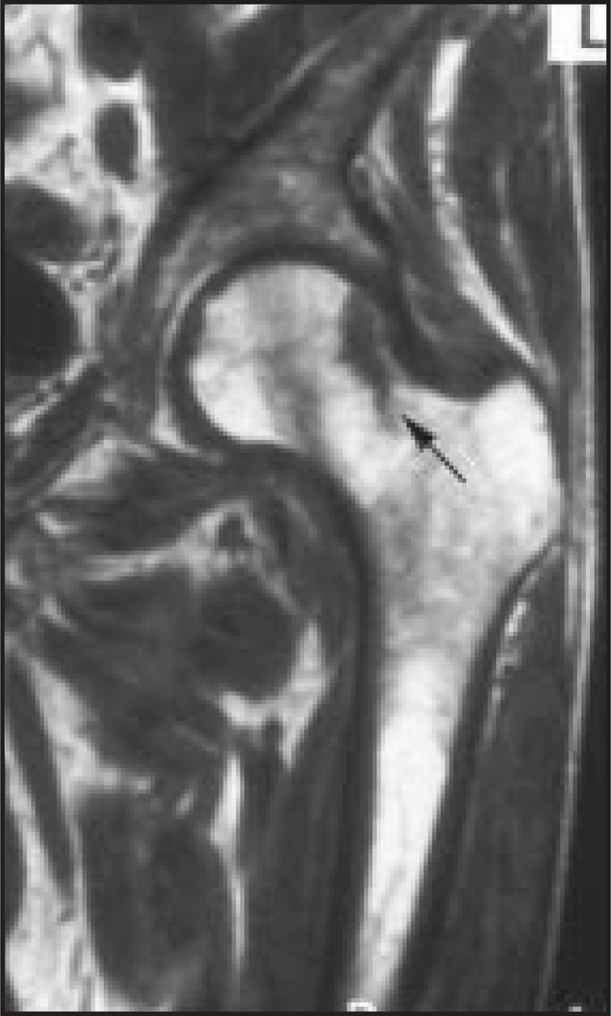
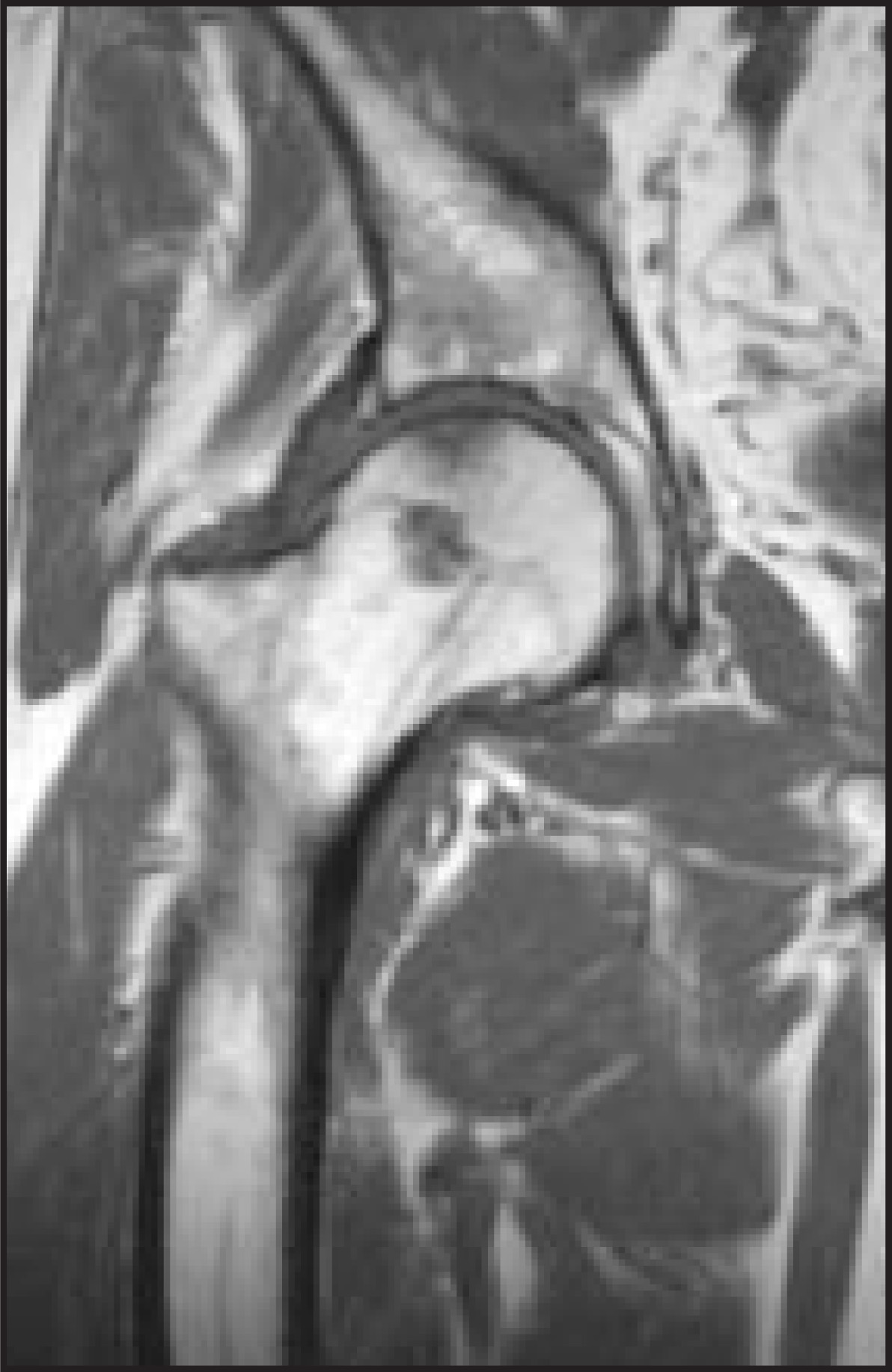
Osteochondritis dissecans or osteochondral fractures are relatively rare in the femoral head, but when they occur, radiographic findings may be subtle. MRI shows the lesion quite well (Figure 7), as well as other important data such as size, location, stability, joint surface congruity, and viability of the fragment can be assessed.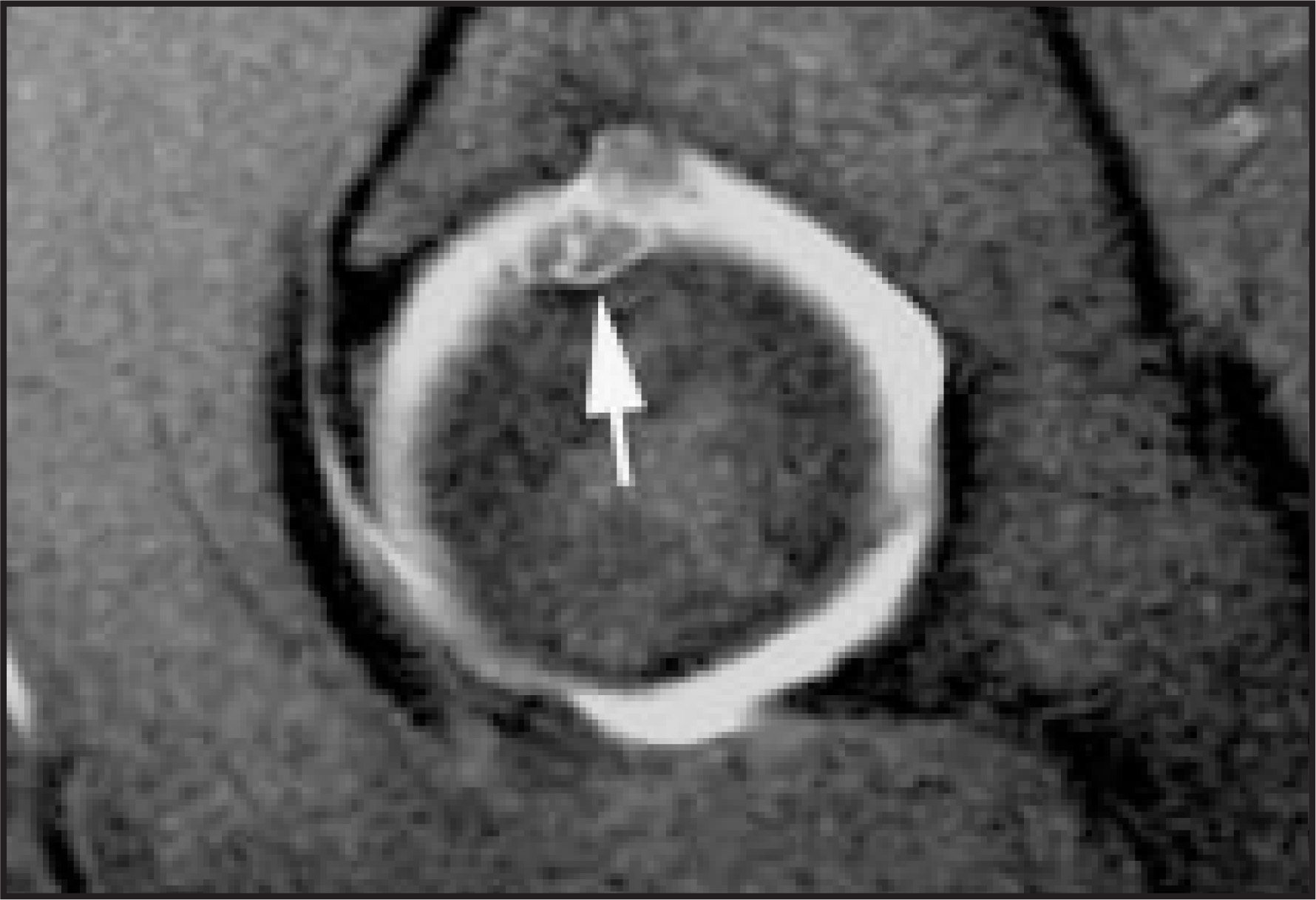
Post-traumatic lesion of the soft tissues can also be studied with MRI. Among the tendon lesions around the hip, hamstring avulsion injuries and tears are frequent in certain sports that involve extreme abduction of the hip. MRI can demonstrate the lesion as well as the degree of tendon retraction (Figure 8).8,9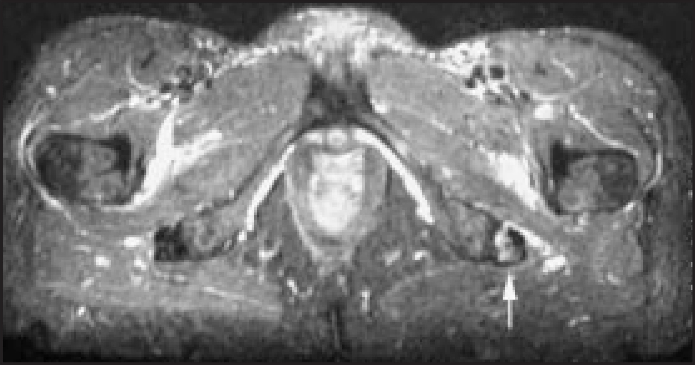
Other soft-tissue lesions that may be related to trauma or degeneration are the labral tears. These tears occur more frequently at the level of the superior aspect of the acetabular labrum and may be difficult to diagnose unless a high-quality MRI is performed or intra-articular or intravenous injection of contrast material is used (Figure 9).10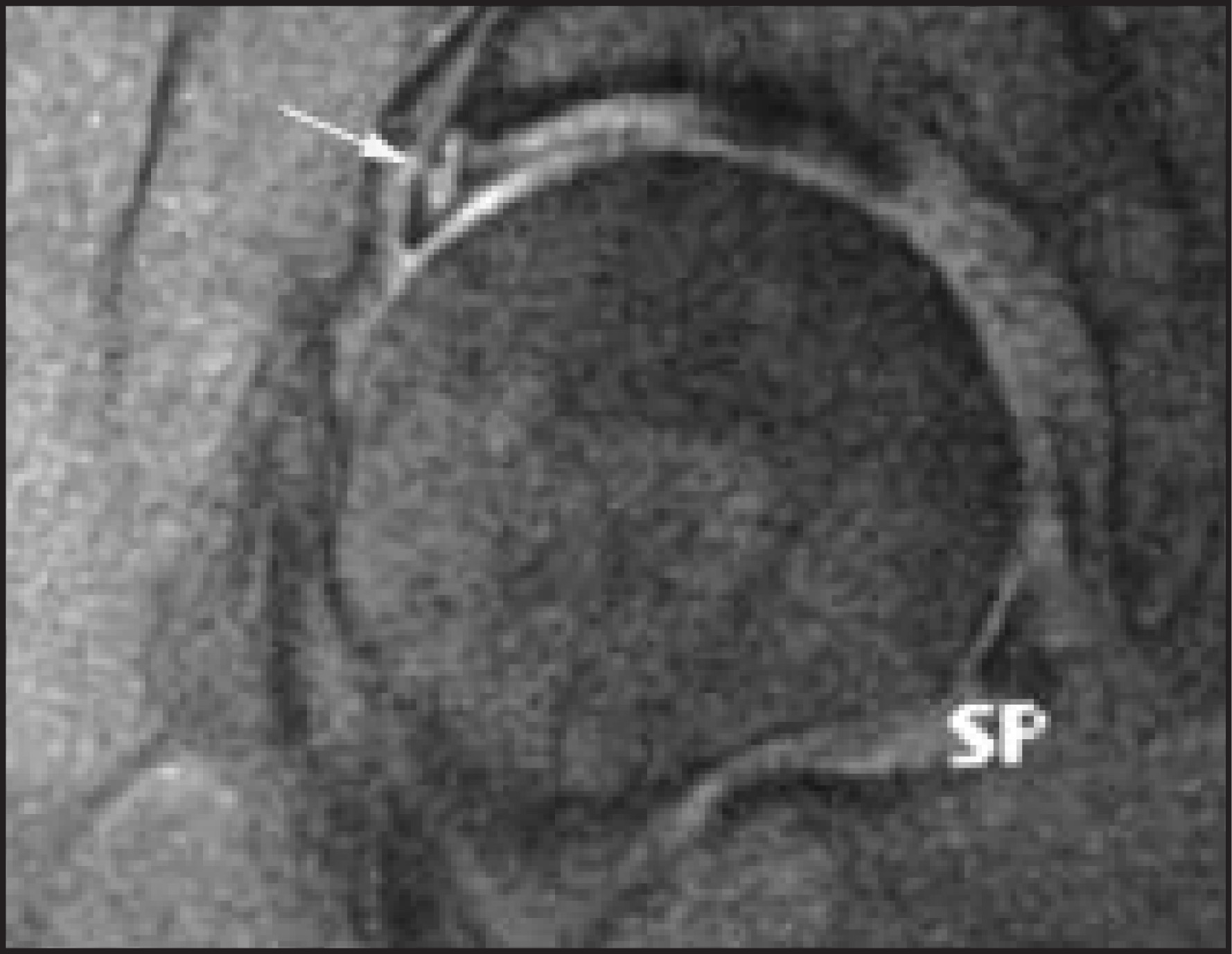
Bone-marrow lesions
Osteonecrosis or avascular necrosis (AVN) of the femoral head is one of the most frequent indications for MRI of the hip. MRI has been proven highly sensitive and accurate for the detection of this condition. The findings include an irregular low-signal intensity line that demarcates the peripheral aspect of the necrotic segment, which histologically correlates with the reactive interphase. A high-intensity line paralleling the hypointense line and termed the “double-line sign” is often seen and has been quite characteristic of AVN (Figure 10).11-13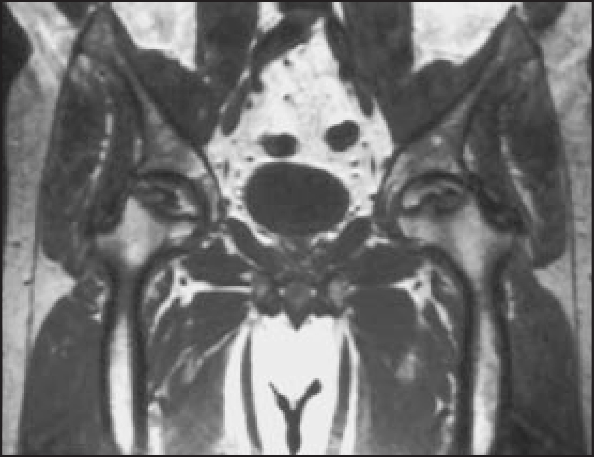
In the recent years, emphasis has been placed in the assessment of the size of the lesion as compared with the size of the weight-bearing portion of the femoral head. This measurement helps to determine the potential outcome if minimally invasive treatment, such as core decompression, is considered. Large areas of AVN will not respond to core decompression and the femoral head will eventually collapse, leading to osteoarthritis and subsequent need for joint replacement. Small lesions (<25% of the weight-bearing portion of the femoral head) have a better chance to remain stable, without collapse, following core decompression.14-17
Transient osteoporosis of the hip is another relatively frequent bone marrow condition seen on MRI. The entity was described in pregnant women during the third trimester of pregnancy, but is now recognized to be more frequent in overweight middle-aged men. The MRI findings include extensive bone-marrow edema of the femoral head and neck, frequently associated with a joint effusion (Figure 11). The condition is generally self-limited, with clearing of the clinical symptoms and MRI changes in 6 to 8 months. Some cases may demonstrate a focal lesion in the subchondral area of the femoral head once the bone marrow edema is clear. This finding suggests that TOH and stress-related subchondral fracture are the same entity, since excessive weight is a common denominator in these patients.18,19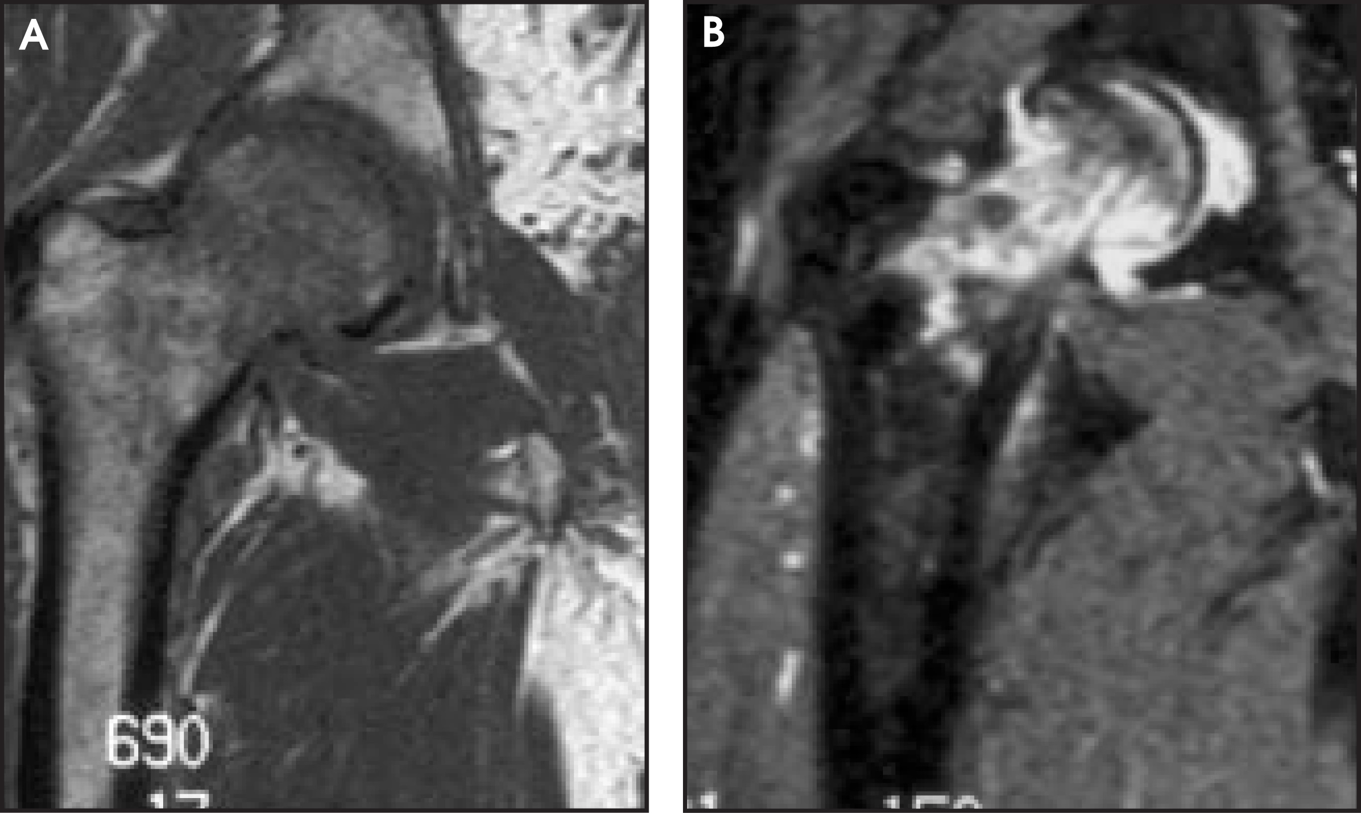
Infection, inflammation
Septic arthritis and osteomyelitis occasionally occur in the hip. The MRI findings of septic arthritis are nonspecific. In the early stages, a joint effusion is present. Periarticular soft-tissue edema may also be noted (Figure 12). The characteristic cartilage loss and surface erosions, also well-demonstrated on plain films, follow these findings. The MRI manifestations of osteomyelitis include bone-marrow edema and periostitis with periarticular soft-tissue edema during the early stages, followed by intraosseous abscess formation (Brodie’s abscess). The value of MRI is the detection of early infection, before the formation of abscess.20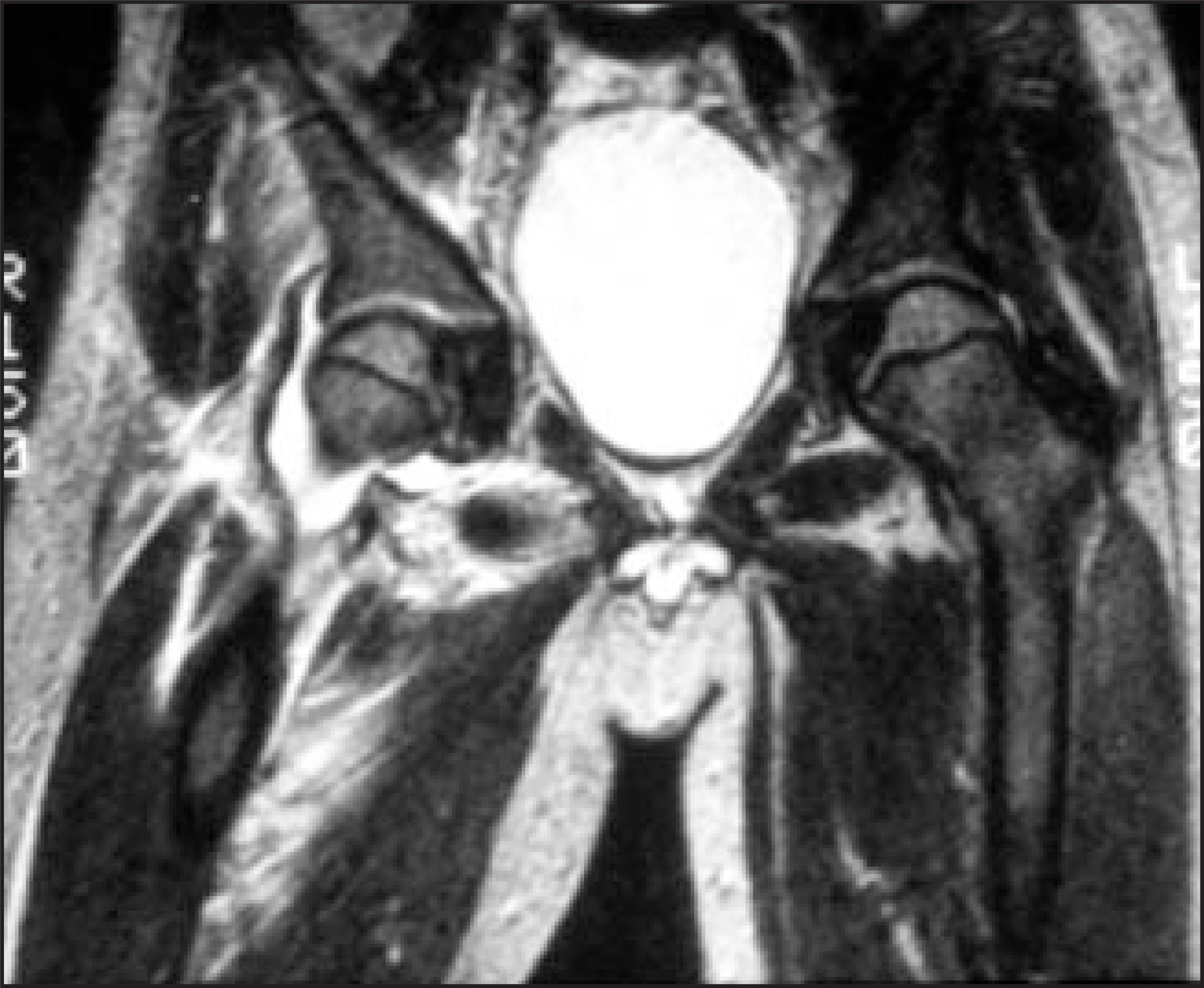
Among the inflammatory conditions, iliopsoas bursitis is a relatively frequent and symptomatic entity. The iliopsoas bursa is not normally seen on MRI since the amount of fluid it contains is not sufficient enough to be detected. Under inflammatory conditions, fluid accumulates in the bursa, which becomes distended, attaining an hourglass shape, in part intrapelvic and in part extrapelvic. This characteristic appearance is easily seen on MRI (Figure 13).21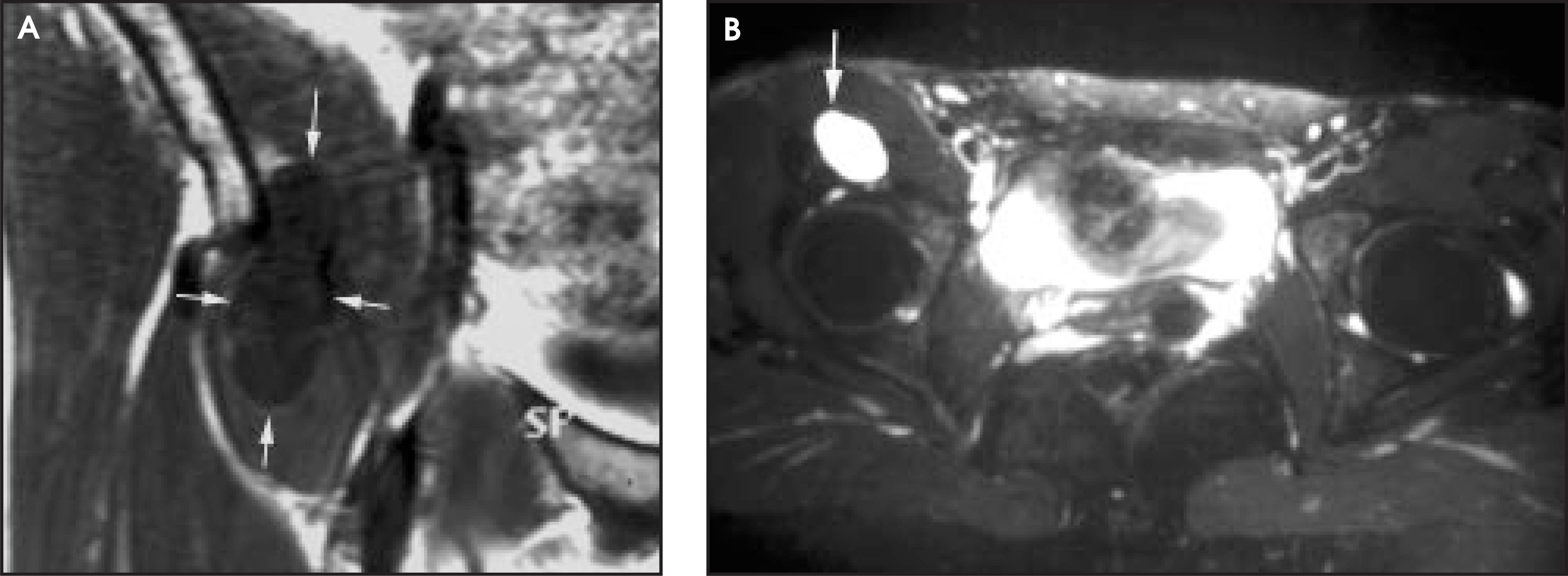
Miscellaneous conditions
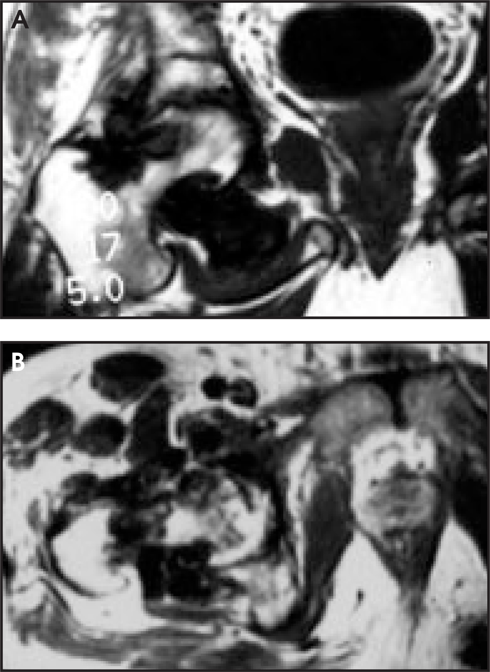
Synovial osteochondromatosis occurs frequently in the hip joint. MRI demonstrates the presence and location of the multiple cartilaginous bodies within the recesses of the capsule, often associated with an effusion and erosions of the surface of the bone, particularly at the level of the femoral neck (Figure 14).22 The differential diagnosis based on the MRI findings includes pigmented villonodular synovitis (PVNS) since both entities can display areas of low signal intensity within the joint capsule. In synovial osteochondromatosis, the hypointense areas are related to ossification of the cartilaginous bodies, whereas hemosiderin deposition is responsible for the areas of decreased signal intensity in various pulse sequences in PVNS (Figure 15).23 Both diseases can also display surface erosions (apple core) of the femoral neck and joint effusion.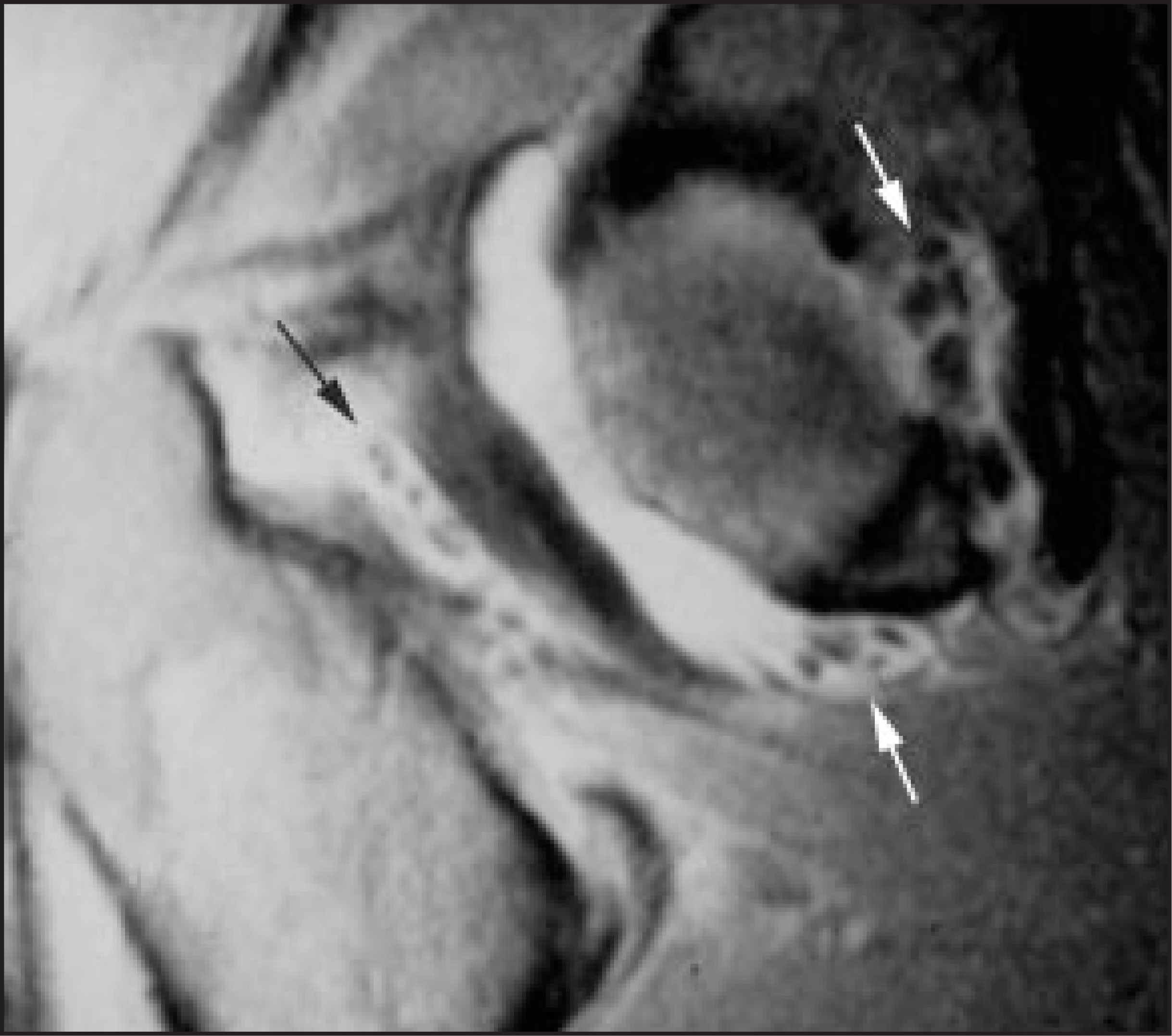
In general, the primary diagnostic approach to bone tumors is made by their radiographic characteristics and MRI is used for evaluation of their intramedullary and soft-tissue extension, articular extension, and neurovascular bundle involvement. Computed tomography (CT) is used for assessment of cortical and periosteal extension and detection of subtle matrix calcifications not seen on plain films. Some tumors, tumor-like conditions, and miscellaneous conditions have a special predilection for the proximal femur. Frequent proximal femoral lesions include osteoid osteomas, osteoblastoma, chondroblastoma, fibrous dysplasia, unicameral bone cyst, chondrosarcoma, and Paget’s disease. Special mention should be made of osteoid osteomas, which are relatively frequent around the hip. Osteoid osteomas are benign bone-forming tumors that characteristically demonstrate cortical thickening surrounding a nidus. These lesions are better evaluated with plain films and CT than with MRI, since the purpose of the imaging is to locate the small nidus prior to surgical removal or percutaneous ablation. However, MRI may be used occasionally if the plain radiographs do not demonstrate the lesion. Unfortunately, MRI findings may be misleading, since these tumors produce extensive bone-marrow and soft-tissue edema to the point that the edema may obscure the nidus.24
Conclusion
Due to the exquisite soft-tissue contrast resolution of MR combined with recent technologic developments, a variety of conditions involving the hip joint and adjacent bony structures can be well evaluated using MRI, with or without contrast material. Among these conditions, hip trauma and AVN are the most common indications. Other entities in which MRI has proven its usefulness include inflammatory and infectious processes, intra-articular lesions (such as synovial chondromatosis and pigmented villonodular synovitis), and a variety of bone-marrow disorders.
References
- Kier R, Wain S, Troiano R. Fast spin-echo MR imaging of the pelvis obtained with a phased-array coil: Value in localizing and staging prostatic carcinoma.. AJR Am J Roentgenol.. 1993;161:601-606.
- Andrews C. Evaluation of the marrow space in the adult hip.. RadioGraphics.. 2000;20:27-42.
- Sadro C. Current concepts in magnetic resonance imaging of the adult hip and pelvis.. Semin Roentgenol.. 2000;35:231-248.
- Bogost G, Lizerbram E, Crues J. MR imaging in evaluation of suspected hip fracture: Frequency of unsuspected bone and soft-tissue iinjury.. Radiology.. 1995;197:263-267.
- May D, Purins J, Smith D. MR Imaging of occult traumatic fractures and muscular injuries of the hip and pelvis in elderly patients.. AJR Am J Roentgenol.. 1996;166:1075-1078.
- Rizzo P, Gould E, Lyden J, Asni S. Diagnosis of occult fractures about the hip. Magnetic resonance imaging compared to bone scanning.. J Bone Joint Surg.. 1993;75-A:395-401.
- Yamamoto T, Schneider R, Bullough P. Subchondral insufficiency fracture of the femoral head: Histopathologic correlation with MRI.. Skeletal Radiol.. 2001;30:247-254.
- Anderson M, Kaplan P, Dussault R. Adductor insertion avulsion syndrome (thigh splints): Spectrum of MR imaging features.. AJR Am J Roentgenol.. 2001;177:673-675.
- Brandser E, El-Khoury G, Kathol M. Hamstring injuries: Radiographic, conventional tomographic, CT, and MR imaging characteristics.. Radiology.. 1995;197:257-262.
- Czerny C, Hofman S, Neuhold A. Lesions of the acetabular labrum: Accuracy of MR imaging and MR arthrography in detection and staging.. Radiology.. 1996;200:225-230.
- Coleman B, Kressel H, Dalinka M. Radiographically negative avascular necrosis: Detection with MR imaging.. Radiology.. 1988;168:525-528.
- Mitchell D, Rao V, Dalinka M. Hematopoietic and fatty bone marrow distribution in the normal and ischemic hip: New observations with 1.5T MR imaging.. Radiology.. 1986;161:199-202.
- Mitchell D, Rao V, Dalinka M. Femoral head avascular necrosis: Correlation of MR imaging, radiographic staging, radionuclide imaging, and clinical findings.. Radiology.. 1987;162:709-715.
- Beltran J, Knight C, Zuelzer W. Core cecompression for avascular necrosis of the femoral head: Correlation between long term results and preoperative MR staging.. Radiology.. 1990;175:533-536.
- Smith S, Fehring T, Griffin W, Beaver W. Core decompression of the osteonecrotic femoral head.. J Bone Joint Surg.. 1995;77-A:674-680.
- Urbaniak J, Coogan P, Gunneson E, Nunley J. Treatment of osteonecrosis of the femoral head with free vascularized fibular grafting.. J Bone Joint Surg.. 1995;77-A:681-694.
- Wassenaar R, Verburg H, Taconis W, Eijken J. Avascular osteonecrosis of the femoral head treated with a vascularized iliac bone graft: Preliminary results and follow-up with radiography and MR imaging.. RadioGraphics.. 1996;16:585-594.
- Guerra J, Steinberg M. Current concepts review-distinguishing transient osteoporosis from avascular necrosis of the hip.. J Bone Joint Surg.. 1995;77-A:616-624.
- Hayes C, Conway W, Daniel W. MR imaging of bone marrow edema pattern: Transient osteoporosis, transient bone marrow edema syndrome, or osteonecrosis.. RadioGraphics.. 1993;13:1001-1011.
- Hammond P, Macnicol M. Osteomyelitis of the pelvis and proximal femur: Diagnostic difficulties.. J Pediatr Orthop B.. 2001;10:113-119.
- Varma D, Richli W, Charnsangavej C. MR Appearance of the distended iliopsoas bursa.. AJR Am J Roentgenol.. 1991;156:1025-1028.
- Hermann G, Abdelawahab I, Klein M. Synovial chondromatosis.. Skeletal Radiol.. 1995;24:298-300.
- Gonzalez Della Valle A, Piccaluga F, Potter H. Pigmented villonodular synovitis of the hip: 2-to 23-year follow-up study.. Clin Orthop.. 2001;388:187-199.
- Kransdorf M, Stull M, Gilkey F, Moser R. Osteoid osteoma.. RadioGraphics.. 1991;11:671-696.
Citation
. MRI of the hip. Applied Radiology. 2002;31(11):33-39. doi:10.37549/AR1139.