Multichannel helical CT of the musculoskeletal system
Applied Radiology — Vol. 32 , Issue 5 , pp. 15 -22
DOI: 10.37549/AR1185
Published: May 1, 2003
Categories
Since its advent, computed tomography (CT) has been an important modality for cross-sectional imaging of the musculoskeletal system, particularly in the setting of trauma. The superior contrast resolution and direct multiplanar capability of magnetic resonance imaging (MRI) led to its dominance over CT for imaging soft tissue, such as meniscus, and cartilage. Slip-ring CT systems offer helical acquisition techniques, providing greater scanning speed and routine multiplanar/three-dimensional (3D) display capability. The latest generation multichannel CT imagers enhance volumetric assessment of anatomy and pathology with ultrathin, isotropic microvoxel data acquisitions, which allow seamless, high-resolution evaluation in any plane. This enhanced volumetric scan capability has led to an expansion of the clinical role of CT as well as significant changes in the way that radiologists interact with the larger amounts of imaging information.
This article will discuss the basic principles of multichannel CT and will demonstrate the impact of the latest generation microvoxel volumetric scan techniques. Optimal methods for data display and interrogation will be suggested, and the expanded clinical role of CT in the evaluation of musculoskeletal disorders will be explored.
Multichannel helical CT
Slip-ring CT systems allow continuous scanner gantry motion, which when coupled with synchronous table feed, provides the basis for helical or spiral CT scanning. As opposed to “step-and-shoot” techniques (the only option on older systems, in which the table moves an increment corresponding to the acquired slice thickness), with helical techniques one can “stretch the spiral” to cover a distance greater than the collimation during each 360° scanner revolution. This “stretch” is generally described in terms of “pitch,” which is defined (for a single-channel system) as the distance the table moves per 360° gantry rotation divided by the scan collimation.
At any given collimation, as pitch increases, the anatomical coverage increases per unit time. Practical limits on pitch exist that relate to the richness of the data supplied to the reconstruction algorithm. As pitch increases, the effective slice profile (thickness) increases. This is less of a limitation than it may seem, since the use of higher pitch values may allow the choice of a thinner collimation acquisition and provide a thinner slice profile than a choice of a thicker slice with a less aggressive pitch. For example, a 1-mm acquisition at a pitch of 3 yields a higher z-axis resolution scan than a 3-mm acquisition at a pitch of 1. The most important practical limit on pitch selection is the incidence of undersampling “helical” artifacts as the data stream gets lean with extended pitch. It is generally accepted that a pitch of 2 (or 2 times the number of imaging channels on a multichannel system) is the practical limit, although on slower single-channel systems this limit could be exceeded in challenging clinical circumstances, such as trauma.
The fundamental advantage of multichannel CT systems is the simultaneous acquisition of multiple helical data streams that contribute to each slice created. This inherently information-rich scan technique permits a proportional increase in the “stretch” of each helical data stream and a proportional increase in table movement/anatomical coverage per unit time while still protecting slice profile.1 The definition of pitch for multichannel CT systems (which reflects the relative effective slice profile) is the distance the table travels per 360° gantry revolution divided by the dimension of exposed detector (defined as collimation times the number of imaging channels). As an example, a 16-channel system collimated to 1.25 mm (at 0.5 seconds per gantry revolution) could cover 1050 mm in 15 seconds. While maintaining a pitch of 1.75, the scanner covers anatomy almost 19 times faster than a single-channel system operating at a pitch of 1.5. The huge boost in speed allows a thinner collimation choice that easily compensates for the slight increase in slice profile broadening.
Isotropic data acquisitions
Multichannel capabilities have led to fundamental changes in the way CT data are acquired, processed, displayed, and interrogated. Typical scan techniques on a 16-channel imager employ slice thicknesses between 0.5 and 1.5 mm, depending on the detector design and the body part evaluated. These thin-slice volume acquisitions provide imaging voxels that are effectively isotropic (equal in size in all dimensions). Isotropic data can be reformatted and viewed in orthogonal and oblique planes without tradeoff in image integrity. It is probably fair to state that the reformatted images possible with an isotropic data set are superior to and higher in inherent spatial resolution than what was once the gold standard (at typically thicker slice) in the direct imaging plane (Figure 1).2 As a result, not only is interrogation of CT studies no longer limited to the acquisition plane, but multiplanar/volumetric scan interrogation is often completed on a 3D workstation or picture archiving and communication system (PACS) interface with multiplanar reformatting (MPR) capability. Such interrogation is becoming more and more routine for primary interpretation purposes, particularly in areas of the body in which multiplanar scanning was the norm previously, such as the musculoskeletal system, and the head and neck.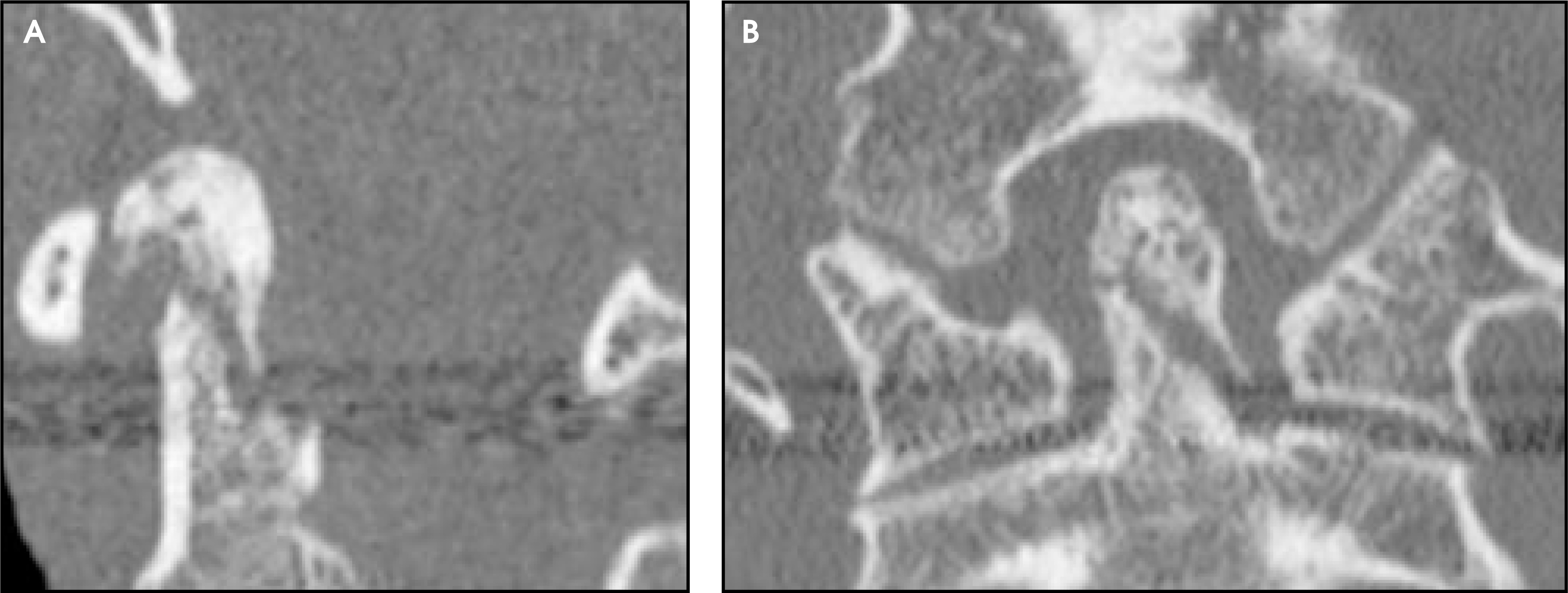
The thinner slices used on today’s scanners bring additional imaging benefits. As the slice thickness used for acquisition decreases, the inherent contrast resolution of the volumetric data increases. This is readily evident in low contrast resolution examinations, such as the posterior fossa of the head, and lumbar spine. Perhaps more important in a discussion of CT of the musculoskeletal system is the fact that as acquired slice thickness decreases, partial volume artifacts, such as those due to metal instrumentation hardware (Figure 2), diminish.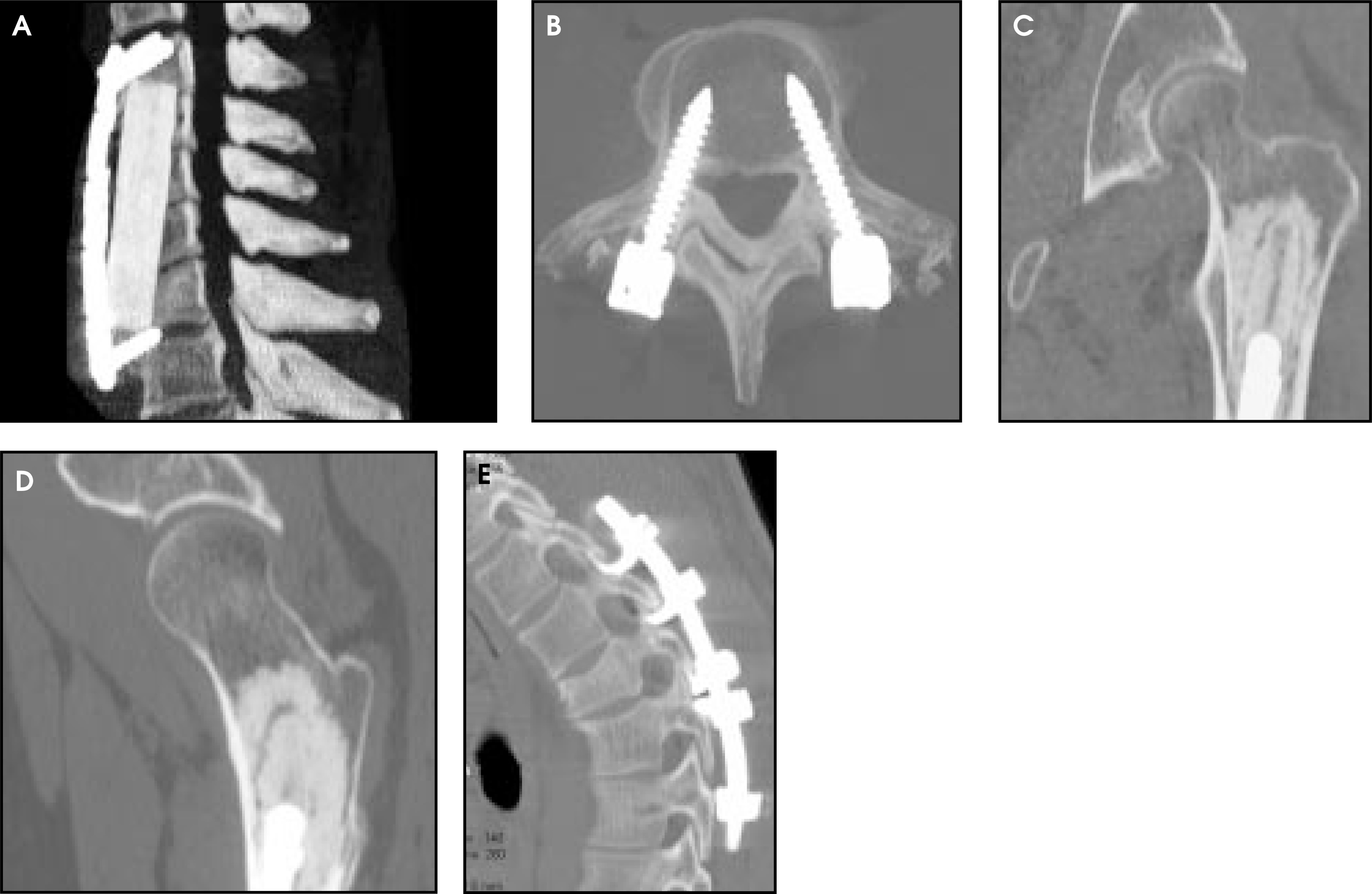
Data management
Microvoxel scan techniques force reconsideration of methods of data recording and archiving. Scan acquisitions produce “raw data,” which, depending on system limits, exist as long as space exists on the system hard disk drives. The oldest case is typically erased as the most recent case is scanned. As long as it remains available, raw data can be reconstructed into slices at novel resolution (thickness and field of view [FOV]), table location, and algorithm. Once raw data have been erased, the slices that have been created can be reformatted in multiple planes and combined in-plane to create a thicker slice, but thinner slices, FOV, and algorithm changes are no longer possible. Raw data are rarely stored in the long-term archive for later manipulation.
To date, the informal standard of care has been to store all reconstructed slices in the patient’s record in the long-term archive. This poses little problem for routine imaging needs, as the number of slices used for interrogation has increased only approximately 20% to 50% over the recent generations of scanners (eg, 80 to 100 slices for a typical body study). Where this strains practicality is in applications for which multiplanar interrogation is critical, as today’s microvoxel data sets may contain up to 3000 slices. These ultrathin slices may never be viewed directly, but might supply the “fuel” for a 3D rendering or MPR interrogation at thicker slice and wider interval. A limited number of axial, coronal, and sagittal images are typically recorded and made available in the patient record (Figure 3). It may be best to shift to a new standard requiring only these images to be stored long-term. The implications for PACS network congestion and archival costs are clear and significant.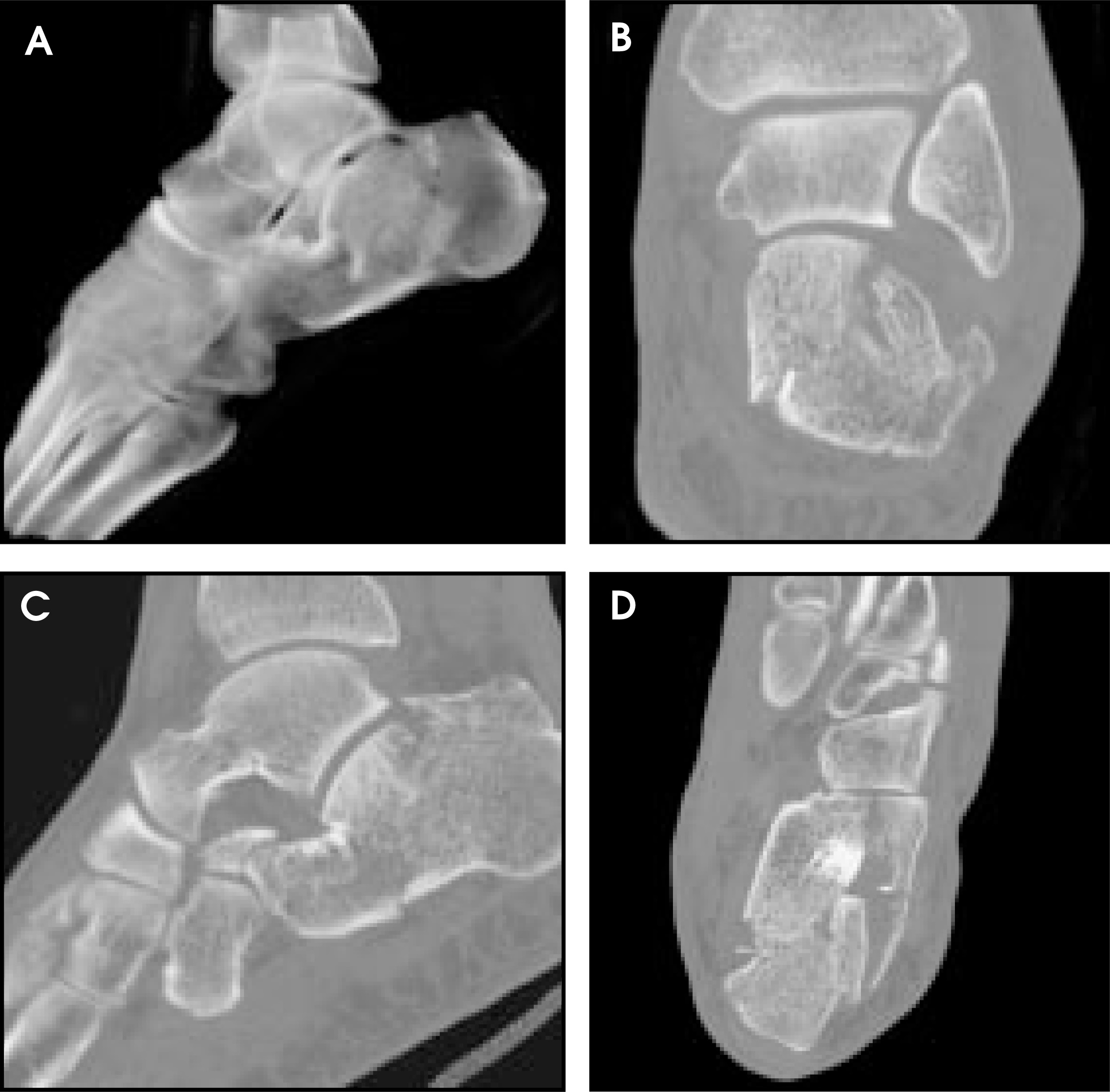
Protocol considerations for musculoskeletal CT
When designing protocols for CT of the musculoskeletal system (MSKCT), it is best to split the applications into axial and appendicular skeleton/spine (Table 1). As bone imaging is generally a high contrast resolution application, low tube current (mAs) is generally employed, particularly for the appendicular skeleton. Noise can limit the quality of studies of the axial skeleton (shoulder, hip, thoracolumbar spine), thus higher mA values and full-second scanning are generally used. In circumstances in which low contrast resolution may be important (eg, soft-tissue evaluations) higher scan techniques are advised.3
Since the primary plane of interrogation is typically along the long axis of patient anatomy (coronal, sagittal) as opposed to the cross-section (axial), the highest z-axis (longitudinal) acquisition resolution (0.5 to 0.75 mm) is best. Moderate pitch values are generally chosen to further maintain the thinnest slice profile. Image noise considerations based on limitations of today’s X-ray tubes and generators limit the slice thicknesses that are practical for scanning the axial skeleton, thus thicker slices (1 to 1.5 mm) may be chosen. Lower pitch values are used to increase the effectiveness of scan exposures and further reduce noise. Imminent advances in tube capacity and generator power should have positive implications and increase technique flexibility in this area.
Without exception, MSKCT studies are obtained in a single microvoxel volumetric acquisition.4 The fuss, discomfort, and additional radiation dose due to direct multiplanar scanning is no longer a feature of the scan experience, which has improved patient satisfaction and department throughput. The superior capabilities and lower dose of today’s multichannel imagers allow an increase in the practical anatomical coverage as well. A typical study of a long bone would include the bone in its entirety, as a plain film radiographic study typically would. A routine lumbar spine examination extends axially and sagittally from T12 through S1, matching the routine coverage of MRI.
Trauma
The exquisite high contrast resolution and seamless multiplanar capability of the latest multichannel CT imagers have their greatest impact in MSKCT in the evaluation of trauma. Thorough assessment requires evaluation in orthogonal (and, occasionally, oblique) planes. For most extremity work, direct sagittal scanning is not feasible. For the coronal scan plane, anatomy has to be positioned parallel to the gantry. It is not at all uncommon for the scanner to impinge on the fractured extremity as the patient passes through, resulting in pain and motion on the examination.
Fractures are now delineated easily in any plane while the patient is situated comfortably on the table. Positioning during the scan is not critical, as any plane can be created from the volumetric data. Studies typically require well under 30 seconds to complete, minimizing the likelihood of patient motion (Figure 4). Lower scan techniques are typically used when the extremity can be scanned away from the remainder of the body. For the axial skeleton and circumstances in which the body must be scanned along with the extremity, high tube current is advised.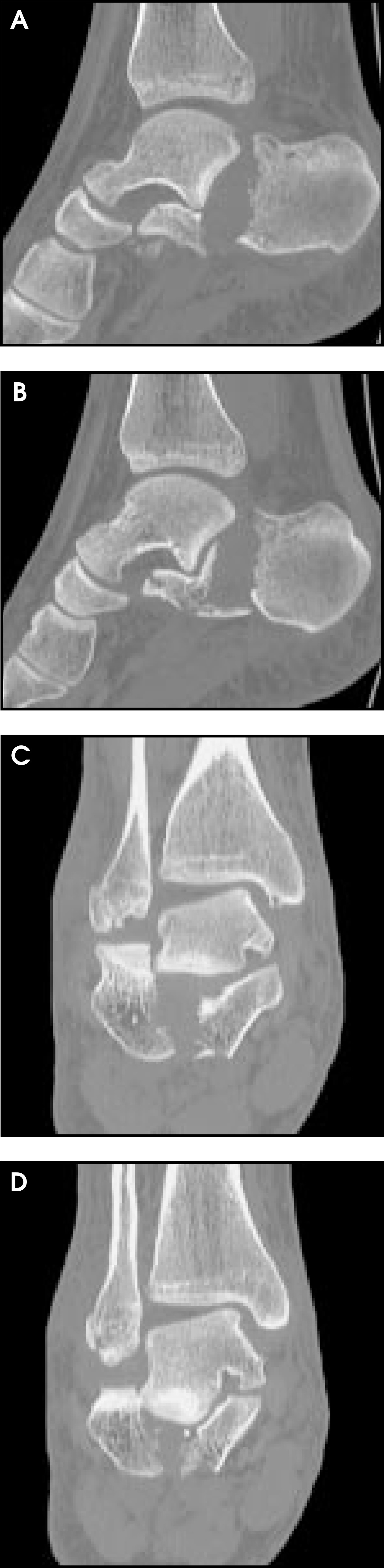
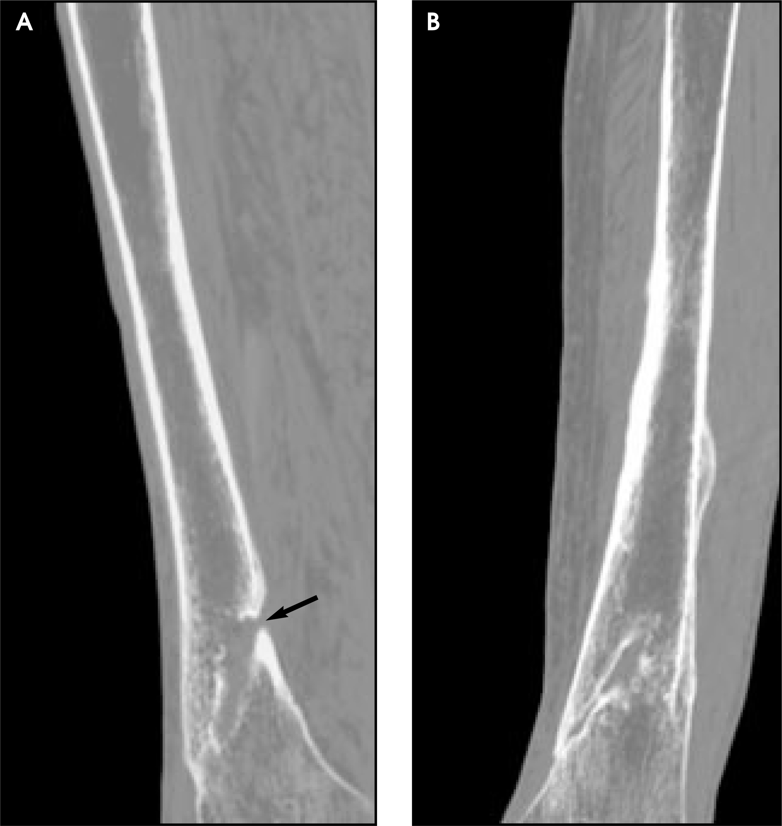
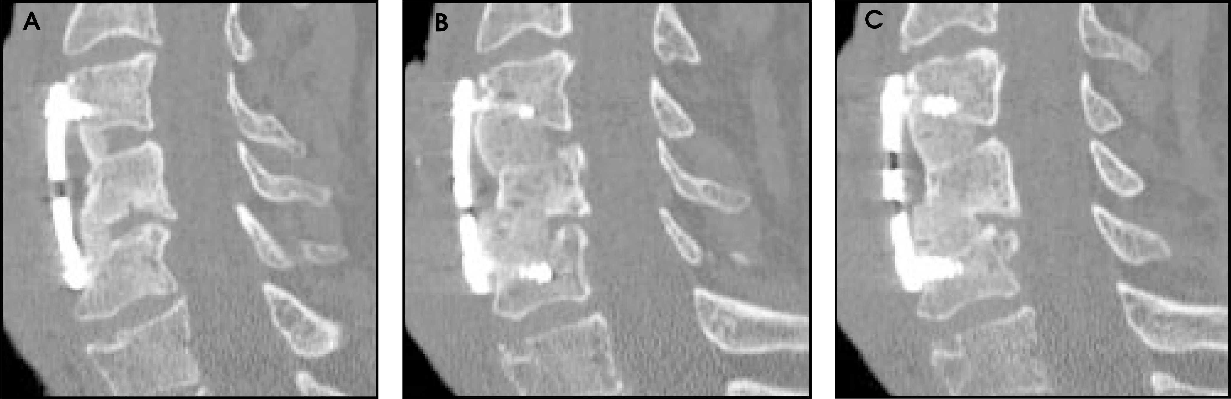
CT is ideally suited to treatment planning in acute trauma, providing delineation of the presence and extent of subtle cortical fractures, fracture fragments, intra-articular loose ossific bodies and articular surface offset/depression (Figure 5). CT is also well suited to following the progress in fracture healing in the skeletal system and spine and detecting pseudarthrosis (Figure 6).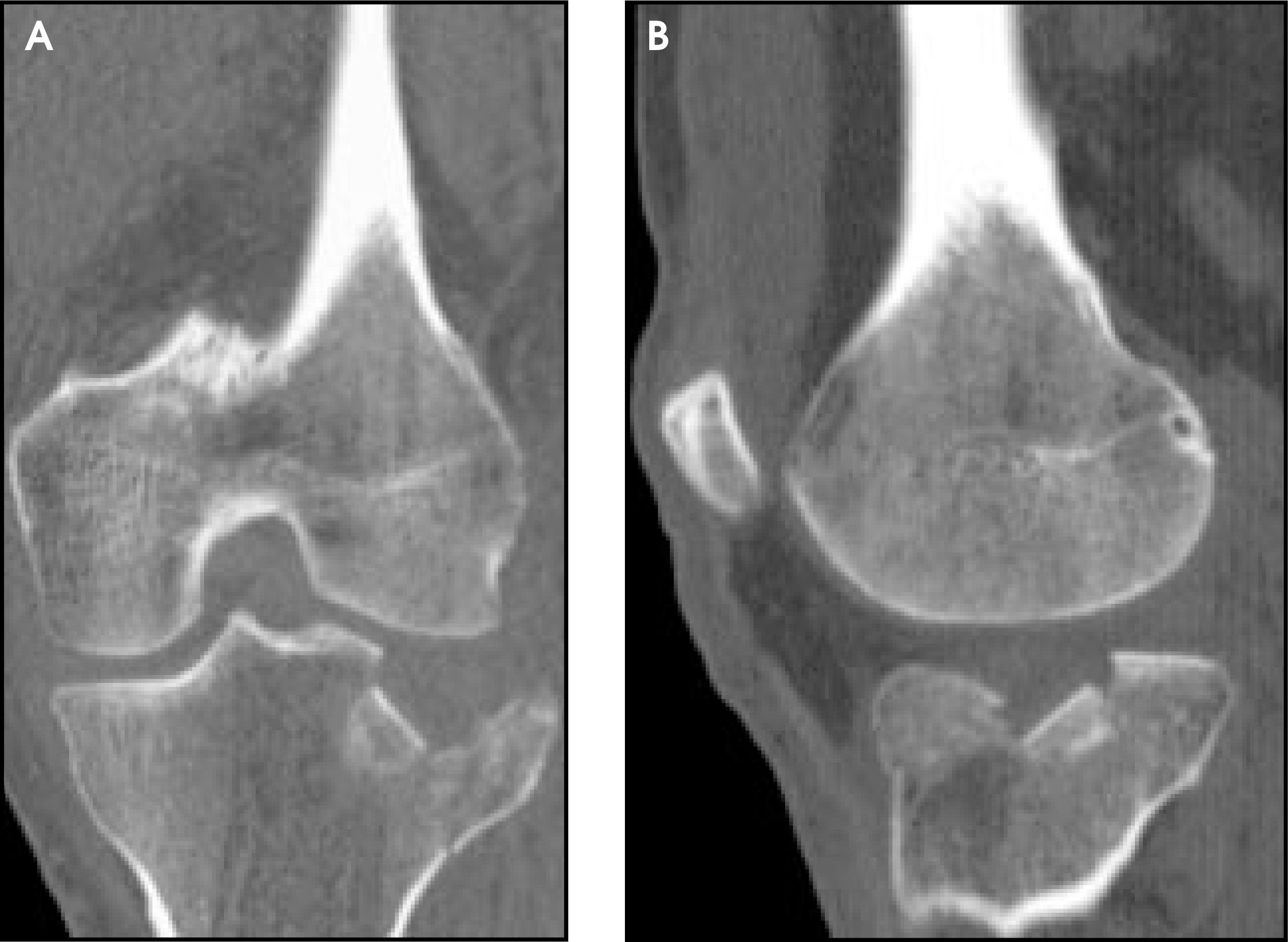
Degenerative disease
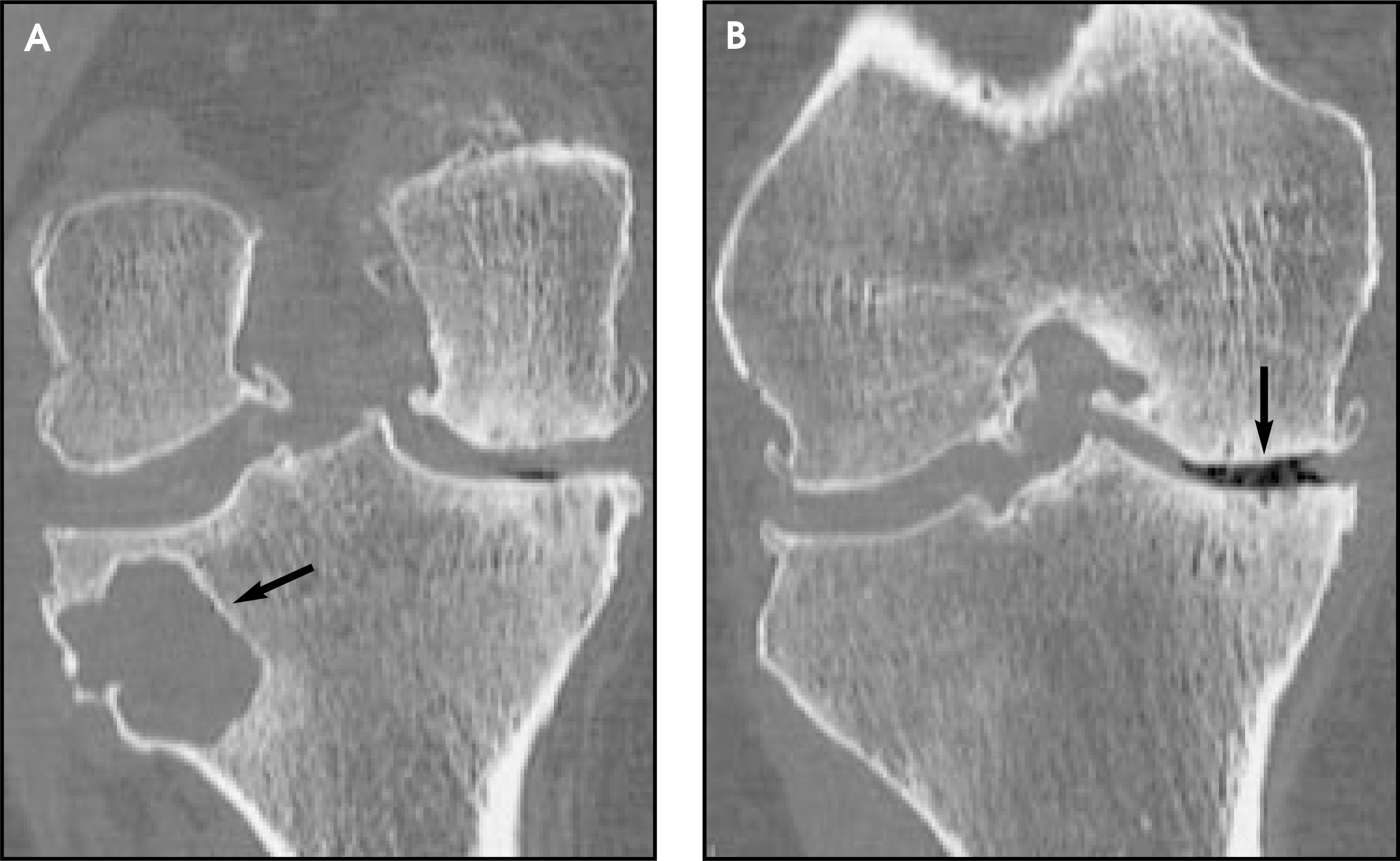
CT is commonly used to delineate the presence and severity of degenerative disease as part of the workup prior to joint replacement surgery. While cartilage is better assessed directly with high-performance MR, CT is superior, particularly to conventional radiography, in displaying the full extent of secondary changes of degenerative disease, such as joint space narrowing, osteophyte formation, cortical eburnation, and subchondral cystic change (Figures 7 and 8). The microvoxel scan techniques employed with current scanners are well-suited to postoperative evaluations, minimizing partial volume artifact and allowing evaluation of the state of the bone surrounding hardware implants, as well as evaluation of possible loosening and migration of prostheses (Figure 2).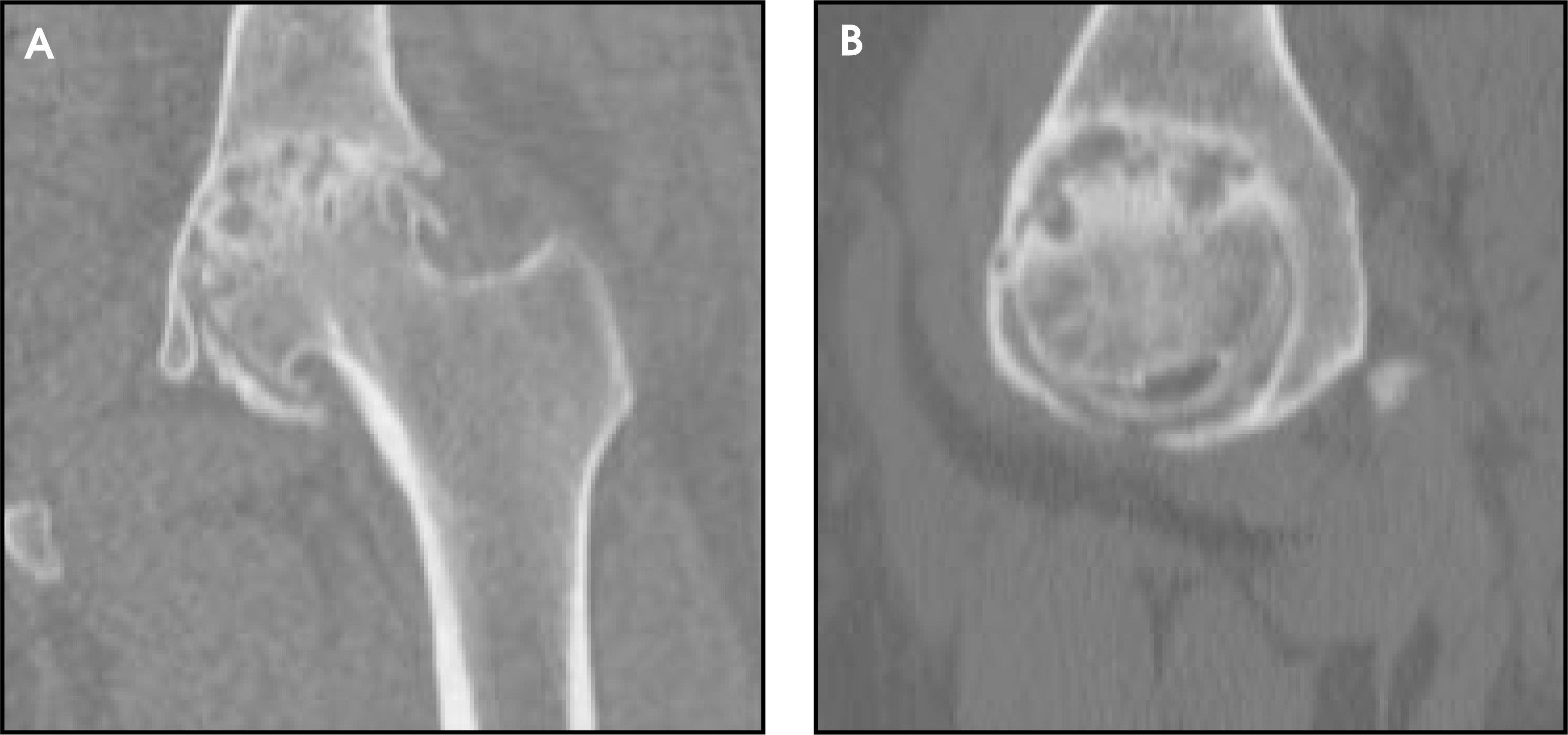
Tumor assessment
CT is often used as a rapid means of assessing possible lesions suggested by conventional radiography. With respect to known tumors, while MR is superior for the delineation of the extent of tumor in medullary bone and offers advantages in the evaluation of extraosseous soft-tissue masses, both situations are well assessed with volumetric multiplanar and 3D CT. Cortical bone (which is less well suited to MR due to the lack of mobile protons), lesion matrix calcification, and periosteal new bone formation are best delineated with CT (Figure 9).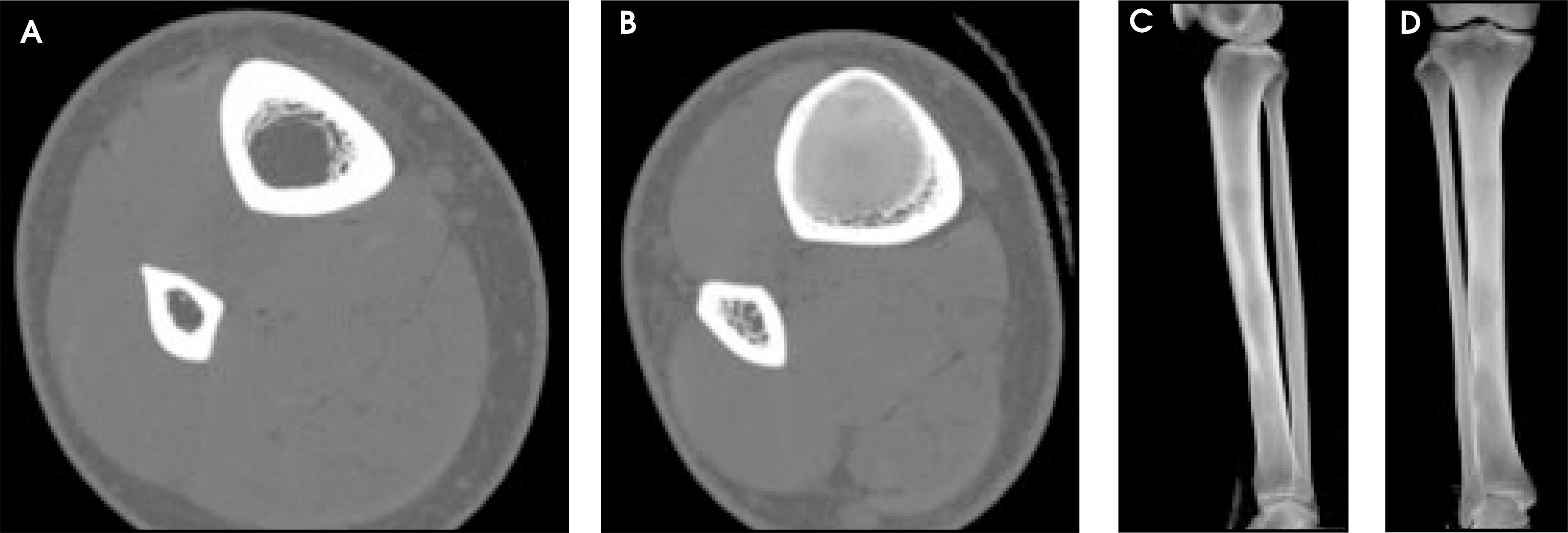
Spine
Multichannel techniques have had a significant impact on the style and quality of spine CT. Coverage is no longer limited by practical concerns. Lumbar exams now routinely extend from T12 through S1 (Figure 10), and cervical exams from C2 through T2 (Figure 11). It is practical to scan continuously through the entire thoracic spine. CT examinations are complete in seconds and are easier for patients to tolerate than are MR examinations. Microvoxel volumetric acquisition techniques provide a significant boost in spatial and contrast resolution, creating exquisite sagittal images from foramen to foramen, which rival MR.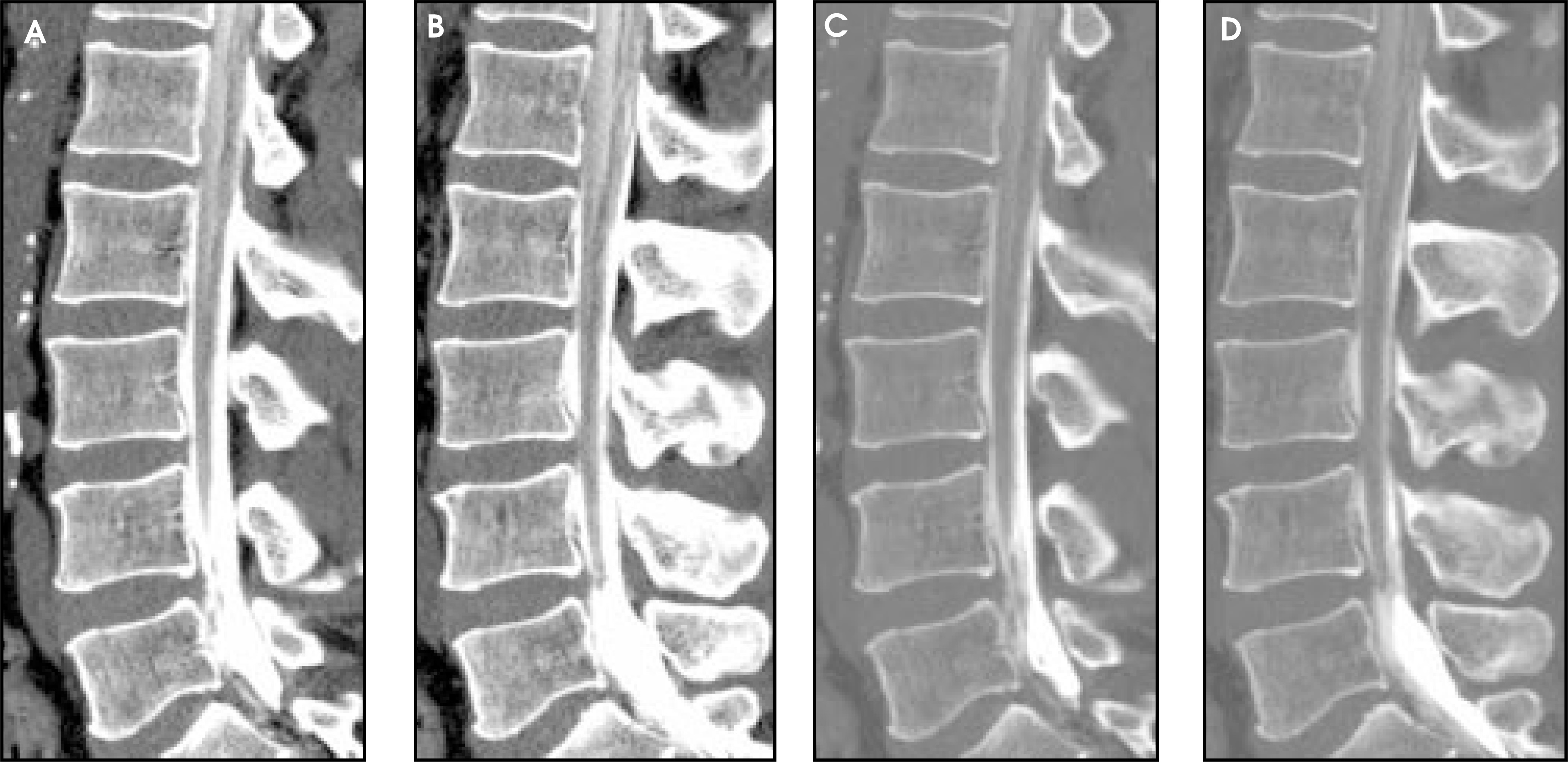
Spinal trauma
CT provides information in the acute trauma setting about alignment, canal compromise, and the presence and extent of fractures (Figure 1). MR, which is often performed in the subacute setting, provides complementary information about the integrity of the spinal cord and ligaments and (at least in the cervical and thoracic spine) provides better delineation of disc pathology than does CT. The whole-body microvoxel volumetric scans used to assess the chest, abdomen, and pelvis in trauma can be reconstructed into ideal spine images, allowing a whole-spine survey at the time of presentation. CT is best suited to the surveillance of fracture healing, with sagittal and coronal images playing a primary role in data interrogation and interpretation.
Degenerative disease of the spine
Due to superior contrast resolution and direct multiplanar scanning capability, MR is the dominant modality for the evaluation of degenerative spine disease, particularly in the cervical and thoracic spine where CT lacks contrast resolution. Microvoxel scan techniques significantly boost low-contrast contrast resolution in both in-plane and in reformatted images, approaching the quality of MR. The spatial resolution of multichannel CT acquired with submilli-meter voxels is superior to that provided by MR in any imaging plane (Figure 12).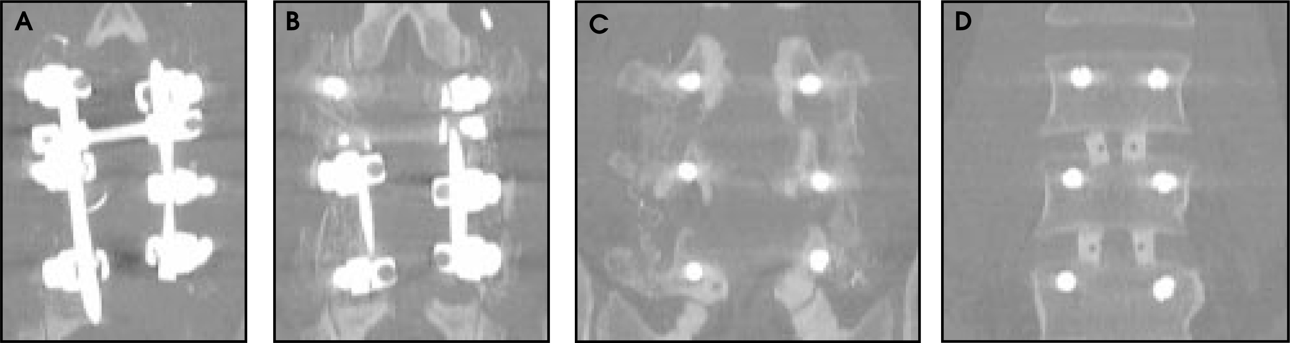
Spinal tumors
Spinal cord tumors are best assessed with MR, although CT and CT myelography continue to play a complementary role (Figure 13). CT can provide exquisite multiplanar and 3D information about the presence and extent of bone involvement.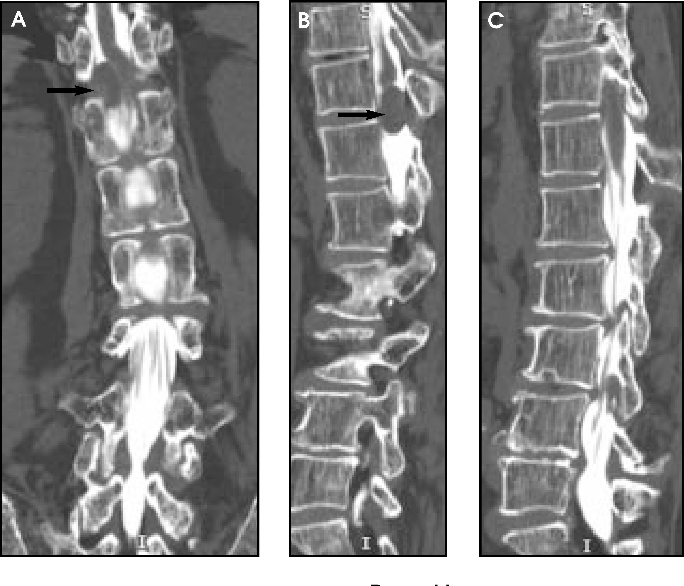
Dysraphism
MR continues to dominate the evaluation of children with suspected dysraphic disorders. The vastly superior speed (and improved contrast resolution) of multichannel CT exams, typically accomplished without sedation, may lead to an increased role in the evaluation of these infants and children. The bone information provided by CT can be very helpful in selected preoperative circumstances.
Conclusion
Continuing developments in multichannel CT technology have had an enormous impact on the style and quality of CT scanning in clinical practice and have led to an increasingly important role in evaluation of disorders of the musculoskeletal system and spine. AR
References
- Fox S, Tanenbaum L, Ackelsberg S. Future directions in CT technology. Neuroimag Clin North Am. 1998;8:497-513.
- Pretorius E, Fishman E. Volume-rendered three-dimensional spiral CT: Musculoskeletal applications. RadioGraphics. 1999;19:1143-1160.
- Fishman E, Silverman P. Multislice Computed Tomography: A Practical Approach to Clinical Protocols. 2002:283-316.
- Buchwalter K, Rydberg J, Kopecky K. Musculoskeletal imaging with multislice CT. AJR Am J Roentgenol. 2001;176:979-986.
Citation
. Multichannel helical CT of the musculoskeletal system. Applied Radiology. 2003;32(5):15-22. doi:10.37549/AR1185.