Multifocal invasive ductal carcinoma and ductal carcinoma in situ
Applied Radiology — Vol. 37 , Issue 8 , pp. 33 -34
DOI: 10.37549/AR1639
Published: August 1, 2008
Categories
CASE SUMMARY
Screening mammography detected a dense, spiculated mass in a 47-year-old perimenopausal woman who had not had a mammogram for 2 years. Although her mother does not have breast cancer, a sister and several cousins do. The patient was later found to be BRCA negative. No other risk factors were present.
IMAGING FINDINGS
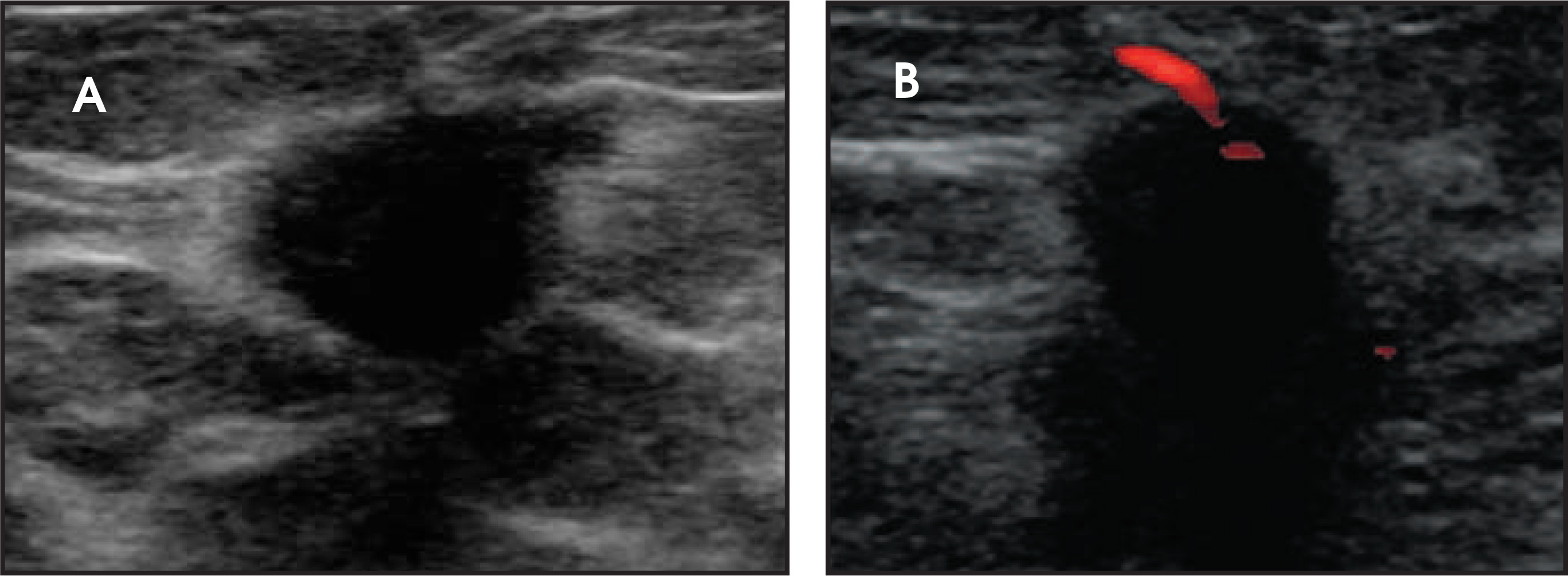
Mammography revealed a solitary high-density speculated mass (not shown). On ultrasound, it was found to be a hypoechoic, hyper-vascular mass with irregular margins, and no additional lesions were seen (Figure 1). The patient then underwent biopsy, which showed an invasive ductal carcinoma with ductal carcinoma in situ (DCIS). Perineural invasion was found. The patient was scheduled for a“golf-ball–sized” lumpectomy pending the results of breast magnetic resonance imaging (MRI).
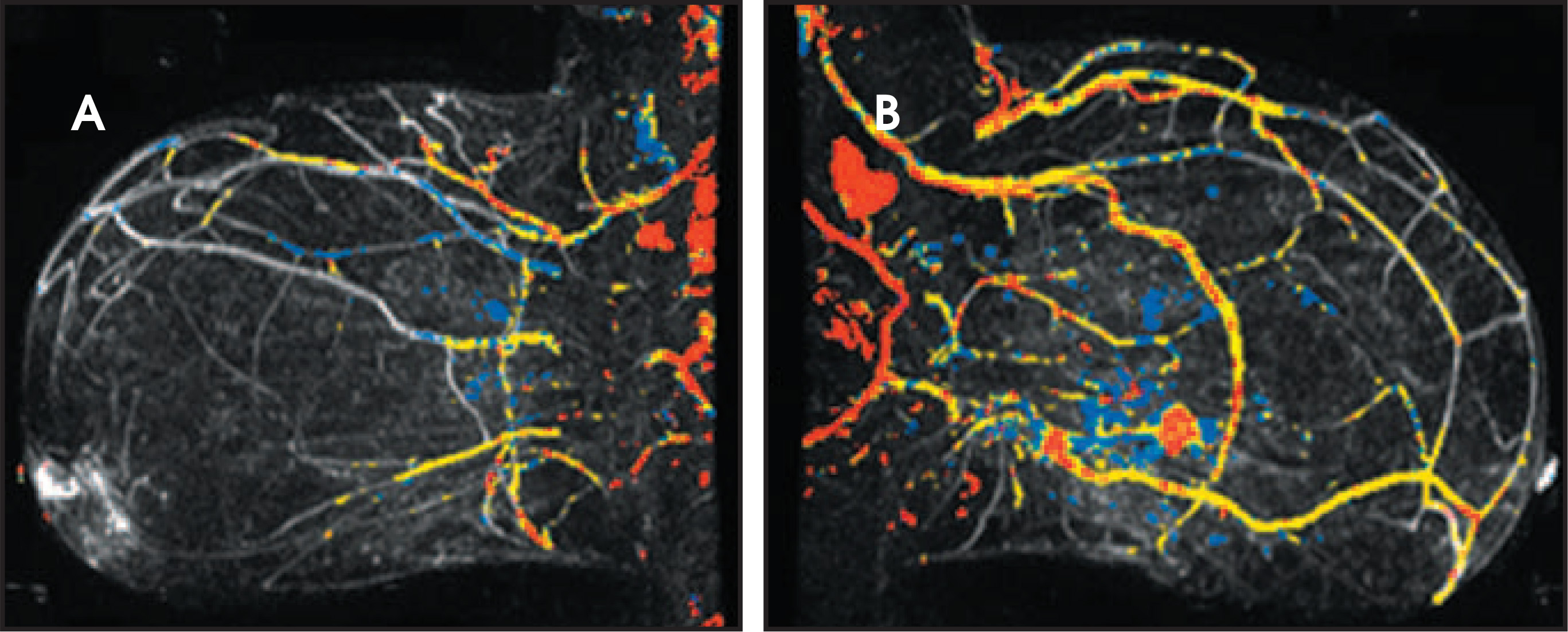
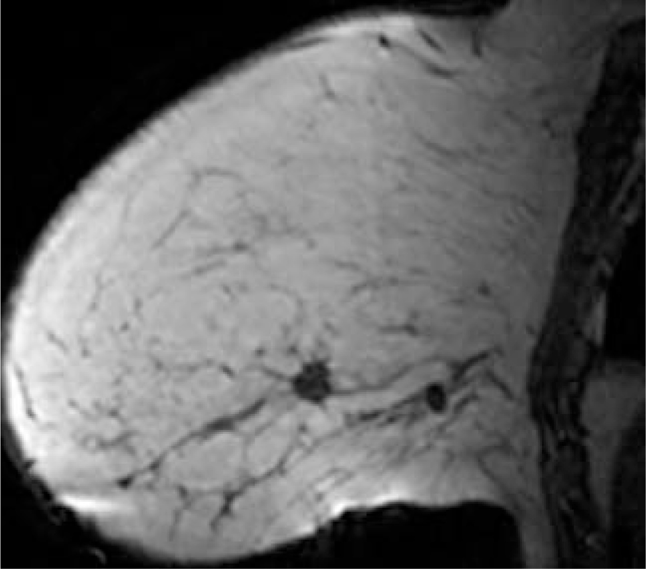
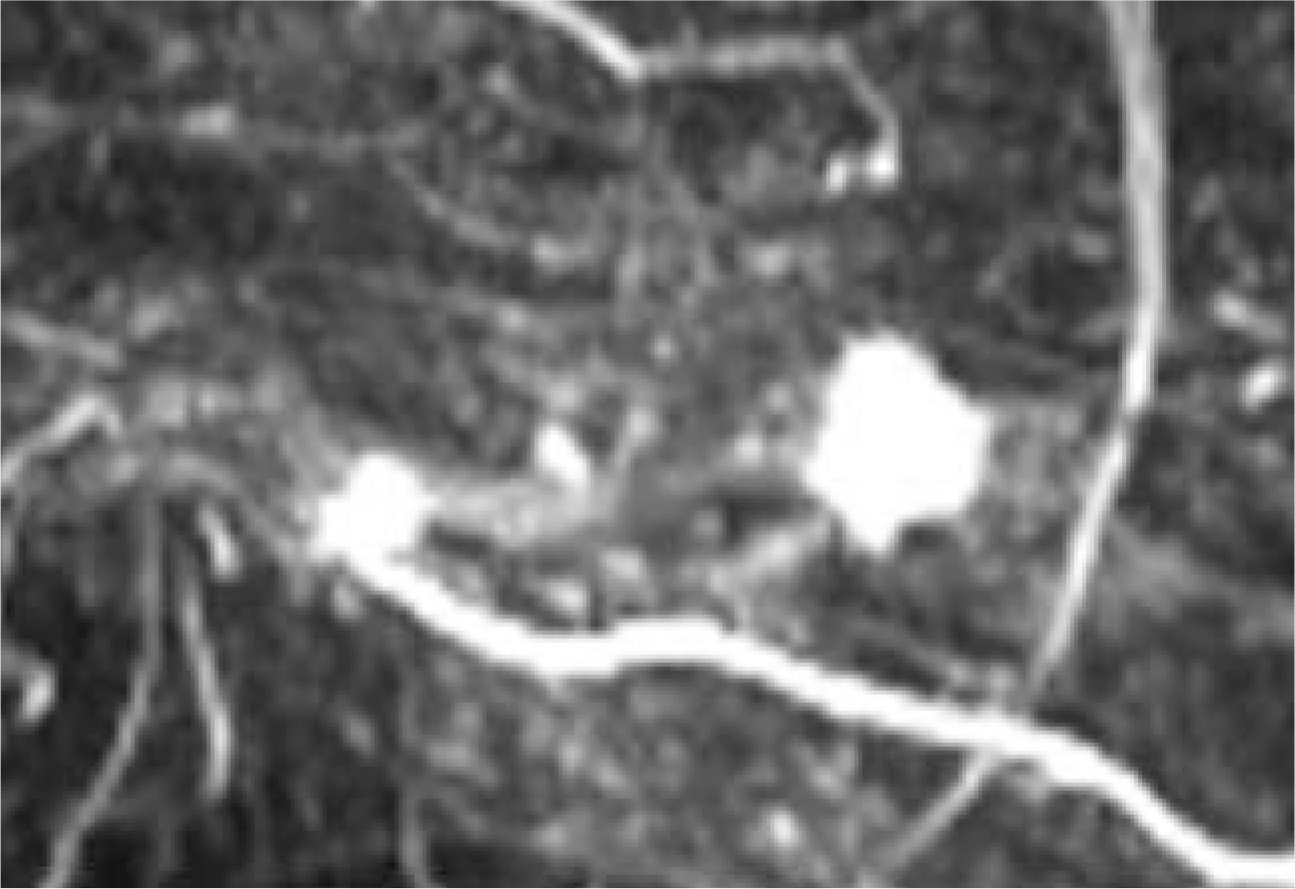
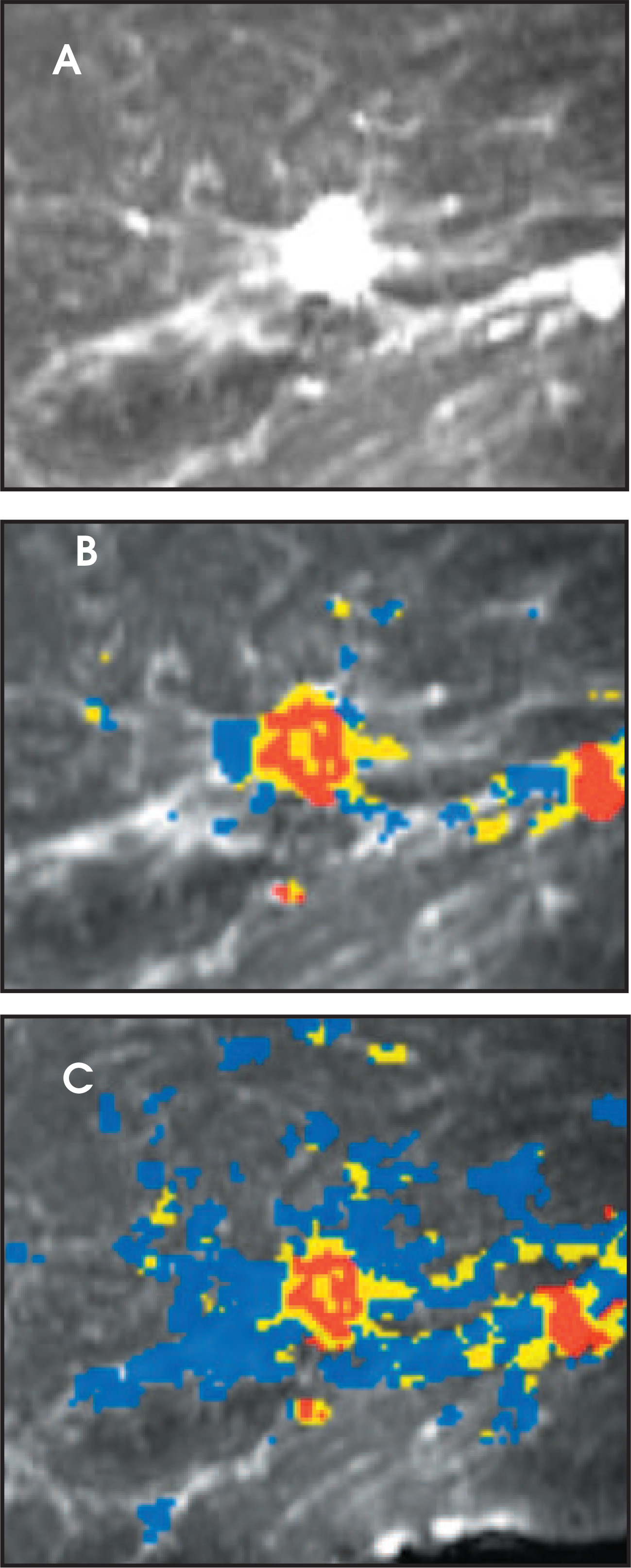
Preoperative MRI identified the cancer as a spiculated heterogeneously enhancing mass but also found a second spiculated heterogeneously enhancing mass with extensive surrounding enhancement, which was indicative of extensive DCIS. The maximum intensity projection (MIP) images show the marked difference between the normal and the abnormal breast (Figure 2). The low-density masses can be seen on the T1-weighted image (Figure 3). The enhancing masses can be seen in Figure 4. Figure 5 shows the use of computer-assisted diagnosis and color mapping of enhancement patterns.
Preoperative MRI changed the surgical management and affected the patient’s and surgeon’s decision making. MRI showed that the disease extended over a 10-cm area and was multifocal. Without the MRI, the “golf-ball–sized” lumpectomy would have resulted inpositive margins, and at least 2 surgeries would have been required to achieve negative margins.
DIAGNOSIS
Invasive ductal carcinoma and DCIS
SURGICAL FINDINGS
The patient opted for a mastectomy. At surgery, in addition to the cancer found by mammography and ultrasound and the additional cancer and DCIS found by MRI, a third invasive cancer was found. The extent of DCIS was marked and correlated with the abnormal enhancement seen on MRI.
DISCUSSION
The role of preoperative breast MRI is still evolving and is controversial. MRI is more sensitive than mammography or breast ultrasound, as has been noted by many authors.1 The American Cancer Society guidelines for breast MRI recognize that despite false-positives MRI is beneficial for those at greater than 20% to 25% lifetime risk of breast cancer.2 Multiple studies have shown that MRI has a sensitivity of 70% to 100%, which is considerably higher than that of mammography or ultrasound. The greater sensitivity of MRI is independent of breast density—ie, even if a woman has fatty breasts, a cancer may be mammographically occult.1,2
In a patient who has been newly diagnosed with breast cancer, MRI is useful in finding additional foci of disease in the same breast and in the contralateral breast. A recent American Roentgoen Ray Society presentation by Dr. Carol Lee reported that breast MRI changed surgical treatment in 28% of patients with 16 unsuspected cancers found in 110 patients (12%).3 Of these, 3 cancers were detected in the contralateral breast. Six patients had more extensive lumpectomies, and 15 patients opted for mastectomy rather than breast conservation.
Preoperative breast MRI is useful in surgical planning. Obtaining negative surgical margins has been shown to improve outcome after breast conservation.4 MRI can show the true extent of disease and decrease the rate of second and third surgeries performed in thequest for negative margins.
CONCLUSION
Breast MRI is an evolving field. Hardware and software advances as well as an increasing experience base are changing the way breast MRI is ordered, performed, and interpreted. Educating patients and referring surgeons about the proper use of breast MRI––its benefits and its limitations––will lead to increased appropriate utilization of this modality.
References
- Pediconi F, Catalano C, Roselli A. Contrast-enhanced MR mammography for evaluation of the contralateral breast in patients with diagnosed unilateral or breast cancer or high-risk lesions. Radiology. 2007;243:670-680.
- Saslow D, Boetes C, Burke W. American Cancer Society guidelines for breast screening with MRI as an adjunct to mammography. CA Cancer J Clin CA Cancer J Clin.
- MRI changes breast cancer treatment choice; increases time to treatment. 2008.
- Leong C, Boyages J, Jayasinghe U. Effect of margins on ipsilateral breast tumor recurrence after breast conservation therapy for lymph node-negative breast carcinoma. Cancer. 2004;100:1823-1832.
Citation
. Multifocal invasive ductal carcinoma and ductal carcinoma in situ. Applied Radiology. 2008;37(8):33-34. doi:10.37549/AR1639.