Multislice computed tomography: 64 and beyond
Applied Radiology — Vol. 33 , Issue 11 , pp. 58 -68
DOI: 10.37549/AR1292
Published: November 1, 2004
Categories
The first computed tomography (CT) scanner was developed and patented in 1972 by Sir Godfrey Hounsfield and quickly became widely used in clinical practice.1 The early axial scanners positioned the patient on a movable table between a stationary X-ray tube and a stationary radiation detector and moved the table to acquire individual sequential image slices. This method often required 15 seconds to obtain a single 10-mm slice and needed 1 minute to reconstruct the data into a clinical image. A simple scan of the head with 10 slices could take 1 hour.2 The long acquisition and reconstruction times caused significant respiratory and motion artifacts and limited accurate imaging to the small intestine and bowel wall. In the 1980s, advances in slip-ring technology allowed the X-ray tube and the radiation detectors to be rotated around the patient.3 Moving the patient through a continuously rotating X-ray beam allowed a larger anatomic area to be imaged in a single breath-hold. The radiation transmitted to the rectangular detector rows produced a spiral pattern of contiguous slices, a characteristic of the helical CT. The acquired image data were now not merely a series of separate slices, but were true volumetric data. However, acquiring very thin slices over large areas caused tube heating problems in early helical CT scanners and thereby limited 3-dimensional (3D) reconstructions.4
The next generation of CT scanners increased the number of rows of radiation detectors and are called multidetector-row, multichannel, or multislice CT (MSCT). Today, an entire organ can be scanned in 1 second, the moving heart within 5 beats, and the whole body in <10 seconds.5 Scanners now routinely acquire 4, 8, and 16 slices per rotation, and 32-, 40-, and 64-slice scanners have recently been introduced. These machines can attain gantry speeds of <0.4 seconds per revolution with 0.35-mm resolution. Sixty-four-slice scanners extend the diagnostic possibilities of CT considerably, especially in cardiology, where the beating heart and all of its blood vessels can be displayed in high resolution (Figure 1). The coronary arteries can be visualized via virtual “arterioscopy”; the colon, via virtual colonoscopy. Multislice CT with volume rendering and stereoscopic display can provide the means for the radiologist and clinician to interactively explore the huge data sets produced by modern scanners and can create a uniquely personalized visual aid to help explain the diagnosis to the patient. However, with an increased number of detector rows, some problems with cone beam angle geometry and increased radiation dose had to be overcome.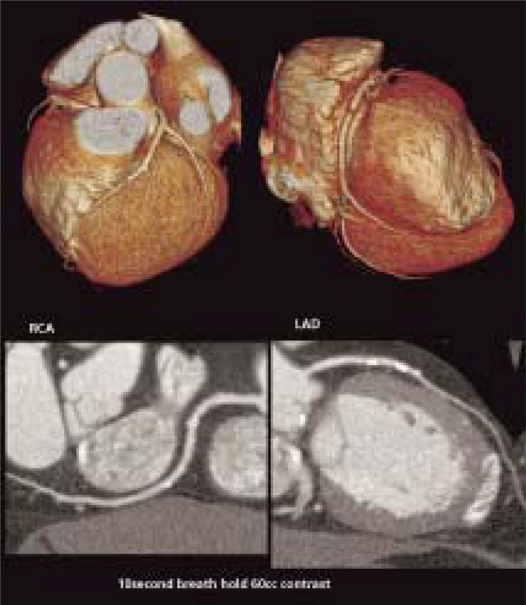
Early problems with MSCT
Cone beam angle geometry
In single-slice systems, each slice is acquired twice: at each end point of a 180° gantry rotation. Because each measurement ray is perpendicular to the z-axis (patient’s longitudinal direction), the data are consistent. However, the measurement rays in the cone beam in spiral CT systems are tilted, and, therefore, the outer rays will not acquire the same image at each end point of a rotation.6 With standard image reconstruction algorithms, these data inconsistencies produce cone beam artifacts and slice blurring. Although these are minimal and can be disregarded in spiral scanners with only a few detector rows, they become a problem when the number of detector rows is >4.
One approach to solving the problems of cone-beam angle geometry in MSCT involves an adaptive multiple plane reconstruction (AMPR) algorithm.6 In this method, the cone pattern is divided into subsections for which multiple double oblique intermediate images are calculated. Next, a z-reformation with appropriate weighting functions is performed on the intermediate voxels in the image plane. This provides optimization for the curvature of the spiral and the shape of the detector and, ultimately, allows full-dose utilization and selection of the spiral pitch. Streak artifacts, which can be a problem in neural imaging with multislice systems, can be eliminated by using the AMPR algorithm.
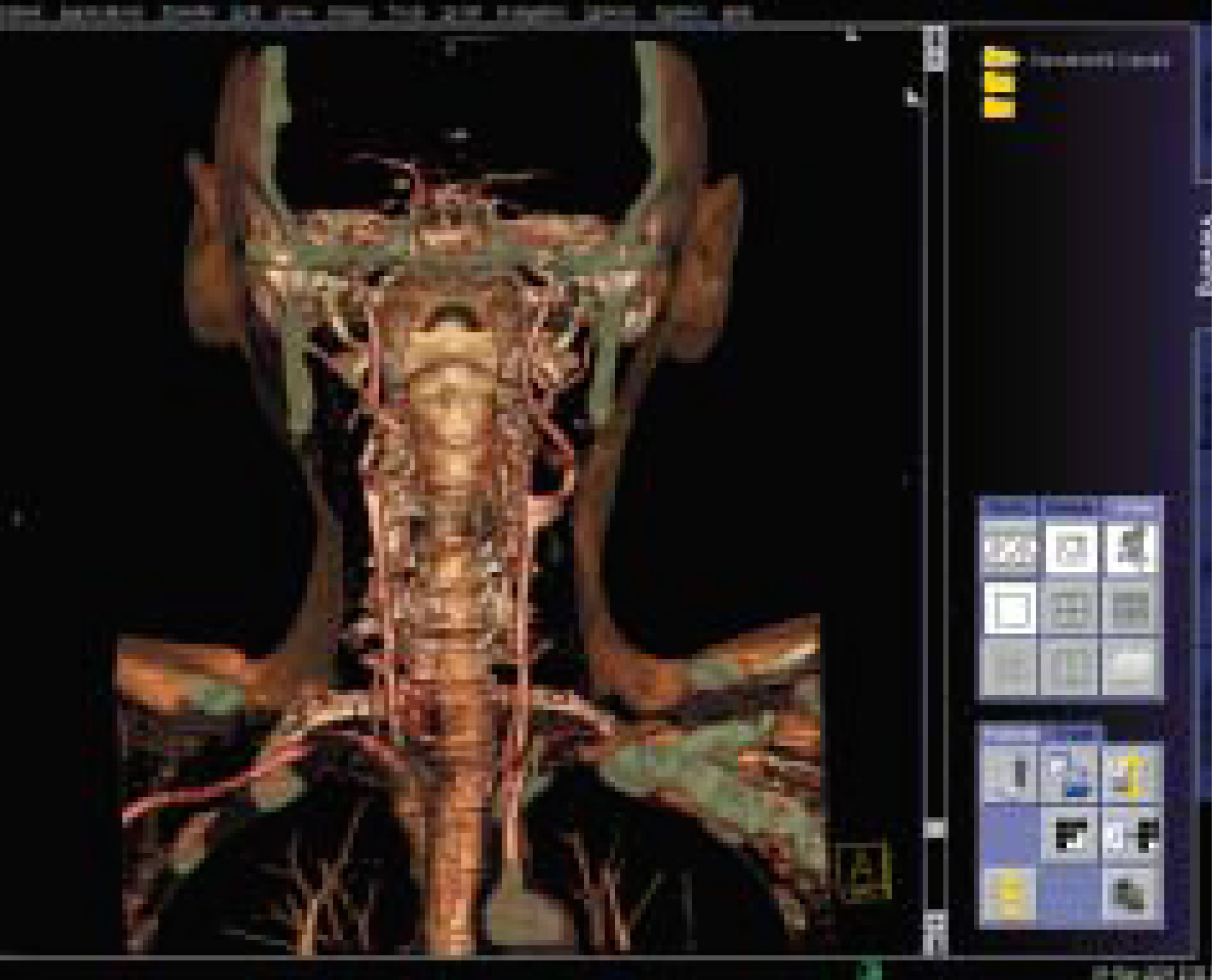
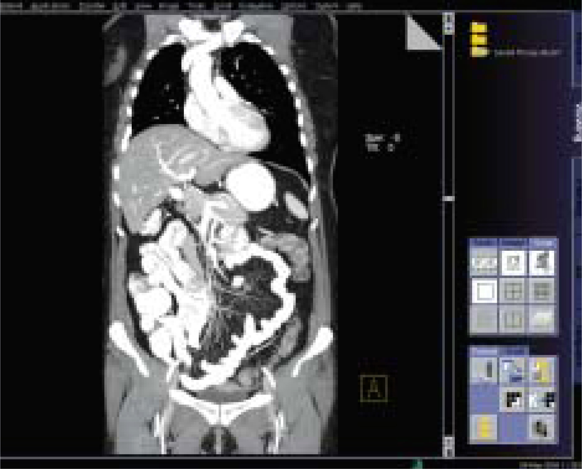
The capability to select slice width and resolution independent of pitch means that MSCT units with advanced reconstruction algorithms can produce true isotropic resolution. Because different slices are possible with the same collimation, 3D, multiplanar, and maximum-intensity projections are all possible in retrospective reconstructions, greatly increasing the utility of MSCT in clinical applications (Figures 2 through 4).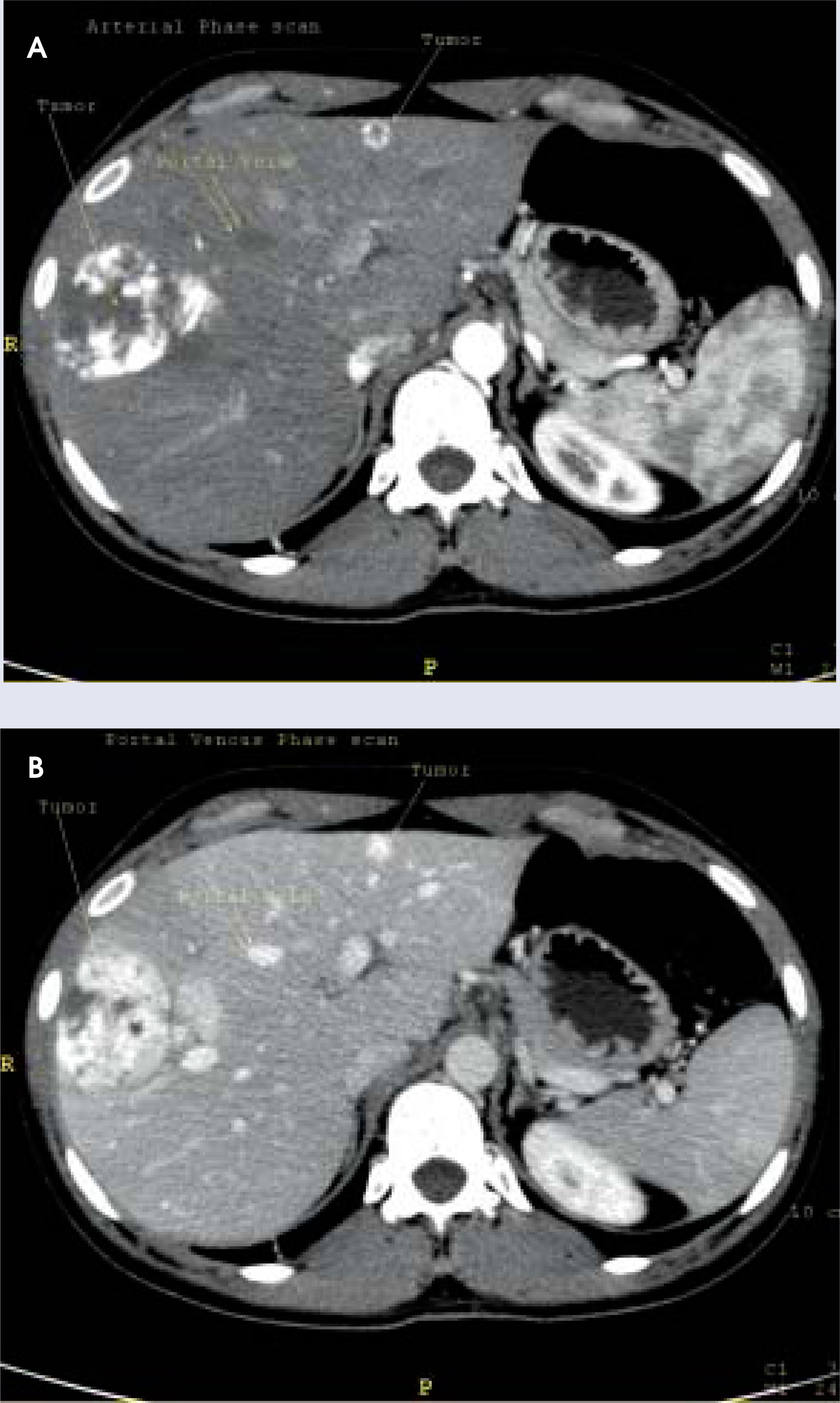
Increased radiation dose
The increasing numbers of simultaneously acquired slices obtained during a CT imaging study can expose the patient to an increasing amount of radiation. According to data from the Center for Radiological Research, a young girl exposed to the radiation from an abdominal helical CT scan has a risk of a fatal cancer later in life of 1 in 1000.7 A recent Lancet article reported that the cumulative cancer risk to age 75 years that could be attributed to diagnostic X-rays was 0.6% in the United Kingdom and >3% in Japan.8
However, radiation exposure does not necessarily increase as the number of detector rows increase. According to Geoffrey Rubin, MD, Chief of Cardiovascular Imaging at Stanford University School of Medicine, radiation dose efficiency for the same collimation increases as the number of detectors increases, giving the 16-slice CT a greater dose efficiency than the 4-slice CT. This is because the penumbra zones at the edges of the detector have an unequal signal level compared with the other detector rows and cannot be used to calculate the image data. Because a 16-detector row CT system has a smaller relative penumbra zone compared with that of a 4-detector row system, the contribution of the “wasted” penumbra zones decreases, and dose efficiency increases.
Many MSCT scanners use a dynamic technique to modulate tube current to maintain image quality while delivering a low overall radiation exposure. For example, a diagnostic scan with a setting of 100 mA, a pitch of 1, and a 1-second scan time will expose the patient to 100 mA. Scanner technology that allows scanning of the same coverage while doubling the tube current to 200 mA and increasing the pitch to 2 will halve the scan time to 0.5 seconds. This approach produces the same overall radiation exposure but with a higher image quality (personal communication, Murat Gungor, Product Manager, Siemens Medical Solutions, Malvern, PA, September 2004). Although there are no known adverse consequences to biologic tissues from a greater intensity, or rate of delivery, of a given diagnostic radiation dose, it is still important to minimize the exact radiation dose that the patient is receiving. Alec Megibow, MD, Professor of Radiology at New York University, thinks it is important to keep patient care and overall radiation dose in mind when considering repeat scanning for follow-up or screening studies. “You can scan faster, but that doesn’t mean you can scan someone 3 or 4 times —it means you have a finer knife.”
Other advanced dose reduction strategies developed by radiologists and equipment manufacturers include pediatric scan modes that use protocols optimized for children, real-time anatomic dose modulation that uses detector signals to control the tube current as a method of delivering the lowest possible dose according to the changing attenuation of the patient’s anatomy, adaptive kilovolt settings in protocols using lower doses for contrast studies, and electrocardiogram-controlled dose modulation of tube current during cardiac studies to reduce the dose during the systolic phase. These methods can reduce the radiation dose by as much as 50% in the shoulder area to 41% for extremities and 26% in the head.6
Advantages of increasing slice capabilities
The decreased scan time offered by multislice scanners is beneficial not just in terms of patient throughput or scheduling. A faster scan means that a shorter breath-hold is necessary, making imaging of trauma, pediatric, or elderly patients much easier and more efficient. For cardiac applications, a faster scan means that the entire heart can be imaged in 5 to 10 seconds. If the scan is delayed for the first 5 seconds of breath-holding, the data will be acquired in the most stable phase of the heart rate, and there should be ample time to complete the scan within the standard 30second breath-hold.9
This capability is critical in cardiovascular evaluation of patients with rapid heartbeats. In a recent study by Giesler and colleagues,10 100 patients underwent evaluation of the coronary arteries with a 4-slice detector using retrospective ECG gating, and results were compared with conventional angiography. Fully 29% (115 of 400 arteries) could not be evaluated by the 4-slice CT because of motion artifacts. The overall sensitivity for detection of coronary stenoses was only 49% (51 of 56 stenoses detected); in accessible arteries, the 4-slice CT had a sensitivity of 91% (51 of 56 stenoses detected) and a specificity of 89%. The optimal window position was during mid- to late diastole in patients with lower heart rates (70 bpm), and during late systole and early diastole in patients with higher heart rates. Because the overall sensitivity for stenosis detection decreased from 62% to 33% when the heart rate increased to >70 bpm, scanning times faster than those provided by the 4-slice machine may be necessary for accurately evaluating stenoses in patients with faster heart rates.
The newer machines bring more than just reduced scanning time to the radiologist’s armamentarium. The 16-slice scanner, for example, can be used for advanced applications that require high-speed, thin-slice scanning of large anatomic areas, and the scanner acquires the volumetric data necessary for exquisite 3D imaging. Results with examination of the coronary arteries with the 16-slice scanner lead to the recognition of CT angiography (CTA) as a less invasive alternative to conventional angiography. In addition to CTA, dynamic perfusion CT (CTP) is now possible with high-speed scanning and volumetric reconstruction. Advanced stroke protocols can combine CTA and CTP to identify cerebrovascular hypoperfusion at its earliest stage.11 Jain and colleagues12 have reported initial favorable results using CTP with acetazolamide challenge during carotid artery balloon occlusion to assess cardiovascular reserve in 8 patients considered candidates for permanent carotid artery occlusion.
The advent of 32-slice CT brought true isotropic volume data further into the realm of diagnostic imaging. The decreased scan times and increased resolution of the newer machines have made coronary CTA effective for preoperative evaluation of plaque extent and characteristics and for postoperative evaluation of stent patency and leaks. CT angiography with volume rendering and sterotactic display provides better visualization and is quicker and more comfortable for the patient than is conventional angiography and avoids both the risks and the extra expense of the invasive procedure. A 32-slice system can scan the circle of Willis in 0.5-mm slices in only 3.2 seconds but can still differentiate the veins and arteries in arterial-phase imaging.9
A 40-channel CT scanner with the added enhancement of an upgrade option to 64 channels has recently become available (Figure 5). This unique upgrade package will bring thin-slice capabilities to the full 40-mm detector width by increasing the number of simultaneously acquired data channels to 64. This scanner was developed in collaboration with 8 hospitals located in the United States, Germany, Israel, The Netherlands, and France.13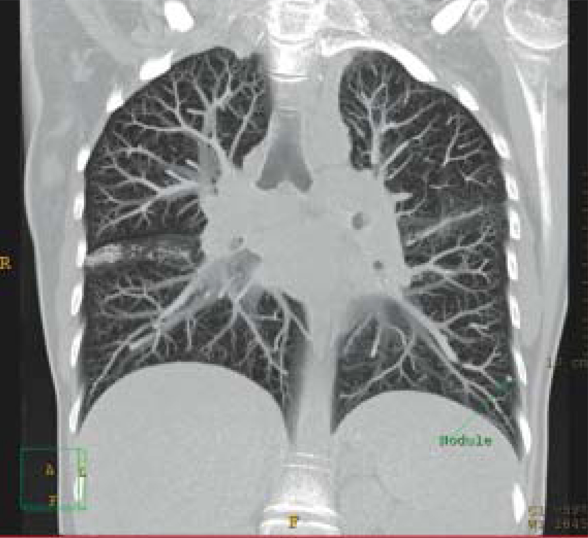
The new 64-slice scanners with ≤0.5 mm slice thickness can scan the circle of Willis in <1.5 seconds. With the 64-slice scanner, the physician can see the vessels that are feeding and draining the lesions; such capability may aid in early detection of malignancy. The fine detail of the vascular arcade of the skin is visible, allowing the plastic surgeon to distinguish branching and superficial vessels in the peripheral circulation and plan the surgical approach. For evaluation of coronary arteries, the higher resolution imaging can go beyond differentiating soft and hard plaque. By categorizing vascular lesions into calcified, fibrous calcified, and noncalcified, radiologists using the 64-slice scanners can determine the risk of plaque rupture and subsequent thrombus. And the higher resolution and faster scan times permit a larger anatomic area to be imaged with the 0.5-mm slice thickness necessary for visualization of much smaller vessels, <2 mm in diameter (Figure 6).14 This is helpful in planning embolization and in evaluating the patency of coronary grafts and stents.
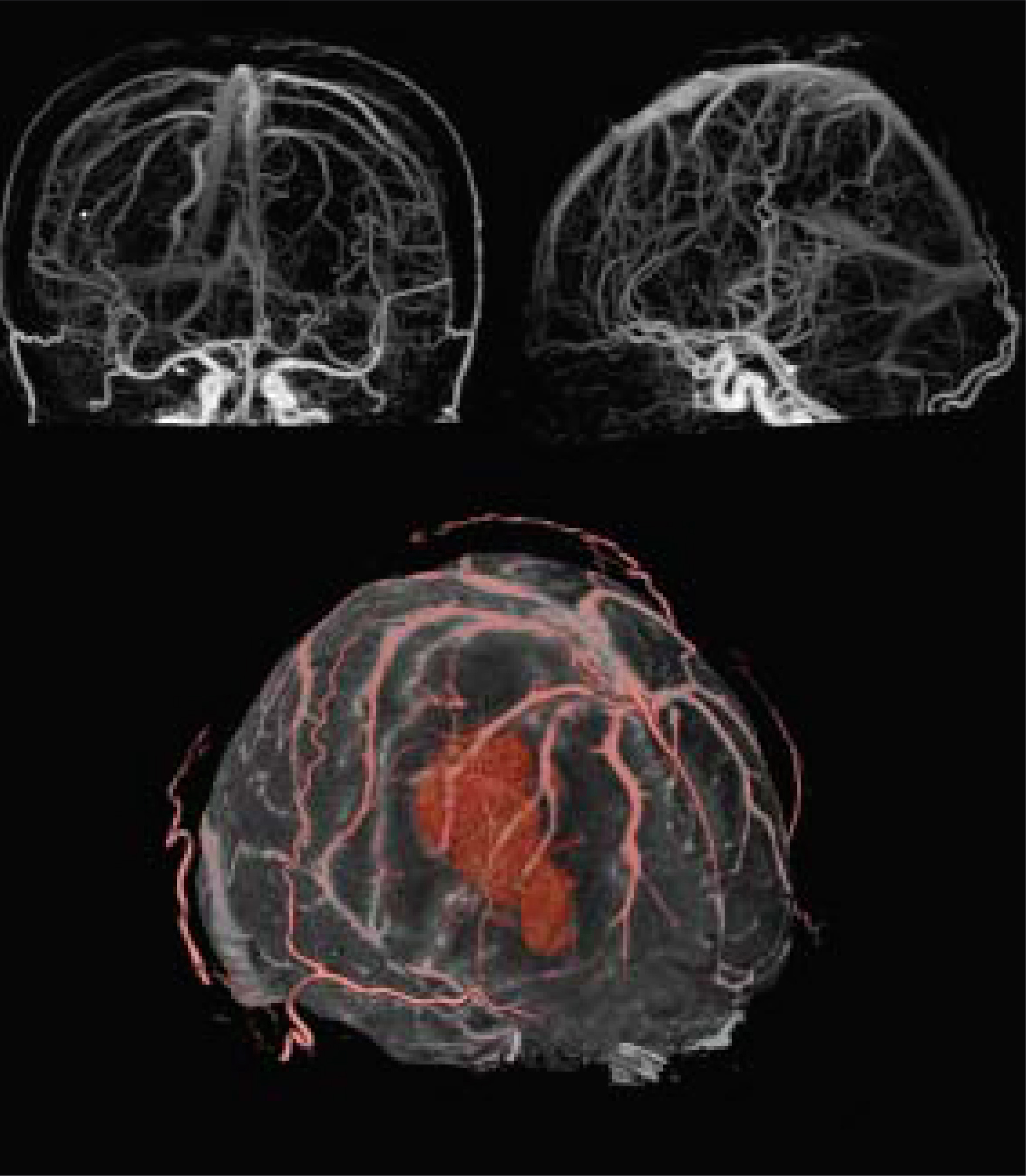
And lastly, visualization of the subtle variations in contrast enhancement of the brain parenchyma that is possible with the 64-slice scanner (Figure 7) will enable assessment of cerebral perfusion, blood fiow, and accurate determination of the cerebral ischemic penumbra for evaluation of early intervention for stroke.11 The wider detector design and greater anatomic coverage in the axial direction provided by the 64slice scanners allow a larger area of the brain to be evaluated in a single continuous rotation.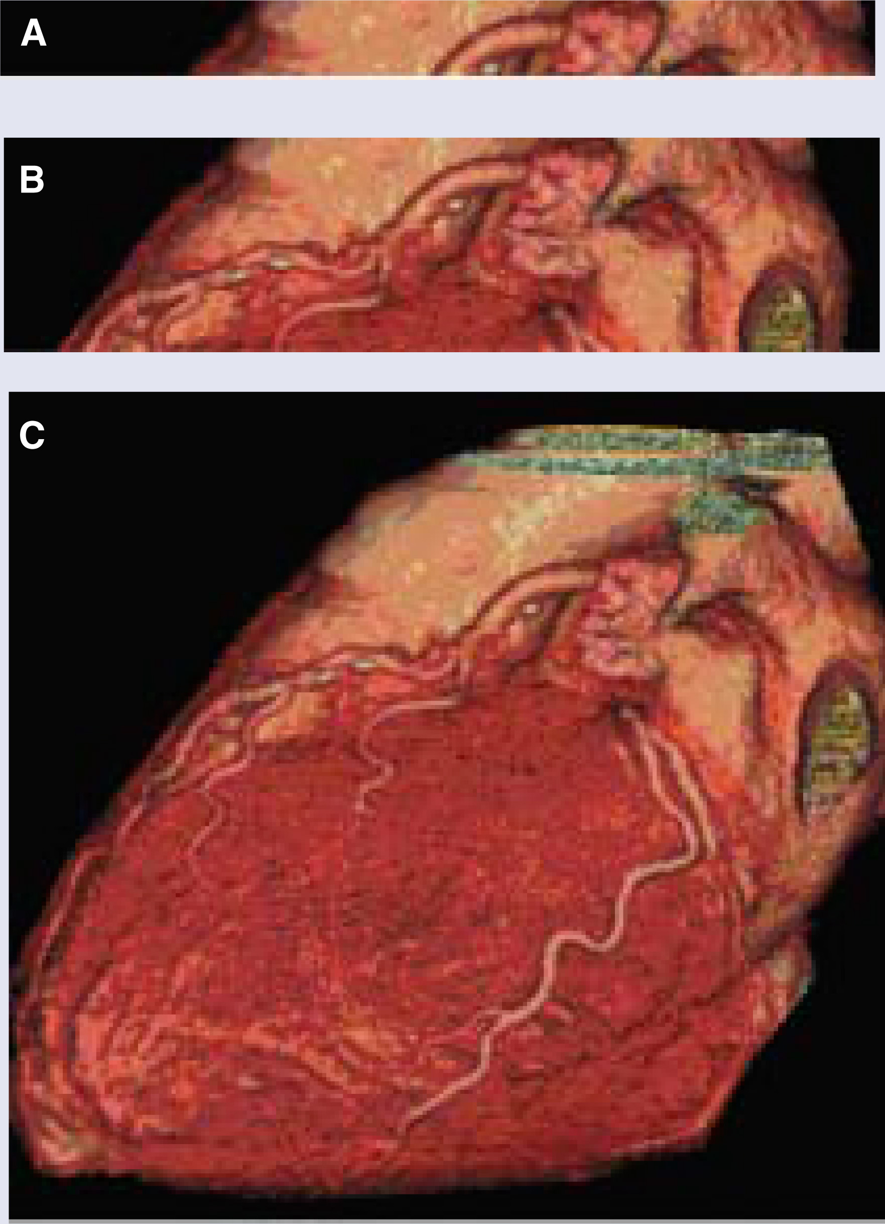
How physicians are adapting scanning protocols to accommodate faster scanning times
The faster scanning times will require some adaptations to the standard CT protocols. “For some types of applications, protocol shifts are apparent and obvious for people who are comfortable with and understand the technology,” noted Dr. Rubin. “For example, a routine pelvis/abdomen CT scan to look for liver metastases or to assess a trauma patient is probably going to be very easy to crosswalk from one technology to another.” But other indications will be more challenging. With the 16-row detectors, scans of the peripheral vasculature may take only 20 seconds, compared with >1 minute for the earlier 4-slice machines. Dr. Rubin found that “patient to patient variations in terms of their circulation time have made us completely rethink how we deliver contrast medium, and it has required some time to evolve our protocols into once again having robust and reproducible results. It wasn’t intuitive.”
The amount of image noise is related to the tube current and the aquisition time. Because “the much-touted capability of routinely getting sub-millimeter-thick sections results in highly noisy images,” Dr. Rubin feels that for noncardiac vascular imaging with the 16-slice scanner, “we are better off with a thicker section that is less noisy.” Some CT vendors have responded to this complaint by increasing the maximum tube currents in the newer scanners to counteract problems with contrast-to-noise ratio in thin sections or fast scans.
Dr. Megibow has had to adjust the delay in image acquisition to accommodate faster scan times. He feels that his department “had to understand the interactions of the detector configurations of the slice outputs, differentiating those slices that we would use for 3D analysis versus those slices that we would archive.” He anticipates that he will be archiving similar images from a 64-slice study as are now stored from a 4- or 16-slice study, but that the major difference will be in how an institution stores the thin slices for 3D reconstructions (Figure 8). He remarked that a site that buys a 64-slice scanner and does not understand the huge number of images that the scanner produces will be overwhelmed if they try to archive all the images.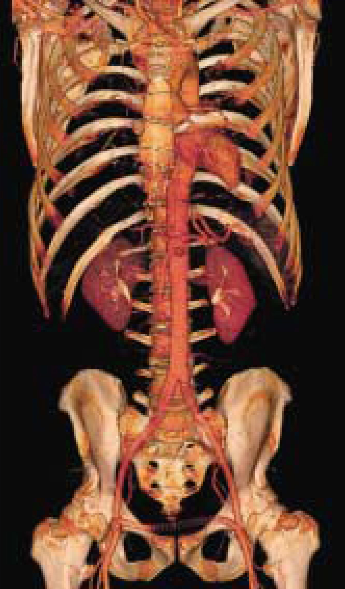
Dr. Megibow believes that the 64-slice scanner would be well suited to perfusion imaging for characterizing a lesion. Another area in which he believes the newer scanners could excel would be in tracking angiogenesis associated with tumor growth. Being able to visualize this process, which is only now possible with the 64-slice scanners, “could have major implications for cancer lesion assessment.” He added that “this could possibly shorten the cycle time, and actually the cost, of bringing new therapeutic agents to market.”
Beyond 64 slices
An experimental 256-slice CT scanner is being tested at the National Institute of Radiological Sciences in Japan. This machine uses a new technology to scan an area as large as an entire heart or head in one rotation. Advanced reconstruction algorithms process the 2-dimensional data from the cone beam into true volume slices. Because data are acquired during continuous rotation over time, this technique is also known as 4-dimen-sional (4D) CT. The scanner is also capable of processing helical cone beam data, a mode called precise 3D, for use in imaging long objects such as the entire thorax or abdomen. The prototype detector consists of 912 × 256 elements, with a 1 × 1 mm element size; at 16-bit transfer speeds, it will create 900 views per second. The reconstruction matrix is 512 × 512 × 512, and the reconstruction area is 25 to 50 cm in diameter × 10 cm in length per rotation.
The novel data aquisition system is closer to that of a flat-panel system than that of a conventional CT scanner. A rotation interface encodes the 912 channels of data, adds the error-correction code, and sends 12 channels of data to the laser diode-photodiode (LD-PD) pairs. Data are transferred by 12 parallel sets of LD-PD pairs at a rate of 622 Mbps. Another interface de-codes these data and sends them to the image processor via a very-high-speed data bus (~5 Gbps). Development of an ultra–high-speed reconstruction processor is ongoing, and a test model of the 4D CT scanner has been undergoing clinical evaluation since 2003.17
Impact on healthcare community
According to Dr. Rubin, “the biggest impact of these newer designs rests upon cardiovascular imaging in particular.” He envisions that as CT scanner technology grows, diagnostic quality and capability will also rise steeply. Dr. Rubin believes that the real issue is when the healthcare community will see widespread adoption of CT as an alternative to cardiac catheterization for evaluating the coronary arteries. He believes that a big determinant will relate to how well cardiologists either interact with radiologists or maintain a level of interpretation quality for CT imaging of the thorax that will enable them to operate effectively independent of radiologists. According to Dr. Rubin, patient demand will have a somewhat smaller role to play in an institution’s selection of the newer scanners because most cardiac catheterization imaging is done on an emergency basis for evaluation of chest pain. “We have not seen a very strong impact of patient preference in imaging, and rarely have patient referrals outside of interventional radiology.”
The classic sequential CT protocol required a predetermined slice thickness from its solitary 1 × 20 mm rectangular bank of detectors. In contrast, each individual bank of detectors in a multidetector-row system can provide a range of slice thickness that can be selected even after the scanning is completed. Very rapid data reconstruction is now possible even with the large data sets from MSCT systems. This very rapid reconstruction time allows the physician to vary the reconstruction algorithms to produce optimum studies for different tissues of the same patient. For example, on a CT scan of the chest, lung parenchyma can be reconstructed with high-resolution technique; soft tissues, with a classic soft-tissue algorithm; and the bony thorax, with a bone algorithm.
However, this means that the radiologist is confronted with hundreds of images from a single CT study. Just doubling the number of detector rows brings a 10-fold increase in the required computational power and necessitates sophisticated data transmission, storage, and display capabilities. According to Doug Ryan, director of the CT business unit at Toshiba America Medical Systems, “the biggest challenge for all vendors is that CT technology is probably growing faster than computer technology.”
One important question that must be asked is whether there is an increased possibility of missing a lesion because there are too many images for the radiologist to review. Radiologists are adapting to this huge volume of graphic information by using workstations, rather than films, to make the diagnosis. Mr. Ryan thinks that with the release of newer 64-bit technology software, there will be a shift toward a diagnostic ability being available directly from the picture archiving and communication system (PACS), which will be integrated with the imaging workstations. It is critical that the site of the newer scanner be equipped with gigabit switches and a cable network that can handle gigabit data transfer.
To further assist the radiologist, some scanner manufacturers offer computer-assisted identification of tumors or nodules, similar to the automated mammography analysis. Krishnamoorthy and colleagues15 found that in CTA, the use of semiautomated, interactive tools for tracking vessels, automated curved and oblique reformations, and advanced 3D visualization decreased the radiologists’ reading time by approximately a factor of 2. In screening mammography, computer-assisted systems have shown a capability to detect findings on prior mammograms that can be attributed to a cancer diagnosed at a later date, suggesting a method for early detection and possibly the ability to estimate of the probability of malignancy for a particular finding.16 Dr. Megibow is currently investigating computer-aided detection of lung lesions and colon polyps and would like to see enhancements in computer-assisted diagnosis that would allow him to just click on the display of a particular lesion and get a measurement. He hopes that the better resolution afforded by the 64-slice scanner will produce the sharper edge and better border delineation that will allow the tedious manual process of measuring small lesions on serial follow-up images to be automated.
Whether or not these new scanners will be more cost effective than the earlier, slower models remains to be seen. There is a limit to the number of patients that can be registered, prepared, and scanned in 1 hour, and the difference between a 1-minute scan or a 20-second scan will likely not make a significant impact on patient throughput. The real cost savings will likely come in terms of elimination of more invasive, riskier, and more expensive alternative tests; reduction in the number of repeat scans needed for uncooperative or difficult patients; and earlier detection and therapeutic intervention for malignancies.
Conclusion
The progression from axial imaging to spiral CT scanning to multidetector/ multislice CT has brought us on a journey whose end is not yet in sight. Scanners with multiple X-ray tube designs that will speed up data acquisition times into the millisecond range may be next! One can imagine a multitube, kilochannel scanner coupled with an ultrafast positron-emission tomographic imaging system and optimized data storage/retrieval systems to create the ultimate in diagnostic imaging.
References
- Hounsfield G. Computerized transverse axial scanning (tomography). Part I: Description of system. Part II: Clinical applications. Br J Radiol. 1973;46:1016-1022.
- Progress in CT. 2004.
- Siemens Medical Solutions celebrates 30 years of computed tomography. 2004.
- Fishman E. 3D postprocessing of multidetector computed tomography data. 2004.
- Schubert S. Volume CT—AGE Healthcare Publication. 2004:4-7.
- Flohr T. CT Lecture Series. 2004.
- Hall E. Lessons we have learned from our children: Cancer risks from diagnostic radiology. Comment. Pediatr Radiol. 2003;33:811-814.
- Berrington de Gonzalez, Darby S. Risk of cancer from diagnostic x-rays: Estimates for the UK and 14 other countries. Lancet. 2004;363:345-351.
- Katada K, Hanafusa K, Furuyashiki M, Burch J. Innervision. 2004:4-8.
- Giesler T, Baum U, Ropers D. Noninvasive visualization of coronary arteries using contrast-enhanced multidetector CT: Influence of heart rate on image quality and stenosis detection. AJR Am J Roentgenol. 2002;179:911-916.
- König M. Brain perfusion CT in acute stroke: Current status. Eur J Radiol. 2003;45:S11-S22.
- Jain R, Hoeffner E, Deveikis J. Carotid perfusion CT with balloon occlusion and acetazolamide challenge test: Feasibility. Radiology. 2004;231:906-913.
- Philips announces global commercial shipments of the Brilliance 40-channel CT scanner and reveals a 64-channel upgrade.
- Hayes J. Diagn Imaging Online. 2004.
- Krishnamoorthy P, Brejl M, Ooijen P. System for segmentation and selective visualization of the coronary artery tree for evaluation of stenosis, soft plaque and calcification in cardiac CTA. Imaging Decis MRI. 2004;8:25-30.
- Yaffe M, Jong R. Digital mammography: Current status. Decis Imaging Econ. 2004.
- Endo M, Tsunoo T, Kandatsu. Four-dimensional computed tomography (4D-CT)—Concepts and preliminary development. Radiat Med. 2003;21:17-22.
Citation
. Multislice computed tomography: 64 and beyond. Applied Radiology. 2004;33(11):58-68. doi:10.37549/AR1292.