Mycosis fungoides involving head and neck mucosal sites: Review of the literature
Applied Radiation Oncology — Vol. 6 , Issue 2 , pp. 11 -19
DOI: 10.37549/ARO1122
Published: June 1, 2017
Categories
Mycosis fungoides is the most common form of cutaneous T-cell lymphoma and comprises 4% of all non-Hodgkin lymphomas. It is characterized by the proliferation of mature, effector-memory T-cells in the skin. Lesions initially present as patches and may progress to plaques, tumors and erythroderma. The disease may spread to the viscera and bloodstream.
Mycosis fungoides presenting in head and neck mucosal sites is exceedingly rare. These lesions most often appear after cutaneous involvement, and progression to these areas often indicates a grave prognosis.1 To date, 57 such cases have been reported in the literature. Here we present 2 additional cases treated in our institution, together with a comprehensive review of the literature.
Case Report One
A 59-year-old Caucasian woman presented to the Cutaneous Lymphoma Clinic of the Department of Dermatology at our institution with a generalized pruritic erythematous rash and lesions consistent with tumors on her right upper extremity and chest. She had a 4-year history of a rash first localized to her palms that had spread to several cutaneous sites, notably the gluteal folds and inframammary areas. Although originally diagnosed as having pustular psoriasis, biopsies of the right upper extremity and right chest lesions were consistent with mycosis fungoides. Hematopathology did not reveal any disease in her blood and she was diagnosed with stage IIB (T3N0M0Bx) disease. Immunohistochemistry of the biopsy specimen revealed an infiltrate that was CD2+, CD3+, CD5+, CD4-/CD8-, CD7-, and CD56-, with TCR-beta F1+ phenotype. T-cell receptor gamma gene rearrangement was positive for a clonal T-cell population.
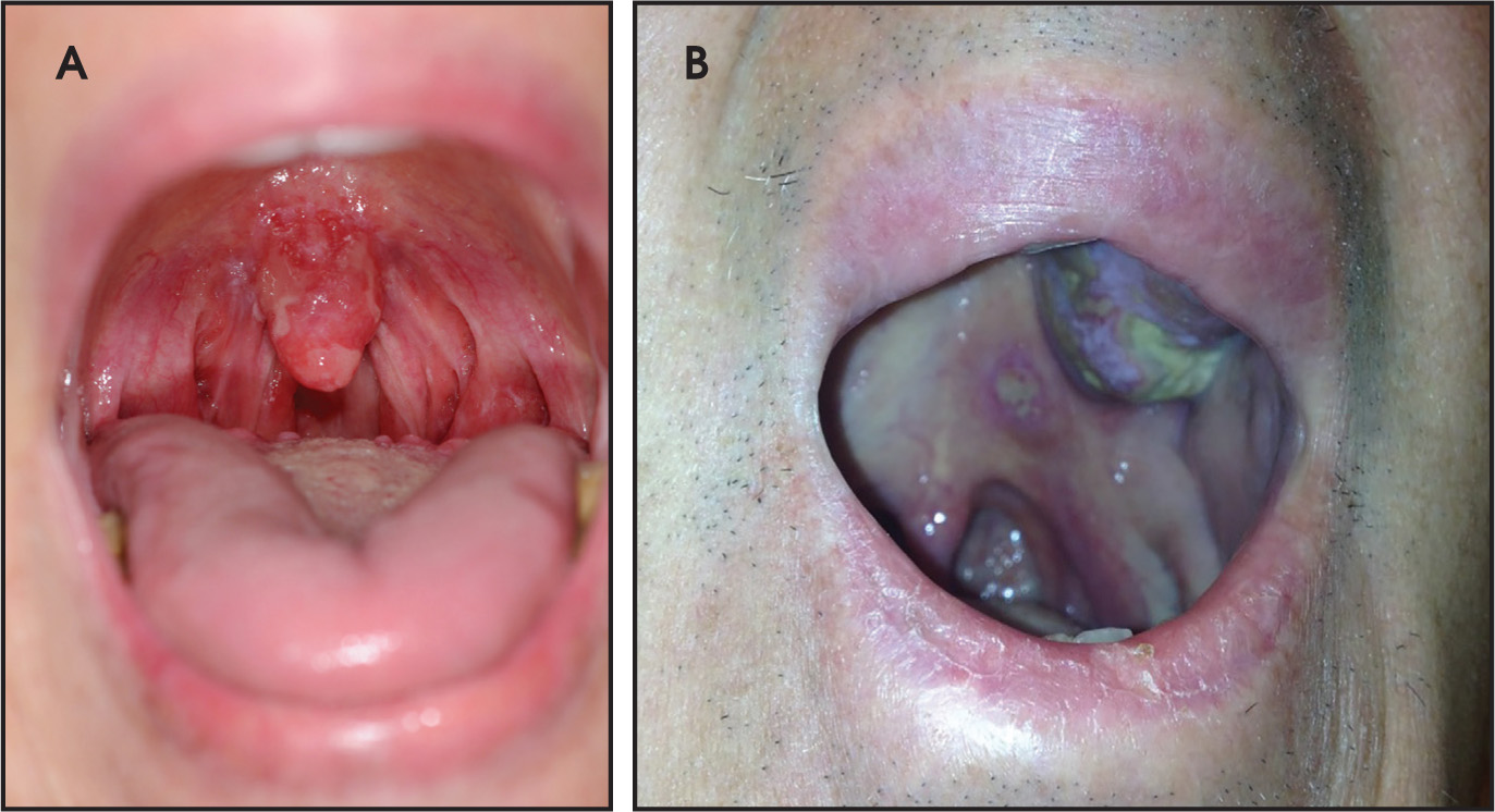
The patient initially underwent total skin electron-beam therapy to a total dose of 36 Gy delivered in 24 fractions of 1.5 Gy each using the 6-dual-field irradiation technique.2 During this period, she reported symptoms of a dry and sore throat as well as odynophagia. Esophagogastroduodenoscopy by an outside gastroenterologist revealed signs of reflux, and she began taking Lansoprazole (Prevacid, Takeda Pharmaceuticals USA, Deerfield, Illinois) 30 mg twice daily, which offered short-lived mild relief. A barium swallow revealed an unremarkable esophagus. Upon further evaluation in the Department of Otolaryngology, areas of inflammation consistent with laryngeal and oropharyngeal candidiasis were identified, and she received treatment with oral fluconazole for 4 weeks. Although the candidiasis resolved at that time, the patient continued to complain of persistent symptoms that were worse after meals and at night. Re-examination of her throat showed an irritated appearance of the posterior soft palate, uvula, and anterior tonsillar pillars (Figure 1A). Flexible nasopharyngolaryngoscopy revealed irritated mucosa diffusely on the epiglottis and arytenoids bilaterally with a similar appearance along the left lateral pharyngeal wall. Biopsy specimens from the uvula and epiglottis revealed lesions consistent with her previously diagnosed mycosis fungoides including an identical TCR-gamma clone. Immunohistochemistry demonstrated an infiltrate positive for CD2, CD3, CD5, CD43 and weakly positive for beta-F1. Infiltrates were negative for CD56, CD30, and CD20. Positron emission tomography and computed tomography (PET/CT) scans showed no involvement of her viscera and mild fluorodeoxyglucose (FDG) uptake of an SUV of 2.3 in her axillary and inguinal lymph nodes. The oropharyngeal and laryngeal lesions were treated with 6-MV photons using an intensity-modulated radiation therapy (IMRT) technique. The initial planned dose was 30.6 Gy to be delivered in 17 fractions. However, she developed confluent fibrinous mucositis (grade III) and severe odynophagia, which required multiple breaks in treatment, significantly prolonging treatment duration. Ultimately, we terminated her treatment at a dose of 20.8 Gy. Two years after treatment completion, the patient has had no local recurrence of her mucosal lesions. She has subsequently undergone external-beam radiation therapy on multiple occasions for localized skin lesions. She continues to follow up with dermatology and radiation oncology.
Case Report Two
A 69-year-old Caucasian man was diagnosed with cutaneous mycosis fungoides 10 years prior to his presentation to our institution. At the time he was seen in the Department of Dermatology, he noted a 2-month history of new leather-like lesions on his bilateral extensor elbow surfaces. He also had multiple exophytic nodules on his bilateral lower and upper extremities, in addition to plaques on his torso and all 4 extremities. The mycosis fungoides lesions on his scalp had been treated with radiation therapy 5 years earlier, with no recurrence at those sites. He had also received intermittent psoralen and ultraviolet A (PUVA) light therapy and methotrexate until 3 years prior to presentation. At consultation, he was taking bexarotene (Targretin, Valeant Pharmaceuticals International Inc., Laval, Quebec, Canada) 75 mg 3 times daily in addition to using triamcinolone 0.1% cream daily. A biopsy of one of the new lesions revealed marked pseudoepitheliomatous hyperplasia with dermal granulomatous inflammation. Culture of the lesions revealed staphylococcus aureus, and he began receiving daily intravenous vancomycin 1 gm for 30 days with minimal improvement. Another biopsy taken from the left forearm lesion demonstrated cutaneous T-cell lymphoma with pseudoepitheliomatous hyperplasia, a positive monoclonal T-cell rearrangement and a CD3+, CD4+, CD8+, and CD30- phenotype. PET/CT showed no metabolic disease at any site.
The patient was treated with total skin electron-beam therapy to a total dose of 36 Gy using the technique described above. Shielded areas, including the palms, soles and buttocks, received a boost of 8 Gy in 1 fraction. This was followed by 4 cycles of weekly pralatrexate 15 mg/m2. Although this treatment initially appeared beneficial, several weeks later the patient presented with new lesions on his torso, extremities, and on the left tonsil and alveoli (Figure 1B). Biopsy of the left soft palate and left superior alveolar ridge again was consistent with mycosis fungoides. The oral lesions were treated with external-beam radiation therapy to a prescribed dose of 30.6 Gy in 17 fractions. However, the patient received only 10 fractions for a total dose of 18 Gy due to severe mucositis, which was not relieved by sucralfate, and eventually required hospitalization due to significant decreased oral intake and failure to thrive. His treatment was discontinued at this dose. Although the oral lesions responded well to the therapy, he eventually began a regimen of cyclophosphamide, hydroxyduanorubicin, etoposide, vincristine, and prednisone (CHEOP) chemotherapy and intrathecal methotrexate due to overall disease progression, including involvement of his bone marrow. Unfortunately, our patient succumbed to disseminated disease after receiving a single cycle of CHEOP.
Materials and Methods
We performed a PubMed search for articles in English containing the following key words: mycosis fungoides, cutaneous T-cell lymphoma, oral cutaneous T-cell lymphoma, oral mycosis fungoides, oropharyngeal mycosis fungoides, and treatment of oral mycosis fungoides. Articles describing patients with only cutaneous manifestations of mycosis fungoides were excluded. Only reports of cases of head and neck mucosal mycosis fungoides were included for analysis. Additional cases were identified by reviewing and evaluating the references in articles retrieved from our PubMed search.
Results
Our literature search uncovered 57 previously reported patients with biopsy-proven mycosis fungoides manifestations in the head and neck mucosal areas,1,3-44 with the first case reported in 1891. We report 2 additional cases treated in our institution. The age of these 59 patients ranged from 12 to 86 years (median 65 years, mean 60.4 years). Of these, 38 were men (64%) and 19 were women (32%) (Table 1). We were unable to verify the gender of 2 patients (3%).7,13
| Authors | Age at H&N presentation/sex | Stage | Diagnostics | Pathological findings | Non H&N lesions (site and maturity) | Site in H&N | Duration before H&N presentation | Treatment | Outcome |
|---|---|---|---|---|---|---|---|---|---|
| Brocq,3 1891 | 55/F | NA | NA | NA | Cutaneous lesions | Tongue | NA | Cocaine suspension | NA |
| Hallopeau and Jeanselme,4 1892 | 72/M | NA | NA | NA | Cutaneous lesions | Tonsils and soft palate | NA | NA | NA |
| Breakley,5 1902 | 33/M | NA | Biopsy | NA | Cutaneous lesions | Tonsils and tongue | NA | NA | NA |
| Corbett,6 1914 | F | NA | Biopsy | NA | NA | Soft palate | NA | Surgery | NA |
| Sequeria,7 1914 | NA | NA | NA | NA | NA | Tongue and buccal mucosa | NA | NA | NA |
| Kren,8 1938 | 86/F | NA | Biopsy | NA | Cutaneous lesions | Tongue | NA | NA | NA |
| Berggreen,9 1939 | 54/M | NA | Biopsy | – | Cutaneous lesions | Tongue | NA | NA | NA |
| Gottron,10 1942 | 57/M | NA | Biopsy | NA | none | Tongue | Initial presentation | Potassium iodine | NA |
| Cheridjian,11 1947 | 45/M | NA | Biopsy | NA | NA | Pharynx, larynx | NA | NA | NA |
| Wertheim and Smith,12 1948 | 36/M | NA | Biopsy | NA | Diffuse cutaneous tumors | Tongue, hard/soft palate | 3 years | Roentgen rays to cutaneous tumors, sodium cacodylate | DFD 2 months after H&N presentation |
| Strauss et al,13 1949 | 56/F | NA | NA | NA | NA | Tongue | NA | NA | NA |
| Strauss et al,13 1949 | NA | NA | NA | NA | NA | Tongue | NA | NA | NA |
| Branscheid,14 1950 | 38/F | NA | NA | NA | NA | Pharynx, larynx | NA | NA | NA |
| Cawley et al,15 1951 | 72/M | NA | Biopsy | Closely packed lymphoblasts and large number of plasma cells | Cutaneous lesions | Tonsils, junction of hard/soft palate | Initial presentation | NA | DFD 2 years after diagnosis |
| Pautrier and Ullmor,16 1954 | 64/M | NA | Biopsy | NA | Cutaneous lesions | Tongue | NA | RT | NA |
| Tillman,17 1965 | 60/M | NA | NA | NA | Face, extremities | Lips | NA | NA | NA |
| Calhoun and Johnson,18 1966 | 43/M | NA | Biopsy | NA | Cutaneous lesions | Tongue, lips, mucosa | NA | NA | NA |
| Kressin and Schoeder,19 1968 | 68/M | NA | NA | NA | NA | Pharynx, larynx | NA | NA | NA |
| Cohn et al,20 1971 | 50/M | NA | Biopsy | NA | Cutaneous lesions | Lips, tongue, mucosa, hard palate | NA | NA | NA |
| Strahan and Calcaterra,21 1971 | 44/F | NA | Biopsy | “Appeared to represent mycosis fungoides” | Eyelid, bridge of nose, upper lip | Upper lip, posterior tongue, oropharynx, hypopharynx | 6 months | RT to face, pharynx | Diffuse disease |
| Laskaris et al,22 1978 | 65/F | NA | NA | NA | Cutaneous lesions | Lips, mucosa | NA | NA | NA |
| Crane/Heydt,23 1979 | 73/F | NA | Biopsy | NA | Cutaneous lesions | Gingiva | NA | Radiation | NA |
| Hood et al,24 1979 | 80/F | NA | Biopsy | Atypical lymphocytes with hyperchromatic cerebriform nuclei | NA | True and false vocal cords bilaterally, arytenoid cartilage, epiglottis, aryepiglottic fold | Initial presentation | 6500 rads in 32 Fx to oral lesions, chemotherapy for cutaneous lesions | DFD 4 years after diagnosis |
| Reynolds et al,25 1981 | 75/F | NA | Biopsy | Large and small cells, irregular nuclei | Diffuse erythroderma | Tongue; hard palate | 15 years | Local RT to symptomatic lesions (hands, inframammary): 2000 rads in 3 Fx, plus 6 meV electron beam; 6 meV electron beam in single dose of 400 rads to tongue lesion | Lesions healed within 1 week; hard palate lesion presented 4 months after treatment of tongue lesion with similar treatment and results; recurrence of tongue lesion at 3 months, received 3000 rads to entire oral cavity in 10 Fx; NEd at 14 months F/U |
| Agarwal et al,26 1982 | 55/M | NA | NA | NA | NA | Aryepiglottic fold, arytenoids, epiglottis, false vocal cords | Initial presentation | NA | DFD within 2 years of diagnosis |
| Damm et al,27 1984 | 68/M | NA | Biopsy, BMB, CT scan | Diffuse dense lympho-reticular cell Infiltrate, scant cytoplasm, Pautrier micro-abscesses, cerebriform lymphoid cells; insufficient amount of cells for B and T-cell typing | Upper arm and back | Hard and soft palate, left nasopharynx and sinus | Initial presentation | Chemotherapy and scheduled RT | Died from complications of chemotherapy; no evidence of disease on autopsy |
| Ferlito and Recher,28 1986 | 78/M | NA | BMB, biopsy | NA | Diffuse lesions | Aryepiglottic fold, arytenoids, epiglottis | 4 years | TSET 40-46 Gy | DFD |
| Gordon et al,29 1992 | 85/M | NA | Biopsy | CD3+, CD45-, negative for UCHL-1 (T-cell lineage marker) | Diffuse involvement | Epiglottis | 4 years | 27 Gy in 9 Fx to larynx, PUVA, nitrogen mustard to cutaneous lesions | Died from disease complications within 1 month of H&N presentation |
| Kuhn et al,30 1992 | 78/M | IV-B | Biopsy, BMB, CT | Atypical lymphoid cells, Pautrier microabscesses, cerebriform nuclei; UCHL+ (T-cell lineage marker) and L-26 negative (B-cell lineage marker) | Skin, liver, bone marrow | Left tonsil, base of tongue, left epiglottis, left aryepiglottic fold, left true and false vocal cords | 6 years | PUVA, TSET, chemotherapy with VP-16, vincristine, doxorubicin cyclophosphamide, alpha-interferon, 2-deoxycoformycin | DFD within 1 month of H&N presentation |
| Kuhn et al,30 1992 | 82/M | IVb | Biopsy, CT | From autopsy: atypical lymphocytes with cerebriform nuclei, no Pautrier microabscesses, UCHL+ (T-cell lineage marker) and L-26 negative (B-cell lineage marker), mycosis fungoides found in Para-aortic and mediastinal lymph nodes, bone marrow, liver, spleen, kidneys, GI tract, lung, heart | Diffuse cutaneous involvement | Aryepiglottic fold, true vocal cords, false vocal cords | 3 years | PUVA, TSET, chemotherapy with VP-16, vincristine, doxorubicin, cyclophosphamide, alpha- interferon, 2-deoxycoformycin, methotrexate | DFD within 1 month of H&N presentation |
| Redleaf et al,31 1993 | 55/M | NA | Biopsy | Leu-4, T4, and LCA+; Irregular nuclei consistent with mycosis fungoides | Plaques and nodules, esophageal involvement | Arytenoids, pharynx, larynx | 9 months | 26 Gy to neck | Developed Sezary syndrome |
| Redleaf et al,31 1993 | 37/M | NA | Biopsy | NA | NA | Posterior and lateral pharyngeal wall | NA | NA | NA |
| Redleaf et al,31 1993 | 74/M | NA | Biopsy | NA | Also had esophageal involvement | Base of tongue, epiglottis, arytenoids | NA | NA | NA |
| Redleaf et al,31 1993 | 38/M | NA | Biopsy | NA | NA | Palate, oropharynx, hypopharynx | NA | NA | NA |
| Sirois et al,1 1993 | 75/M | IVa | Biopsy | NA | Cutaneous tumors | Gingiva, palate, tongue, lip, buccal mucosa, tonsil | 4 years | RT-responded, then had recurrence | Died from other causes 2 years after H&N presentation |
| Sirios et al,1 1993 | 57/M | III | Biopsy (Sézary syndrome) | CD2-, CD3+, CD4+, CD8-, CD7-, CD30- | Skin lesions | Tongue | 13 years | RT-responded fully | Died from other causes, 1 year after H&N presentation |
| Sirios et al,1 1993 | 49/M | IVa | Biopsy | CD2-, CD3+, CD4-, CD8+, CD7+, CD30- | Skin lesions | Gingiva, tongue | 3 years | alpha- interferon had no effect | Died from other causes 1 year after H&N presentation |
| Sirios et al,1 1993 | 74/M | NA | Biopsy | NA | Skin lesions | Gingiva, palate | 3 years | RT-partially responded | Partial remission at 1 year F/U |
| Sirios et al,1 1993 | 66/F | IIb | Biopsy | NA | Skin lesions | Gingiva, palate | 2 years | RT-complete response | Died from other causes 3 years after H&N presentation |
| Sirios et al,1 1993 | 53/F | IVa | Biopsy | CD2-, CD3+, CD4-, CD8+, CD7+, CD30- | Skin lesions | Gingiva | 2 years | RT-complete response | DFD 3 years after H&N presentation |
| Sirios et al,1 1993 | 73/F | Ib | Biopsy | NA | Skin lesions | Tongue | 6 years | RT-complete response | Died from other causes 8 years after H&N presentation |
| Sirios et al,1 1993 | 51/M | III | Biopsy | NA | Skin lesions | Tongue | 8 years | RT-complete response | DFD 2 years after H&N presentation |
| Harman,32 1998 | 57/M | NA | Biopsy, normal chest x-ray and abdominal ultrasound | NA | Scaling, plaques, and tumors | Gingiva, palate | 4 years | NA | Died from other causes 7 months after H&N presentation |
| de la Fuente et al,33 2000 | 45/F | NA | Polymerase chain reaction of blood and lesions showed clonality; eosinophilia =1 x 10⁹; ESR = 40 mm/h; normal CT and x-ray thorax; normal BMB | atypical lymphocytes, focal exocytosis, numerous mitosis and eosinophils; CD3+, CD4+, CD8-, CD30- | Many cutaneous plaques and tumors | Tongue, uvula, oropharynx | 10 years | PUVA, interferon, methotrexate, carmustine, electron beam therapy, photophoresis; polychemotherapy and BMT after developing Hodgkin’s; corticosteroids, methotrexate, etoposide after recurrence | DFD 6 months after development of H&N lesions |
| de la Fuente et al,33 2000 | 66/F | NA | Biopsy of lesions; CT revealed enlarged soft palate and tonsils with no extra cutaneous involvement | Atypical lymphocytes, irregular nuclei, many mitotic figures, exocytosis, microabscesses; CD3+, CD4-, CD8+, CD30- | Head, neck, trunk, leg-plaques, papules and nodules | Uvula | 4 years | Interferon then carmustine for original disease; CHOP for recurren disease | Alive with NED at 5 years |
| Chua and Veness,34 2002 | 81/M | NA | NA | CD3+, CD8+, CD4+, CD5-, CD15-, CD30-, no Pautrier microabscesses | Erythematous plaques | Hard palate, upper gingivae | 1-2 years | PUVA+psoralen, external beam radiation | NED at 12 month F/U |
| Lippert et al,35 2002 | 75/F | NA | Biopsy | CD3+; Pautrier microabscesses | Multiple lesions on arms/legs | Left larynx, paranasal sinuses | 1 year | Initial treatment of cutaneous lesions with TSET 6 x 5 Gy, cylophosphamide, vincristine, steroids; 6 Gy boost to larynx with total dose to larynx and maxillary sinuses of 40/46 Gy planned doses | DFD 4 months after H&N presentation |
| Wain,36 2003 | 12/M | Ib | Biopsy; PCR | Enlarged lymphocytes, Pautrier microabscesses; CD2+, CD3+, CD4+, CD8-, CD30- | Papules on upper/lower extremities, trunk | Right soft palate, tongue | 8 years | UVB (6 weeks), emollients, topical steroids, patient declined oral PUVA and RT | H&N disease but no systemic or cutaneous disease at 3 years |
| Viswanathan et al,37 2004 | 69/M | NA | Biopsy; BMB | CD3+, CD20-, cytokeratin negative | Multiple skin lesions | Base of tongue, left lateral pharyngeal wall, soft palate | NA | Steroids, Levamisole | NA |
| Le et al,38 2006 | 36/M | IIb | Biopsy | CD4+, CD8-; residual lesions after therapy found to be CD4- and CD8+ | Diffuse patches and plaques including on eyelids | Tonsils | 4 years | Bexarotene interferon, PUVA, 6 cycles of doxyrubicin | NA |
| Wahie et al,39 2006 | 69/M | NA | Biopsy, CT scan identified inguinal and left iliac nodes; CBC, LFT, LDH all WNL | CD2+, CD3+, CD4+, CD5+, CD8-, CD20- | Diffuse involvement | Epiglottis | 7 years | PUVA, gamma interferon, RT to oropharynx 24 Gy in 12 Fx | NED at 6 months F/U |
| Gruson,40 2007 | 60/M | Ib-IIb | NA | large atypical lymphocytes, Pautrier microabscesses; CD4+, CD30-, CD56- | Patches/plaques over 40% surface area, knee | Left nostril | 4 years | Original: PUVA then UVB; acitretin and alpha interferon, narrow UVB; radiation for nasal lesion | Nasal lesion healed with radiation, continued treatment for other lesions at 12 months |
| May et al,41 2007 | 40/F | NA | CT, PET, BMB, BM aspiration all normal | CD2+, CD3+, CD4+, CD5+, CD43+, CD8-, CD10-, CD20-, CD23-, CD30-, CD56-, CD57-, cyclin D- | Left fingers, after H&N involvement | Tongue | Initial presentation | Cyclophosphamide, vincristine, dexamethasone, then cytarabine and methotrexate | NED at 13 months F/U to stem cell transplant |
| May et al,41 2007 | 44/M | NA | CT, PET, BMB, BM aspiration all normal | CD3+, CD4+, CD8-, CD20-, CD30-, CD56- | Finger | Tongue | 4 years | Chemotherapy and stem cell transplant, radiation after cutaneous relapse | NED 21 months after cutaneous relapse |
| Maleki and Azmi,42 2010 | 69/M | NA | Biopsy | D3+, CD4+, CD 7-, D8-, CD20- | Diffuse involvement | Left true vocal cord | 21 years | Nitrogen mustard, total body irradiation, photopheresis, methotrexate and alpha-interferon surgical debulking of tonsillar mass | NA |
| Goldsmith et al,43 2014 | 64/F | NA | Biopsy of lesions | Sheets of atypical large mononuclear cells with nuclear indentations; CD3+, CD4+ CD8+; CD56-, CD68-, FoxP3-, IL-17- | Diffuse involvement | Posterior right palate | 20 years | Full body radiotherapy; PUVA; topical corticosteroids; received 36 Gy in 15 Fx to oral lesion | NED at 2½ year F/U |
| Postorino et al,44 2016 | 60/M | NA | Biopsy | CD3+, CD2+, CD4+, CD8+, CD7 | Tumor on leg | Buccal mucosa | Initial presentation | Alemtuzumab and CHEOP | Relapsed at 6 months, treated with alemtuzumab and gemcitabine |
| Our case report 1 | 59/F | Ilb | Biopsy | CD2+, CD3+, CD4-, CD8-, CD5+, CD56-; TCR-beta F1 + immunophenotype | Diffuse involvement | Epiglottis, uvula | < 1 year from beginning of treatment | Initially with UVB, bexarotene and prednisone, then TSET 36 Gy in 12 Fx + 12 Gy boost to soles, 20.8 Gy/30.6 Gy to oral lesions | TSET relieved all areas aside from inframam-mary folds, infraglutial region, palms and soles |
| Our case report 2 | 69/M | NA | Biopsy | NA | Patches, plaques keratotic nodules; upper and lower extremities, trunk, scalp | Oral cavity and tonsils | 11 years | TSET 36 Gy in 24 Fx, 8 Gy in 1 Fx to soles, palms, ventral penis, and buttocks, HDR 8 Gy in 1 Fx to left knee, PUVA, bexarotene, methotrexate, triamcinolone; 18Gy/30.6 Gy to oral lesions and later, systemic pralatrexate and CHEOP | Initially responded well to TSET; DFD within 1 year of oral presentation |
Since many case reports provide only the age of the original diagnosis, we estimated the exact age of a given patient at the time of head and neck mucosal manifestations using the time frame given in the reports.
Duration Before Oral Cavity or Oropharyngeal Involvement
All but 6 patients were previously diagnosed with cutaneous mycosis fungoides prior to head and neck mucosal presentation. Based on the limited information from prior publications, we were able to estimate the duration from initial cutaneous presentation of mycosis fungoides to mucosal involvement in 30 of the 53 patients (this excludes the 6 patients who presented initially with oral/oropharyngeal disease). These periods ranged from 1 month to 21 years (Table 1). The mean time was 5.7 years, with a median time of 4 years.
Histology and Sites of Disease
Atypical lymphocytes exhibiting cerebriform and indented nuclei were described in most biopsy specimens. Pautrier microabscesses were also noted in some reports. When examining cellular markers, 12 patients were CD4+, a common finding in mycosis fungoides.1,33,34,36,38-42 Of these, 7 were CD4+/CD8-1,33,38,39,41,42 and 3 were CD4+/CD8+.34,43,44 Three were CD4-/CD8+1,33 and only 1 patient was CD4-/CD8- (case 1 of this report), a rarity.
Although head and neck mucosal mycosis fungoides was noted in a wide array of anatomical sites, the most common sites for disease presentation were the tongue (n = 25, 42%) and hard or soft palate (n = 18, 31%). Other sites of disease were (in descending order of frequency) gingiva, epiglottis, buccal mucosa, tonsils, and lips.
Treatment Modalities
The treatment modalities of 36 patients were described. Of these, 26 patients received some form of radiation, including total skin electron-beam therapy and/or local field radiation therapy. Based on the information in the published reports, at least 18 patients received external-beam radiation therapy to their mucosal lesions. An additional 2 patients were prescribed radiation, but either declined or died before receiving radiation therapy. Prescribed doses to mucosal lesions included 65 Gy in 32 fractions, 36 Gy in 15 fractions, 30.6 Gy in 17 fractions, 27 Gy in 9 fractions, 24 Gy in 12 fractions, and 4 Gy in 1 fraction. We initially prescribed 30.6 Gy in 17 fractions to both of our patients; however, neither one could complete the entire treatment due to severe toxicities. Only 2 patients were reported to have undergone surgical tumor debulking.
Survival
Information regarding follow-up was available for 35 (59%) patients. Of these, 24 died; however, it should be noted that not all deaths were related to mycosis fungoides. The time from head and neck mucosal presentation to death in these patients ranged from 1 month to 8 years, with a mean of 1.7 years and a median of 1 year. Fourteen patients were alive at last known follow-up, which ranged from 1 month to 5 years. Of these, 7 had no evidence of oral or cutaneous disease at follow-up.
Discussion
Mycosis fungoides is the most common form of cutaneous T-cell lymphoma. Although it typically presents with cutaneous manifestations, the viscera and bloodstream may become involved. Rarely, mycosis fungoides is found in head and neck mucosal sites, generally in the context of previously diagnosed disease.1
The most common area for disease manifestation in the oral cavity is the tongue, with 50% of reported patients having such lesions. The palate and gingiva are the next most common areas, followed by the buccal mucosa, lips and oropharynx.43
Histological changes that may be seen on biopsy include Pautrier microabscesses and large, convoluted or indented nuclei. Immunostaining typically will show CD4+ and CD8- phenotypes. CD8+ cells are unusual; we report a patient with lesions found to be both CD4- and CD8- and another with CD4+ and CD8+ phenotype. In a single-center departmental review of 140 patients presenting with mycosis fungoides, 18 were found to have CD4- and CD8- staining.45
Treatment of oral manifestations is typically external-beam radiation therapy prescribed at doses ranging from 30-40 Gy in 15-17 fractions (ie, 1.6 to 2.5 Gy per fractions).43 Postorino et al, however, reported successfully treating a patient with a combination of weekly alemtuzumab 15 mg, a monoclonal CD52 antibody, once per week and 6 monthly cycles of CHOEP. Although that patient relapsed after 6 months, further treatment of alemtuzumab and gemcitabine stabilized the disease with maintenance photopheresis.44
Our patients both experienced severe mucositis during their treatment regimens. Reynolds et al similarly reported a patient with mucosal irritation after receiving 3000 rads to the entire oral cavity; this resolved with conservative therapy.25 Other publications in our review did not report these or other similar treatment side effects. It is, however, important to consider these toxicities during treatment planning and to recognize them as possible impediments to treatment completion.
Conclusion
Predominantly a cancer of the skin, mycosis fungoides presenting in the oral cavity and oropharyngeal mucosa is uncommon. A review of the previously reported 57 cases and 2 new cases at our institution shows that head and neck mycosis fungoides is a rare and late manifestation of the disease and carries a poor prognosis. External-beam radiation therapy is the most common treatment modality. Toxicities can include severe mucositis and can delay or prevent full treatment regimens.
References
- Sirois D, Miller A, Harwick R. Oral manifestations of cutaneous T-cell lymphoma. A report of eight cases. Oral Surg Oral Med Oral Pathol. 1993;75:700-705.
- AAPM Report No. 23: Total Skin Electron Therapy: Technique and Dosimetry. 1987.
- Brocq L. Cas de mycosis fungoides. Ann Dermatol Syph.. 1891;2:583-597.
- Hallopeau H, Jeanselme E. Un case de mycosis fungoides avec gangrene progressive. Bull Soc Franc Dermal Syph.. 1892;3:496-502.
- Breakey W. A case of mycosis fungoides. J Cutan Dis.. 1902;20:316-334.
- Corbett D. A case of mycosis fungoides. Brit J Dermatol.. 1914;26:366-367.
- Sequeira J. Discussion on mycosis fungoides. Proc R Soc Med.. 1914;7:190-204.
- Kren. Mycosis fungoides, a ile Stadien zeigende Zungenknoten. Derma tol Wochenschr.. 1938;106:169.
- Berggreen P. Verlaufsweisen der Mycosis fungoides. Archiv für Dermatologie und Syphilis.. 1939;178:501-549.
- Gottron H. lsolierte Mycosis Fungoides der Mundschleimhaut. Zentralbl Hautkrkb. 1942;68:614-615.
- Cheridjian Z. Mycosis Fungoide du pharynx et rhino-pharynx lateral chez un sclerodermique. Schweiz Med Wschr.. 1947;77:366.
- Wertheim L, Smith G. Mycosis fungoides. Arch Derm Syphilol.. 1948;57:625-635.
- Strauss M, McCarthy F, Kennedy B. Discussion of cases of mycosis fungoides. Arch Derm Syphilol.. 1949;60:929-930.
- Branscheid F. Mycosis fungoides des Verdauungskanals ohne Miterkrankung der Haut. Dermatol Wochenschr.. 1950;121:319-324.
- Cawley E, Curtis A, Leach J. Is mycosis fungoides a reticuloendothelial neoplastic entity?. Arch Derm Syphilol.. 1951;64:255-272.
- Pautrier L, Ullmo A. Mycosis fungoide, s’accompagnant de tumeur linguale. Bull Soc Franc Derm.. 1954;61:67.
- Tillman H. Malignant lymphomas involving the oral cavity and surrounding structures. Report of twelve cases. Oral Surg Oral Med Oral Pathol.. 1965;19:60-72.
- Calhoun N, Johnson C. Oral manifestation of mycosis fungoides: report of a case. Oral Surg Oral Med Oral Pathol.. 1966;22:261-264.
- Kressin J, Shoeder H. Mycosis Fungoides der Schleimhaut. Z Hals Nasen Ohren Heild.. 1968;16:261-264.
- Cohn A, Park J, Rappaport H. Mycosis fungoides with involvement of the oral cavity. Arch Otolaryngol.. 1971;93:330-333.
- Strahan R, Calcaterra T. Otolaryngologic aspects of mycosis fungoides. A case report. Laryngoscope.. 1971;81:1912-1916.
- Laskaris G, Nicolis G, Capetanakis J. Mycosis fungoides with oral manifestations. Oral Surg Oral Med Oral Pathol. 1978;46:40-42.
- Crane R, Heydt S. Gingival involvement in mycosis fungoides: report of case. J Oral Surg.. 1979;37:585-587.
- Hood A, Mark G, Hunt J. Laryngeal mycosis fungoides. Cancer.. 1979;43:1527-1532.
- Reynolds W, Feldman M, Bricout P. Mycosis fungoides in the maxillary sinus and oral cavity: report of two cases. J Oral Surg.. 1981;39:373-377.
- Agarwal N, Gupta S, Gupta O. Mycosis fungoides of the larynx. Asian Med J.. 1982;25:180-184.
- Damm D, White D, Cibull M. Mycosis fungoides: initial diagnosis via palatal biopsy with discussion of diagnostic advantages of plastic embedding. Oral Surg Oral Med Oral Pathol.. 1984;58:413-419.
- Ferlito A, Recher G. Laryngeal involvement by mycosis fungoides. Ann Otol Rhinol Laryngol.. 1986;95:275-277.
- Gordon L, Lee M, Conley J. Mycosis fungoides of the larynx. Otolaryngol Head Neck Surg.. 1992;107:120-123.
- Kuhn J, Wenig B, Clark D. Mycosis fungoides of the larynx. Report of two cases and review of the literature. Arch Otolaryngol Head Neck Surg.. 1992;118:853-858.
- Redleaf M, Moran W, Gruber B. Mycosis fungoides involving the cervical esophagus. Arch Otolaryngol Head Neck Surg.. 1993;119:690-693.
- Harman M, Akdeniz S, Arslan A. Mycosis fungoides with involvement of the oral cavity. J Eur Acad Dermatol Venereol.. 1998;10:253-256.
- de la Fuente E, Rodriguez-Peralto J, Ortiz P. Oral involvement in mycosis fungoides: report of two cases and a literature review. Acta Derm Venereol.. 2000;80:299-301.
- Chua M, Veness M. Mycosis fungoides involving the oral cavity. Australas Radiol.. 2002;46:336-339.
- Lippert B, Hoft S, Teymoortash A. Mycosis fungoides of the larynx: case report and review of the literature. Otolaryngol Pol.. 2002;56:661-667.
- Wain E, Setterfield J, Judge M. Mycosis fungoides involving the oral mucosa in a child. Clin Exp Dermatol.. 2003;28:499-501.
- Viswanathan N, Nair R, Nair R. Cutaneous T cell lymphoma with multiple oral lesions. Indian J Otolaryngol Head Neck Surg.. 2004;56:311-314.
- Le B, Setlur J, Sikora A. Mycosis fungoides: a case of tonsil involvement. Arch Otolaryngol Head Neck Surg.. 2006;132:794-796.
- Wahie S, Lucraft H, Hartley C. Oropharyngeal mycosis fungoides. Clin Exp Dermatol.. 2006;31:821-822.
- Gruson L, Heller P, Hemmerdinger S. Mycosis fungoides involving the nasal mucosa. J Am Acad Dermatol.. 2007;56:S112-114.
- May S, Jones D, Medeiros L. Oral-cutaneous CD4-positive T-cell lymphoma: a study of two patients. Am J Dermatopathol.. 2007;29:62-67.
- Maleki Z, Azmi F. Mycosis fungoides of the true vocal cord: a case report and review of the literature. Arch Iran Med.. 2010;13:429-431.
- Goldsmith S, Seo B, Kumara de Silva R. Oral mycosis fungoides: report with immune profile. Oral Surg Oral Med Oral Pathol Oral Radiol.. 2014;118.
- Postorino M, Pupo L, Provenzano I. A case of oral mycosis fungoides successfully treated by combination of alemtuzumab and chemotherapy. Ann Hematol.. 2016;95:153-154.
- Hodak E, David M, Maron L. CD4/CD8 double-negative epidermotropic cutaneous T-cell lymphoma: an immunohistochemical variant of mycosis fungoides. J Am Acad Dermatol.. 2006;55:276-284.
Citation
. Mycosis fungoides involving head and neck mucosal sites: Review of the literature. Applied Radiation Oncology. 2017;6(2):11-19. doi:10.37549/ARO1122.