Noninvasive imaging evaluation of carotid artery occlusive disease
Applied Radiology — Vol. 33 , Issue 8 , pp. 16 -24
DOI: 10.37549/AR1272
Published: August 1, 2004
Categories
For many years after C. Miller Fisher’s1 groundbreaking clinico-pathologic studies in 1951, physicians continued to erroneously believe that the majority of strokes were caused by primary thrombosis of the circle of Willis arteries supplying the ischemic territories. Although there was some speculation that extracranial carotid stenosis might play a role in the development of stroke, this was thought to be on the basis of low flow through a stenotic artery, rather than from emboli formed at the site of stenosis. After a single autopsy session of just 9 brains and 9 internal carotid arteries, Fisher was able to provide the first pathologic evidence of a link between embolic stroke and extracranial carotid artery stenosis. Between 1953 and 1955, Fisher subsequently studied 1100 pairs of carotid arteries, helping to establish conclusively the clinicopathologic link between internal carotid artery occlusive disease and stroke.1 Techniques for carotid endarterectomy soon followed, beginning in 1954.
Although there are many causes of anterior circulation stroke, emboli from carotid occlusive disease account for a high overall percentage. A study of 189 consecutive stroke patients, performed at Massachusetts General Hospital from 1998 to 1999, showed that approximately 30% of ischemic stroke was due to extracranial carotid atherosclerotic disease (eg, “large vessel” stroke).2 Approximately 30% of cases were attributed to cardioembolic disease (eg, atrial fibrillation). An additional 20% were from small-vessel disease (eg, lacunar infarction) in patients with hypertension and diabetes. The remaining 20% were from other causes, the most significant being aortic arch atherosclerotic disease. Furthermore, more than any of the other causes, stroke due to large vessel disease or cardioemboli accounts for the highest rate of morbidity and mortality2 and most often results in either paralysis, loss of language ability, or death. Of note, although carotid dissection, an entity for which imaging plays a key role, is another significant source of anterior circulation stroke, a discussion of this important topic is beyond the scope of this article.
Since the NASCET (North American Symptomatic Carotid Endarterectomy) trial in 1991 demonstrated strong evidence that, for symptomatic patients with >70% stenosis, the reduction in ischemic stroke risk is greater for surgical than for medical treatment, imaging has played a pivotal role in defining which patients may benefit from carotid artery revascularization.3 Carotid revascularization now includes both carotid endarterectomy (CEA) and carotid angioplasty with stent placement (CAS).4,5 Another key role for carotid imaging is to track progression of known stenoses, as well as for assessment of potential restenosis following revascularization.6
In this article, we review the strengths and limitations of current noninvasive imaging techniques—including ultrasound (US), magnetic resonance angiography (MRA), and computed tomographic angiography (CTA)—in guiding triage of patients with carotid occlusive disease to medical (aspirin, coumadin, lipid and anti-platelet agents, and modification of risk factors) or surgical (CEA or CAS) intervention aimed at preventing future stroke.
Indications for CEA
Initial results of the NASCET trial strongly supported a role for CEA, over that of medical therapy, for treatment of symptomatic patients with >70% internal carotid artery (ICA) stenosis.3 “Symptomatic” is defined as a recent ipsilateral stroke or transient ischemic attack (TIA). The greater the percent stenosis, the higher the stroke risk for patients treated with medical therapy alone.6
Further analysis of the NASCET patients, published in 1998, addressed the risks and benefits of CEA for more moderate stenoses.4 This study found more modest benefits, which outweighed the risks of surgery only if the rate of serious complication (disabling stroke or death) was <2% at the center where the procedure was performed. Per NASCET, CEA is not indicated for mild (<50%) stenoses, even in symptomatic patients.
Although the controversial Asymptomatic Carotid Atherosclerosis Study (ACAS) suggested a modest benefit in risk reduction for asymptomatic male patients with stenoses of ≥60% (5.8% over 5 years),7,8 several other trials, including the CASANOVA (Carotid Artery Surgery Asymptomatic Narrowing Operation Versus Aspirin)9 and the MACE (Mayo Asymptomatic Carotid Endarterectomy Trial)10 suggest there may be no significant risk reduction from CEA over medical therapy for asymptomatic patients with ≥60% stenosis. Critical opinion remains mixed regarding which, if any, asymptomatic patients should be treated surgically, no matter how severe the stenosis.
Carotid angioplasty and stenting is an alternative to CEA. Its efficacy and relative risks are currently being evaluated in multiple trials, the largest of which is the CREST Trial (Carotid Revascularization Endarterectomy versus Stent Trial), a multicenter prospective study that, when complete, will ultimately enroll 2500 patients randomized to the two treatments.5,11,12 Although some investigators have suggested that the indications for CAS should be the same as for CEA, there is little consensus on patient selection criteria.11 The Collaborative Panel of the American Society of Interventional and Therapeutic Neuroradiology (ASITN), the American Society of Neuroradiology (ASNR), and the Society of Interventional Radiology (SIR),11 proposed recent guidelines for patient selection for CAS. Currently, CAS is recommended only for patients with severe symptomatic stenoses, for whom surgery would be either technically difficult or at significantly higher risk due to significant medical comorbidities.11 The reader is encouraged to review the full article for a thoughtful, detailed discussion of the scientific rationale for guidelines recommended by the panel.
Indications for imaging
Carotid imaging is performed on asymptomatic patients with carotid bruits, patients who have experienced TIAs, and patients who have experienced ischemic stroke. Carotid bruits are easy to detect clinically, but are neither sensitive nor specific for hemodynamically significant carotid atherosclerotic disease. Approximately one-third of patients with bruits have no clinically relevant carotid disease. Conversely, a third of patients with advanced stenoses have no detectable bruits. Moreover, asymptomatic patients with carotid stenoses identified by bruit typically don’t require revascularization, although they will often be followed with serial ultrasound examinations in order to assess for disease progression.6
All patients with TIAs should be evaluated for carotid stenosis—ideally, as soon as possible after their event—before significant irreversible ischemic damage occurs. There is an 11% overall stroke risk at 90 days for patients with TIA; however, this risk exceeds 25% if there is a >70% ipsilateral carotid stenosis.1 Such patients should also be evaluated for a potential cardiac source of emboli.
Of note, although TIA has historically been defined as a focal neurologic deficit of vascular origin lasting <24 hours, stroke neurologists have long since informally discarded this definition in favor of a definition with a shorter time frame. A recent position paper in the New England Journal of Medicine proposed a new formal definition for TIA: a focal neurologic deficit of vascular origin with symptoms lasting <1 hour, and without evidence of acute infarction by imaging (when available).13
All symptomatic patients with imaging evidence of embolic infarction should be referred for carotid imaging to assess degree of stenosis, to determine if they qualify for revascularization.
Measuring carotid stenosis
Considerable debate exists about how best to measure carotid stenosis. NASCET used percent stenosis, based on the ratio of lumenal diameter at the point of maximal narrowing (typically the carotid bifurcation), to the “normal” distal lumen diameter. In distinction, the European Symptomatic Carotid Trial used a ratio of the maximally stenotic lumen diameter, to a presumed normal diameter of the native carotid bulb.4,14
Although most practitioners in North America continue to use the NASCET method of measuring stenosis, percent stenosis may not necessarily be the strongest correlate of a hemodynamically significant carotid lesion. One problem with percent stenosis is that distal lumenal diameters vary considerably among normals. Thus, a 1.5-mm residual lumenal stenosis might be interpreted as a 75% stenosis in a patient with a 6mm distal lumen, but only as a 66% stenosis in a patient with a 4.5-mm distal lumen—despite similar stroke risk due to the same underlying physiology with regard to essential factors such as turbulent flow, plaque morphology, and hemato-crit (related to viscosity). Also, because arteries are muscular and elastic, distal lumen diameters may also collapse in the presence of a severe proximal stenosis, due to reduced perfusion pressure. The bottom line is that there is considerable potential variation in the clinical significance of a “70% stenosis” between different individuals.15
Residual lumen diameter may therefore provide a more meaningful and reproducible measure for the detection and follow-up of hemodynamically significant carotid stenoses than does percent stenosis.15,16 Support for this approach comes not only from transcranial Doppler studies showing hemodynamically significant changes in intracranial flow when the proximal internal carotid artery lumen is <1.5 mm, but also from our own decades-long experience of our neurovascular laboratory.15,16 Residual lumen diameter measurements correlate well with studies showing increased stroke risk with worsening stenosis, and are well suited for following small increments of progression in patients who receive serial US or CTA for clinical indications such as post-CEA follow-up.6
Data from our institution has shown that an internal carotid lumenal diameter of approximately 1.5 mm should be considered the breakpoint for a hemodynamically significant stenosis in most individuals.15,16 This diameter correlates roughly with both an ultrasound peak systolic velocity of >250 cm/sec, and with a NASCET measurement of approximately 70% stenosis (for individuals with “average”-sized distal lumenal diameters).
Ultrasound
By virtue of its low cost and availability, US is most often the first-line noninvasive imaging modality used to screen patients for carotid artery occlusive disease. It is also the method of choice for serial evaluation of the carotid arteries—either for monitoring progression of medically treated disease, or for assessing for restenosis following revascularization—due to its ability to define small increments of change more precisely as compared with CTA or MRA (Figure 1).6,17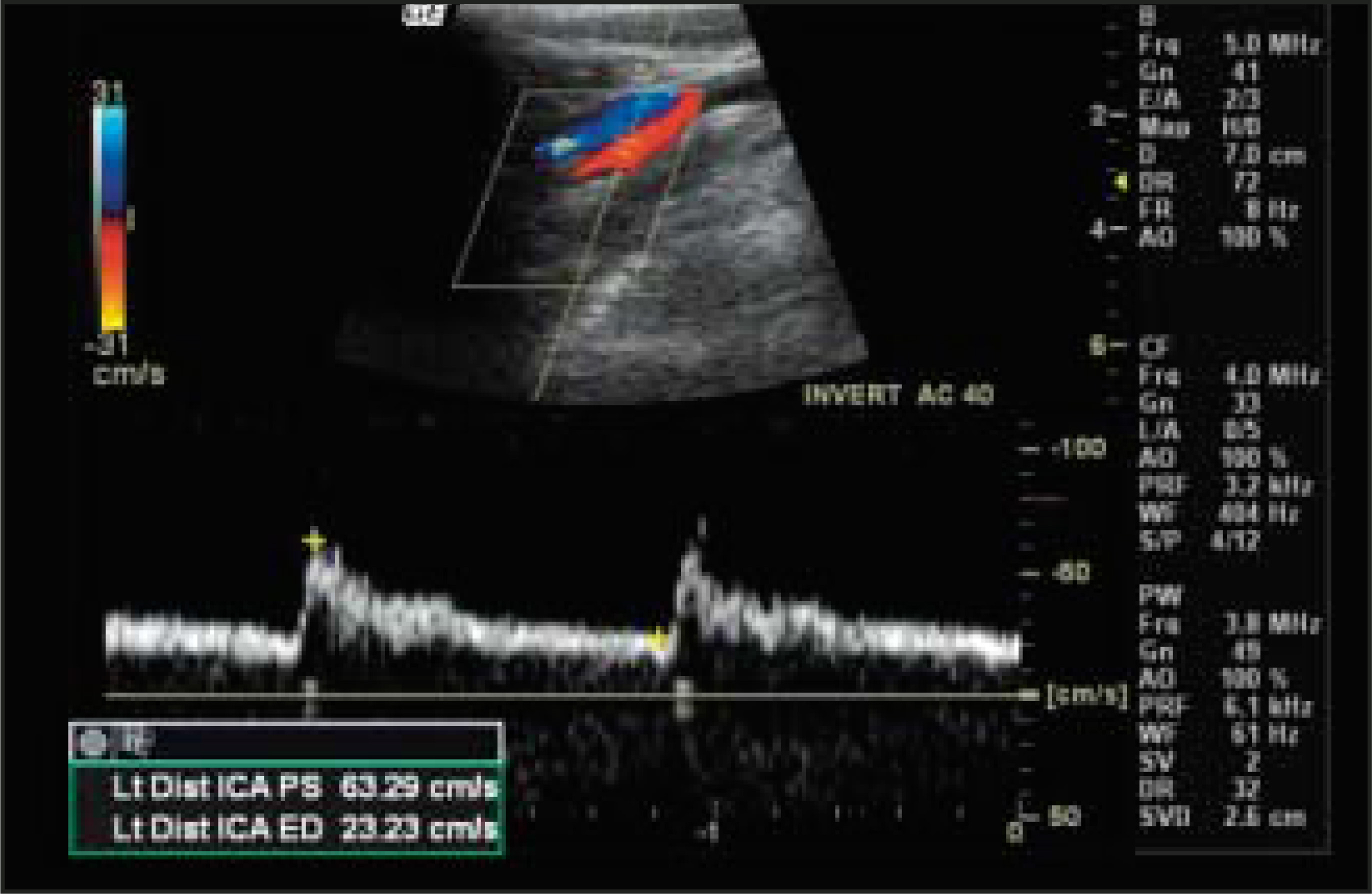
Criteria for determining the degree of carotid stenosis differ widely among various neurovascular laboratories.18 What all of these criteria have in common is that, because blood flow increases as lumenal diameter narrows, stenosis is assumed to be proportional to Doppler velocity measurements. In our facility, a peak systolic velocity of 250 cm/sec has been shown to approximately correlate with a residual lumen diameter of 1.5 mm, or what, in many centers, would be considered a 70% stenosis.17 Using this cutoff value, in a study comparing ultrasound and MRA with catheter arteriography, investigators at Brigham and Women’s Hospital reported a sensitivity, specificity, and accuracy of ultrasound of 94%, 83%, and 86%, respectively, for the detection of a 70% stenosis.19
An important caveat to the assumption that the degree of lumenal narrowing is proportional to the increase in peak systolic velocity is the phenomenon of “velocities falling off.” Although uncommon, this phenomenon is not rare. It occurs in the setting of a critical ICA stenosis, typically at residual lumen diameters <0.5 to 0.7 mm, where the narrowing is so severe that there is reduction, or “pseudonormalization,” of the peak systolic velocity to a normal range.17 Although inspection of the B-mode (grayscale) and colorflow findings will sometimes help to avoid such a false-negative “normal” reading, examination of the Doppler waveform patterns is often more revealing.20 Specifically, waveforms immediately distal to a hemodynamically critical stenosis may have a damped appearance, whereas those immediately proximal to it may have a high resistance pattern. It is noteworthy that B-mode US images are not typically relied upon for determining the degree of ICA stenosis; rather, they guide qualitative assessment of plaque morphology (which may be useful in the follow-up of post-CEA patients at risk for intimal media hyperplasia) (Figure 2).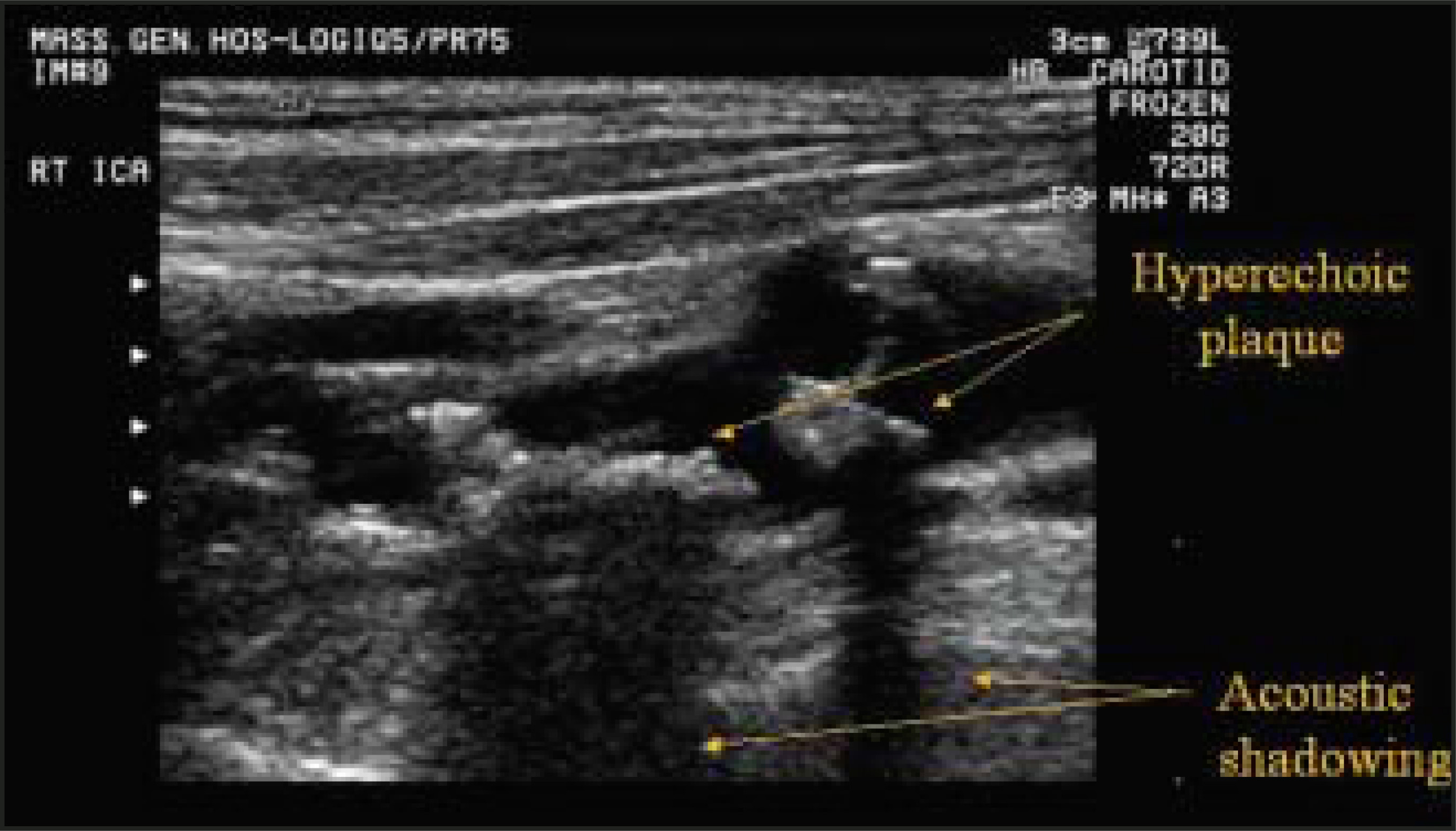
Preliminary results from our neurovascular laboratory have also suggested that there are several distal waveform patterns, which are highly specific for confirming a severe (but not necessarily a critical) proximal stenosis.20 An example of one such pattern is provided in Figure 3.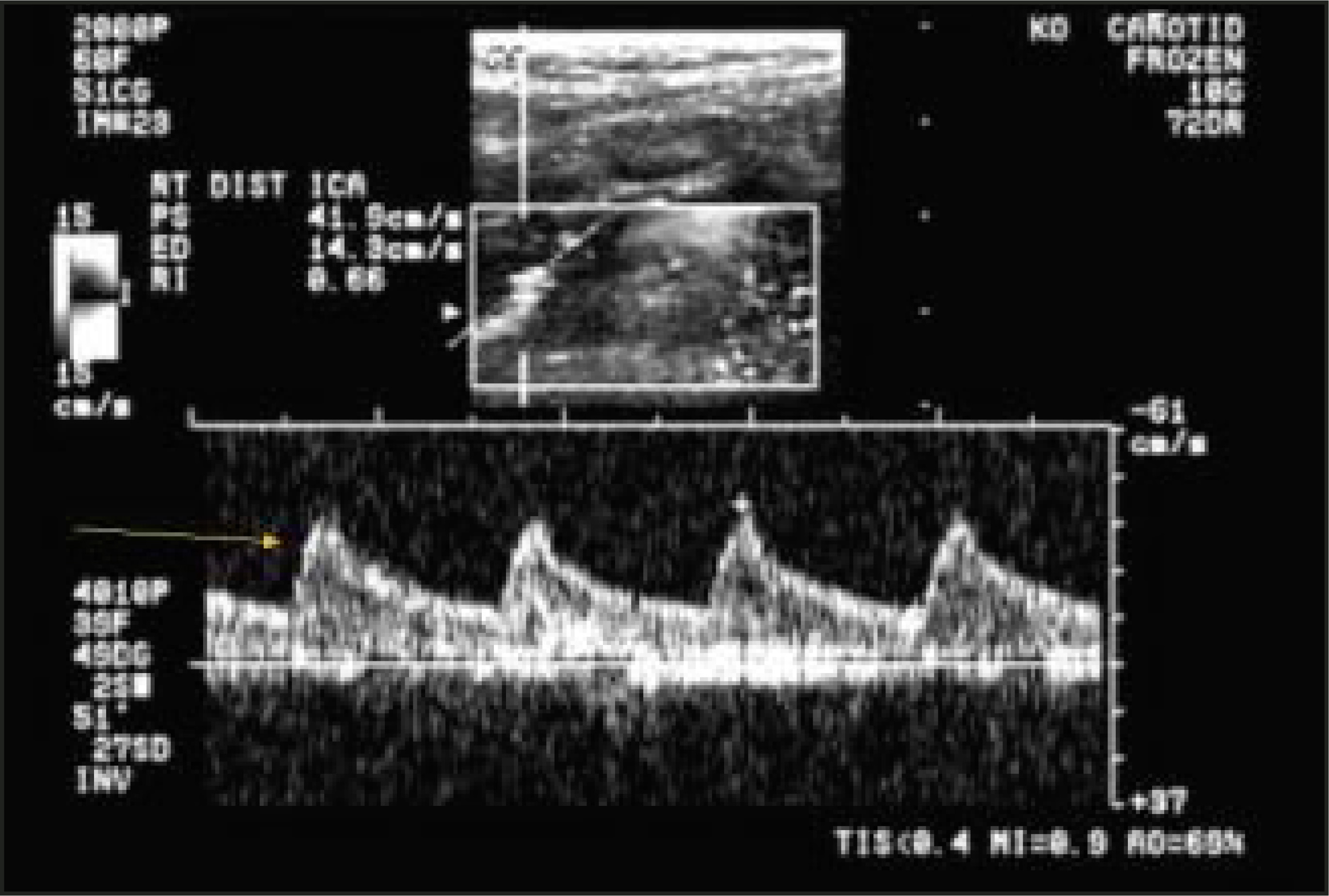
MRA
In addition to providing an alternative screening technique for the detection of clinically significant carotid artery occlusive disease, MRA also permits simultaneous acquisition of parenchymal brain MR images. Although a comprehensive discussion of carotid MRA is well beyond the scope of this review, it is noteworthy that, of the unenhanced methods, it is the time-of-flight (TOF)—and not the phase-contrast (PC) techniques—that are most clinically applicable. Bolus timing with elliptic centered k-space acquisition has become the most widely used technique for gadolinium-enhanced MRA and is the specific gadolinium-enhanced technique to which we will refer later in this article.
Defining reliable criteria for quantification of carotid stenoses with MRA—whether using TOF or gadolinium-enhanced imaging—has proved problematic. Although both narrowing and reduced signal intensity of the flow-related MRA signal column are suggestive of a severe stenosis, determination of a precise percentage narrowing is unreliable. While some studies have suggested that the presence of a surgically severe lesion correlates with complete, segmental, signal dropout on two-dimensional (2D) TOF sequences,21 the accuracy of this finding has been questioned.22 Indeed, there is a broad range of percent stenoses for which one can observe signal dropout on a TOF MRA.8 Moreover, because signal dropout is caused by intravoxel dephasing in regions of turbulent flow, it is highly dependent on the echo time (TE) used for scanning. The shorter TEs and more homogeneous magnetic fields common on newer generation MR scanners therefore imply that a more severe degree of stenosis is required to produce signal dropout. At our institution, we performed a comparison of 2D-TOF MRA using two different TE values. At a TE of 8.7 msec, 95% of patients with peak systolic velocity (PSV) >250 cm/sec showed complete signal dropout at the site of maximal stenosis. At a shorter TE of 4.7 msec, however, the proportion of patients with complete signal dropout at PSV >250 cm/sec dropped to only 50%22 (Figure 4).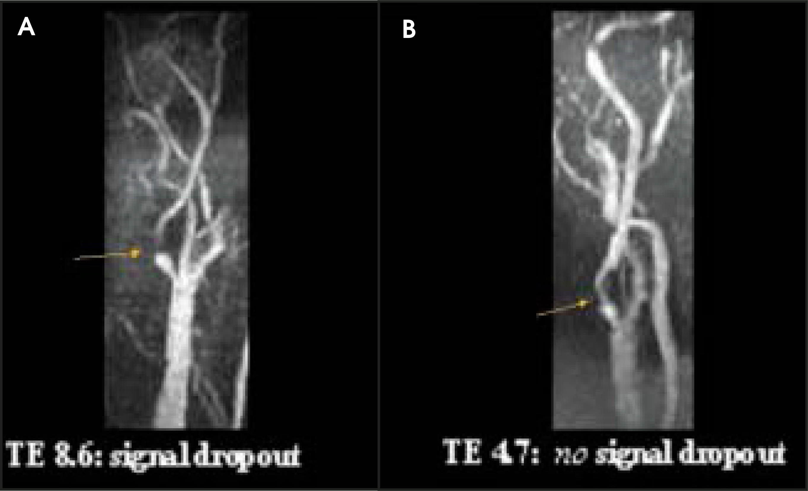
Contrast-enhanced MRA (CE-MRA) has shown promise in being able to distinguish higher-grade stenoses than can TOF MRA.23,24 Compared with 2D-TOF MRA, CE-MRA likely has better spatial resolution, increased coverage (including the great vessel origins and the intracranial segments of the internal carotid arteries, two common sites of clinically significant stenosis17), and less susceptibility and motion artifact. Early results have shown a good correlation with digital subtraction angiography (DSA), which is still widely con- sidered the gold standard for carotid stenosis measurement.25,26 A recent meta-analysis by Nederkoorn et al,26 comparing CE-MRA with catheter arteriography, revealed a pooled sensitivity of 95% and specificity of 90% for CE-MRA in distinguishing 70 to 99% stenoses from <70% stenoses (Figure 5).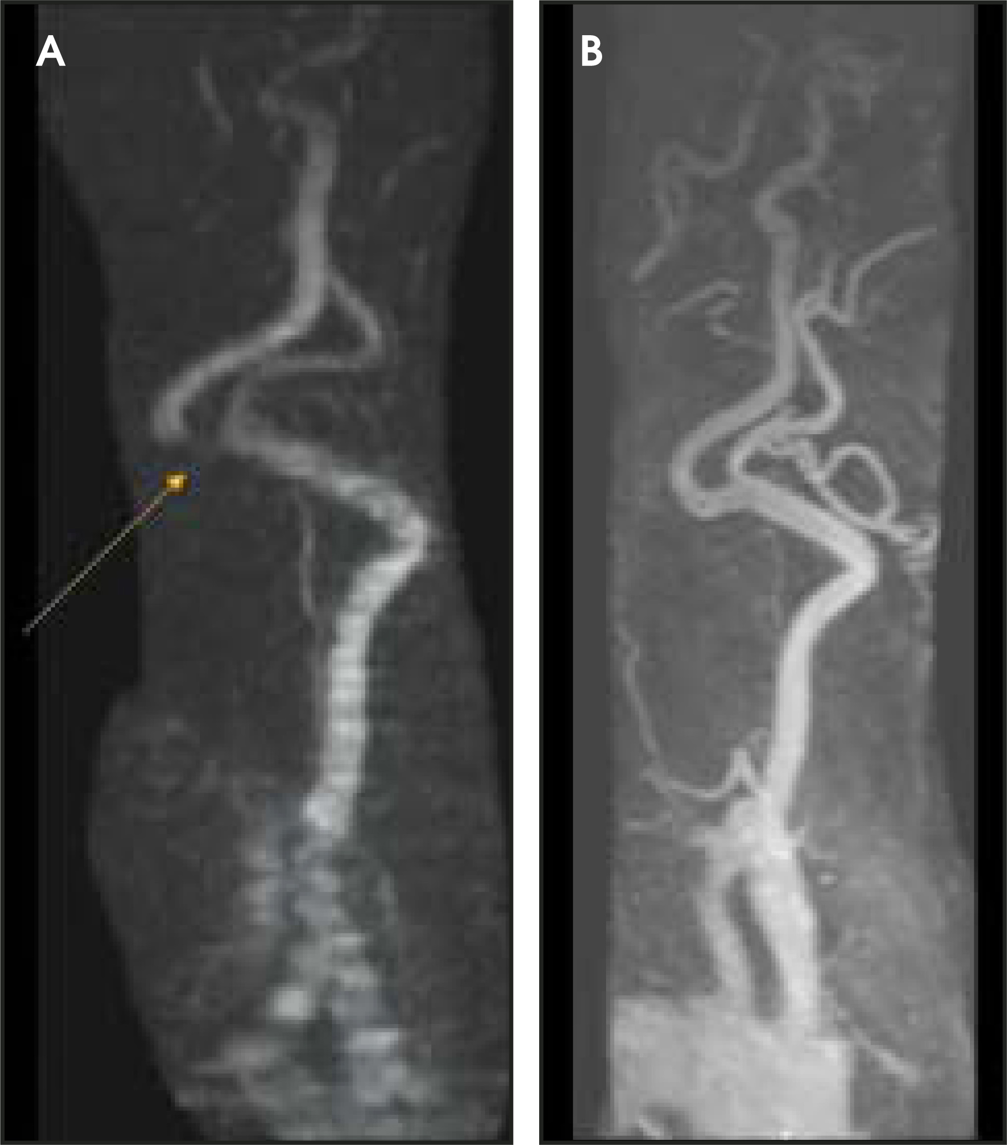
Even given the increased spatial resolution afforded by gadolinium-enhanced MRA, however, the voxel size in CE-MRA is larger than that of CTA, and significantly larger than that of catheter arteriography. Because, however, a clinically significant ICA narrowing is approximately 1.5 mm in length, an error of even a single-pixel diameter—which can be due to image noise, artifact, or vessel pulsation—has the potential to change the category of stenosis from moderate to severe, thus potentially altering the decision for CEA or CAS versus medical therapy unless additional studies are used to confirm the findings. Indeed, the reader is referred to a recent editorial by Turski27 in the American Journal of Neuroradiology, which discusses sources of variability in CE-MRA measurement of carotid stenosis. As with 2D-TOF MRA, turbulence just distal to a severe stenosis on CE-MRA can result in intravoxel dephasing, resulting in overestimation of the degree of stenosis, which can sometimes be worse than that occurring with the TOF technique.28
The diagnostic accuracy for determining percent stenosis is dramatically improved when the results of both gadolinium-enhanced MRA and carotid US are concordant.28-30 Some investigators have suggested that this combination of diagnostic tests can obviate the need for preoperative conventional catheter angiography.31 Whether or not conventional catheter arteriography is occasionally still required in some clinical situations prior to surgery remains controversial.31-33
CTA
With the advent of newer, faster, multislice CT scanners, CTA may ultimately supplant DSA for cases in which CE-MRA and US provide discordant findings. An inherent advantage of CTA over both TOF and CE-MRA techniques is that it is an anatomic, rather than a physiologic, imaging modality, and it is therefore relatively insensitive to flow-related signal dropout—which can result in overestimation of stenosis. With CTA, pixel intensity is directly proportional to the density of contrast within the vascular lumen, and therefore more directly correlates with residual lumen diameter on surgical pathologic specimens.34
Other advantages of CTA over MRA include a lower likelihood of motion artifact (patients should be instructed to neither breathe nor swallow at the start of the acquisition!), higher spatial resolution (including slice thickness of from 0.5 to 1 mm, reconstructed at arbitrarily small intervals), and the ability to distinguish calcified from noncalcified atherosclerotic plaque. CT angiography can depict the course of a vessel with very slow or absent flow—something that cannot be reliably accomplished by other techniques, including catheter angiography (Figure 6). CT angiography can also depict tandem stenoses of the intracranial vasculature, a finding present in 9% of patients with carotid bifurcation stenoses.17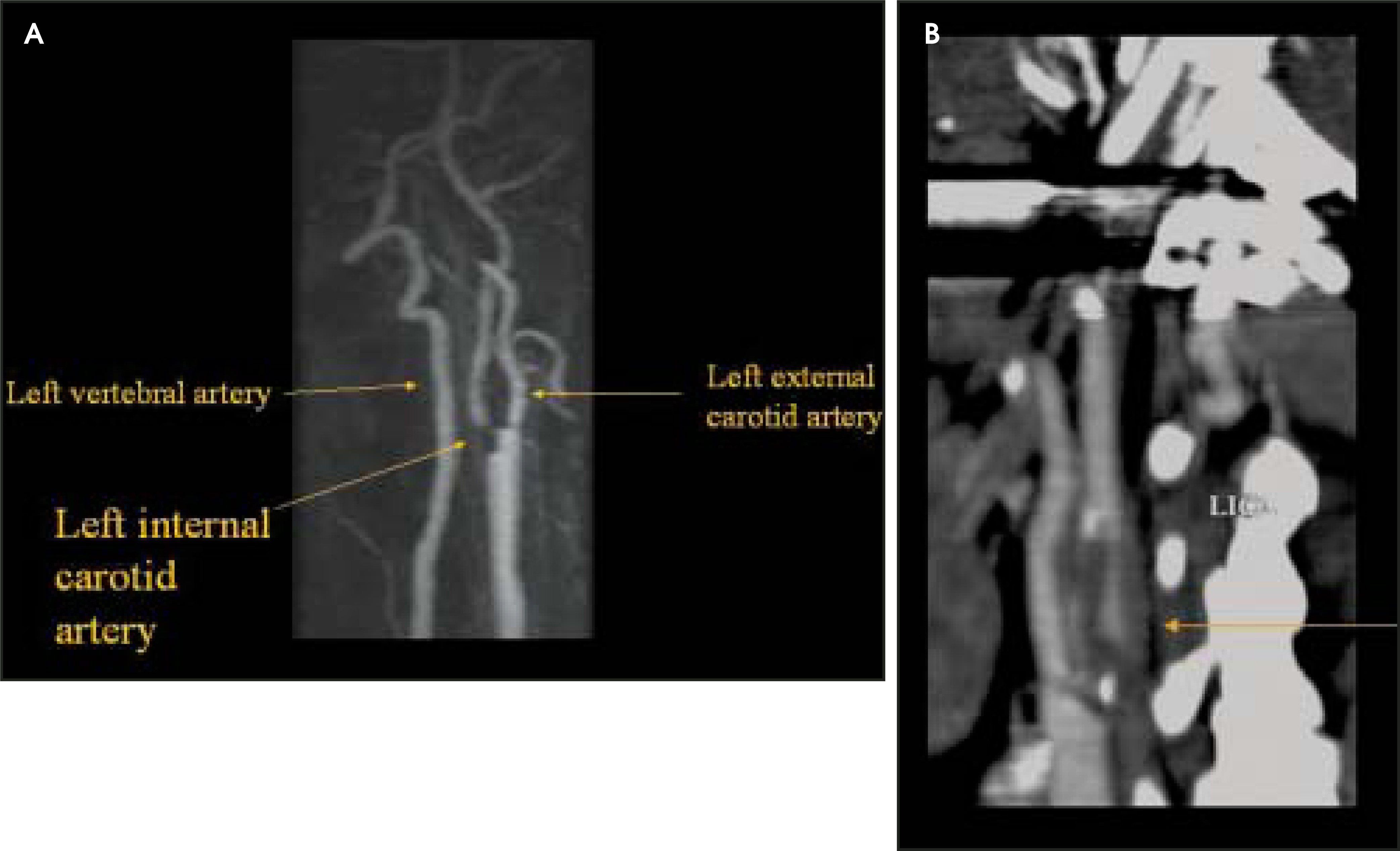
Perhaps most important is that CTA has the ability to distinguish—with a high degree of accuracy—a hairline residual lumen from complete ICA occlusion (Figure 7). This distinction can sometimes, in symptomatic patients, make the difference between operative revascularization (CEA or CAS) and conservative medical therapy. In a recent study by Lev et al35 of patients with US and/or MRA findings suggesting complete ICA occlusion, CTA was found to have a 90% overall sensitivity for detecting a hairline residual lumen when compared with catheter angiography. The potential importance of distinguishing a hairline residual lumen from a complete ICA occlusion should not be underestimated. In a recent editorial in Stroke, Rothwell31 explains that if initial imaging fails to reveal a hairline residual lumen, “for every three patients who are not operated on, . . . one stroke will go unprevented.”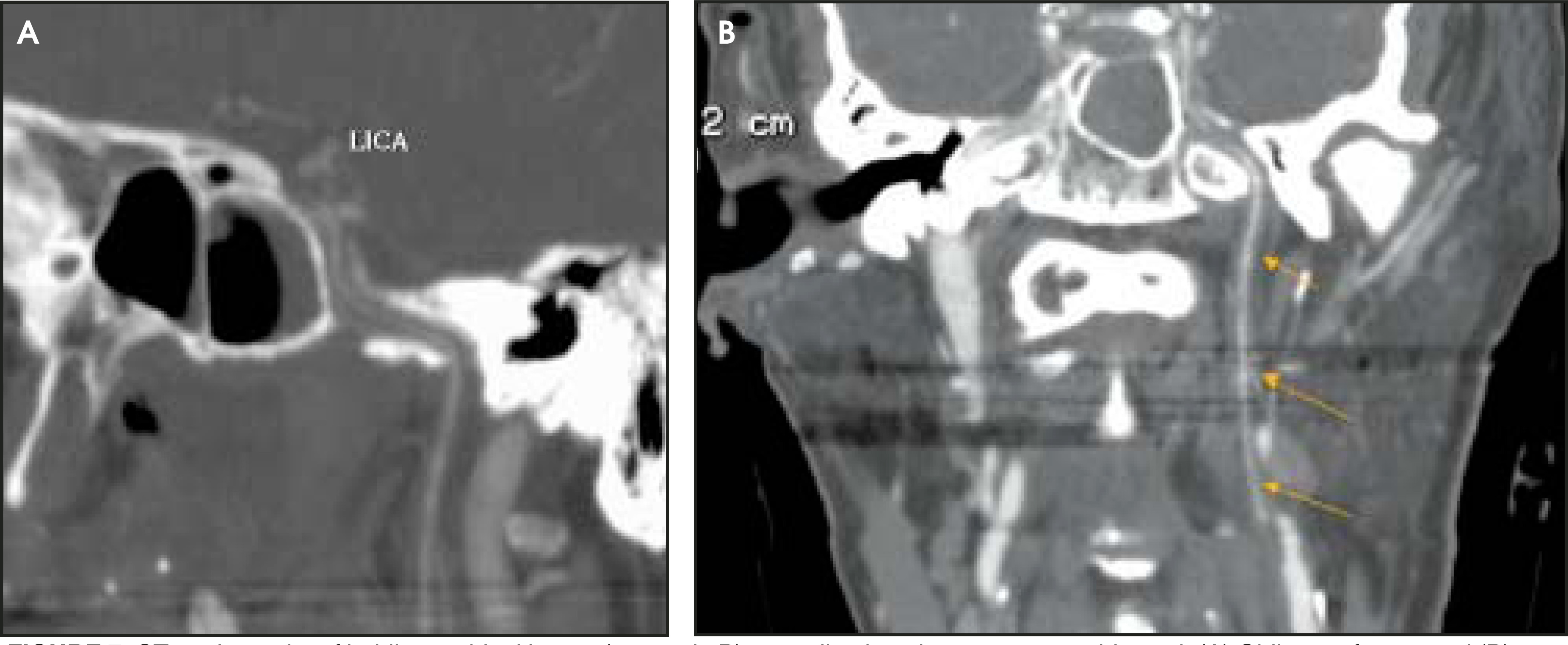
In addition to its high sensitivity for distinguishing hairline lumen from occlusion, CTA can also be highly specific, provided one avoids certain pitfalls. First, one must make sure the “string” is not, in fact, the ascending pharyngeal artery. Although this vessel follows a similar path to the extracranial ICA, it does not extend into the petrous ICA canal. The hypoglossal artery, which will enter the hypoglossal canal, can also potentially mimic a proximal internal carotid string sign (Figure 8).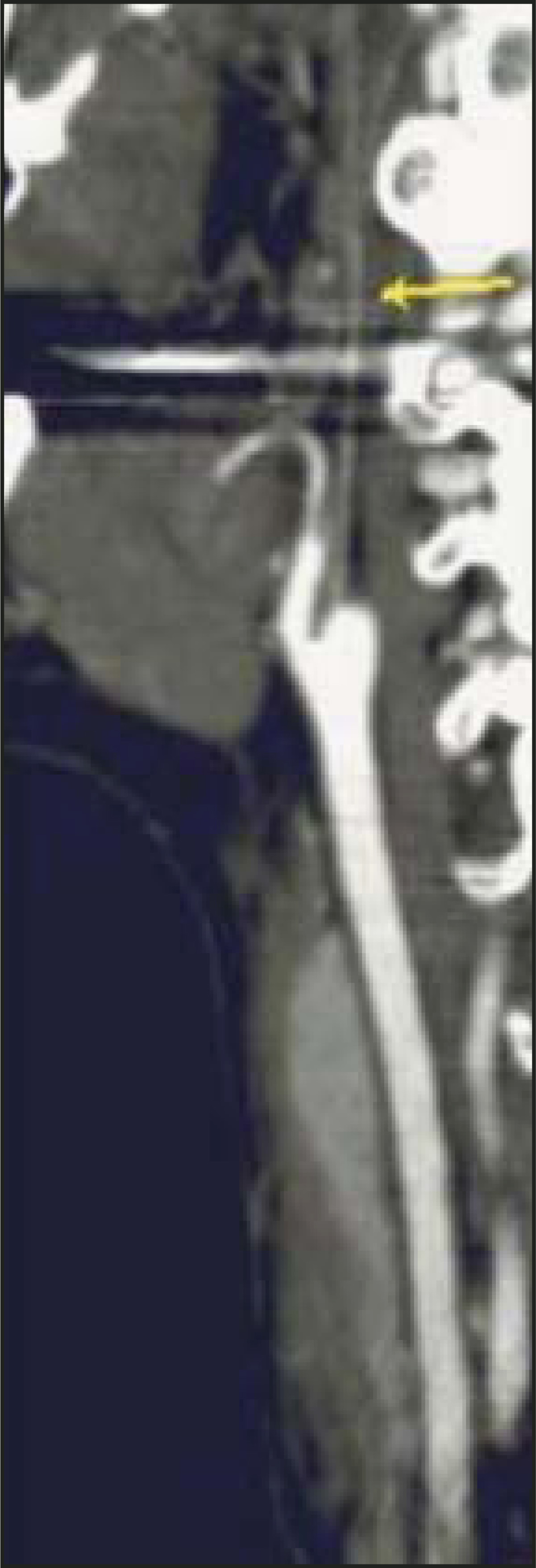
Another important technical point is the importance of choosing appropriate window and level settings for CTA image review. This is especially relevant for measuring the most severe degrees of stenosis. In this regard, the reader is referred to an excellent paper by Liu et al,36 which provides a chart for optimal window and level settings based on the HU attenuation of the contrast within the vessel lumen. The bottom line is that, like the brightness and contrast settings on a television set or computer monitor, the dynamic range of the CTA image display should include the entire grayscale of the contrast-filled lumen and the surrounding vessel wall (Figure 9).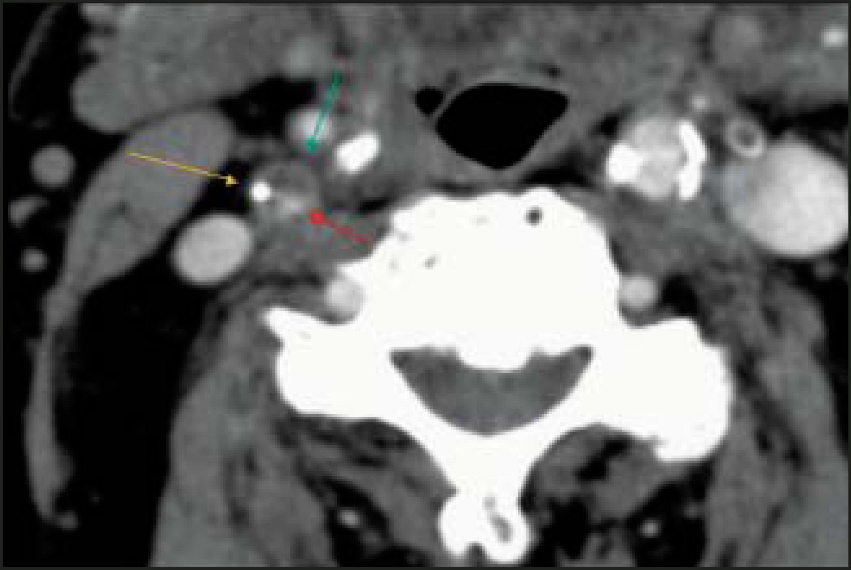
Future directions
Although it is well-established that stroke risk increases in direct proportion to the degree of carotid stenosis, the fact remains that not all critical stenoses will result in a stroke, and that some patients with <70% stenosis will have a stroke. This observation has resulted in in-creased recent interest in the topic of “vulnerable plaque.” The concept of vulnerable plaque, which is commonplace in the cardiology literature, has become something of a “hot topic” among neuroimagers. “Vulnerable plaque” refers to the morphologic/histologic characteristics of an atherosclerotic plaque that may or may not predispose to platelet-fibrin clumping, and hence the formation of embolic material.
Specifically, recent pathologic-MR imaging correlation studies have revealed that, within a given category of stenosis, certain characteristics may help to distinguish “vulnerable” plaque (ie, that more likely to result in stroke) from “stable” plaque (ie, that less likely to result in stroke).37,38 The histopathologic features suggestive of a vulnerable plaque include high lipid content (fatty appearance), increased macrophages (infiammation and enhancement), and a thinner overlying collagen cap (thin or absent plaque wall) (Figure 10).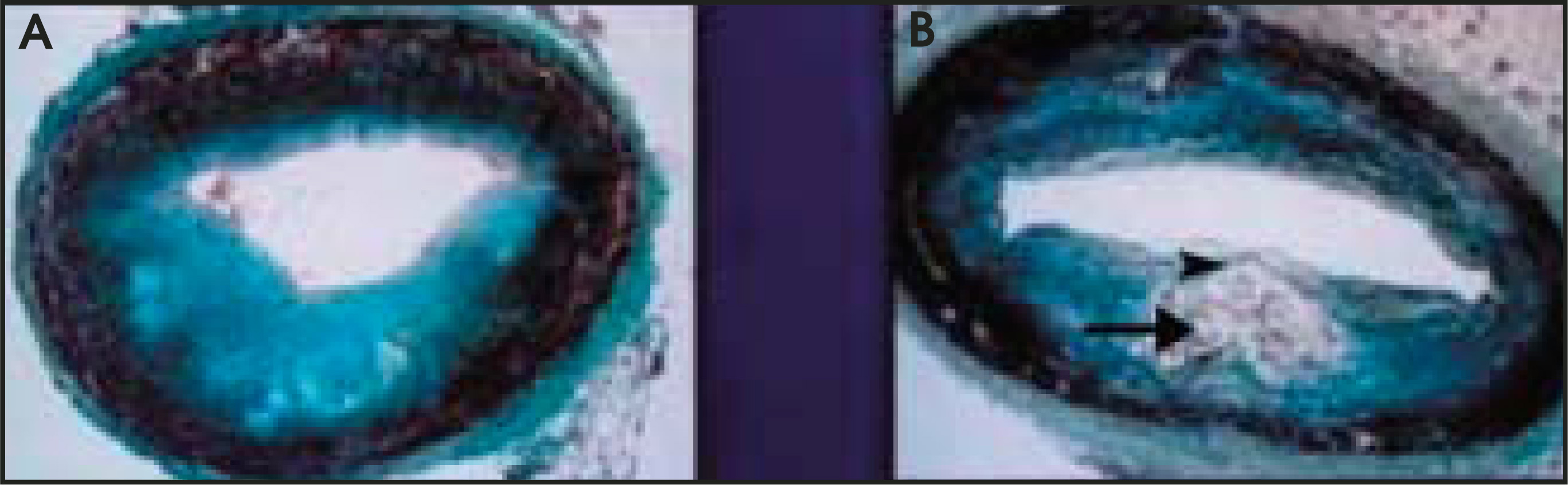
At least two groups of investigators thus far have reported a good correlation between these pathologic features and the appearance on MR using very thin slices.37,38 Future imaging efforts directed at identification of “vulnerable” plaque may help in further stratifying stroke risk for a given category of stenosis.
Conclusion
With continued advances, noninvasive imaging techniques are likely to fully supplant catheter arteriography for helping to triage patients with carotid artery occlusive disease toward medical therapy, CEA, or CAS. Due to its accuracy, low cost, and availability, US is often the first imaging study obtained and is useful for screening, initial quantification of stenoses, and assessment of small amounts of incremental progression of stenosis over time. In cases in which US and MRA results are discordant, or in which there is a possibility of occlusion, and hence the need to exclude a potential hairline lumen, CTA provides a higher degree of accuracy, approaching that of catheter arteriography. Further defining the characteristics of “vulnerable” plaque may prove to be the next frontier for carotid artery imaging, in the ongoing effort to prevent stroke.
References
- Fisher C. A career in cerebrovascular disease—A personal account. Stroke. 2001;32:2719-2724.
- Gleason S, Furie K, Lev M. Potential influence of acute CT on inpatient costs in patients with ischemic stroke. Acad Radiol. 2001;8:955-964.
- Beneficial effect of carotid end-arterectomy in symptomatic patients with high-grade carotid stenosis. N Engl J Med. 1991;325:445-453.
- Barnett H, Taylor D, Eliasziw M. Benefit of carotid endarterectomy in patients with symptomatic moderate or severe stenosis. N Engl J Med. 1998;339:1415-1425.
- Hobson R. Update on the Carotid Revascularization Endarterectomy versus Stent Trial (CREST) protocol. J Am Coll Surg. 2002;194(1 suppl):S9-S14.
- Ackerman R, Lev M, Romero J, Latchaw R, Kucharczyk W. Neuroradiology. 2004.
- Endarterectomy for asymptomatic carotid artery stenosis. JAMA. 1995;273:1421-1428.
- Biller J, Thies W. When to operate in carotid artery disease. Am Fam Physician. 2000;61:400-406.
- Carotid surgery versus medical therapy in asymptomatic carotid stenosis. Stroke. 1991;22:1229-1235.
- Results of a randomized controlled trial of carotid endarterectomy for asymptomatic carotid stenosis. Mayo Clin Proc. 1992;67:513-518.
- Barr J, Connors J, Sacks D. Quality improvement guidelines for the performance of cervical carotid angioplasty and stent placement. Am J Neuroradiol. 2003;24:2020-2034.
- Hobson R. CREST (Carotid Revascularization Endarterectomy versus Stent Trial): Background, design and current status. Semin Vasc Surg. 2000;13:139-143.
- Albers G, Caplan L, Easton J. Transient ischemic attack—proposal for a new definition [editorial]. N Engl J Med. 2002;37:1713-1716.
- MRC European Carotid Surgery Trial: Interim results for symptomatic patients with severe (70–99%) or with mild (0–29%) carotid stenosis. Lancet. 1991;337:1235-1243.
- Ackerman R, Candia M. Identifying clinically relevant carotid disease. Stroke. 1994;25:1-3.
- Suwanwela N, Can U, Furie K. Carotid doppler ultrasound criteria for internal carotid artery stenosis based on residual lumen diameter calculated from en bloc carotid endarterectomy specimens. Stroke. 1996;27:1965-1969.
- Ackerman R, Taveras J, Ferrucii J. Radiology: Diagnosis Imaging Intervention. 1995:1-28.
- Grant E, Benson C, Moneta G. Carotid artery stenosis: Gray-scale and Doppler US diagnosis—Society of Radiologists in Ultrasound Consensus Conference. Radiology. 2003;23:340-346.
- Patel M, Kuntz K, Klufas R. Preoperative assessment of the carotid bifurcation. Can magnetic resonance angiography and duplex ultrasonography replace contrast arteriography?. Stroke. 1995;26:1753-1758.
- Romero J, Lev M, Chan S. US of neurovascular occlusive disease: Interpretive pearls and pitfalls. RadioGraphics. 2002;22:1165-1176.
- Heiserman J, Zabramski J, Drayer B, Keller P. Clinical significance of the flow gap in carotid magnetic resonance angiography. J Neurosurg. 1996;85:384-387.
- Lev M, Romero J, Gonzalez R. Flow voids in time-of-flight MR angiography in carotid artery stenosis? It depends on the TE [letter]. AJNR Am J Neuroradiol. 2003;24:2120.
- Enochs W, Ackerman R, Kaufman J, Candia M. Gadolinium-enhanced MR angiography of the carotid arteries. J Neuroimaging. 1998;8:185-190.
- Nederkoorn P, Elgersma O, van der Graaf Y. Carotid artery stenosis: Accuracy of contrast-enhanced MR angiography for diagnosis. Radiology. 2003;28:677-682.
- Remonda L, Senn P, Barth A. Contrast-enhanced 3-D MR angiography of the carotid artery: Comparison with conventional digital subtraction angiography. AJNR Am J Neuroradiol. 2002;23:213-219.
- Nederkoorn P, van der Graaf Y, Hunink M. Duplex ultrasound and magnetic resonance angiography compared with digital subtraction angiography in carotid artery stenosis: A systematic review. Stroke. 2003;34:1324-1332.
- Turski P. Sources of variability in measuring carotid stenosis on time-resolved contrast-enhanced MR angiograms [editorial]. AJNR Am J Neuroradiol. 2002;23:178-179.
- Townsend T, Saloner D, Pan X, Rapp J. Contrast material-enhanced MRA overestimates severity of the carotid stenosis, compared with 3D time-of flight MRA. J Vasc Surg. 2003;38:36-40.
- Johnston D, Eastwood J, Nguyen T, Goldstein L. Contrast-enhanced magnetic resonance angiography of carotid arteries—utility in routine clinical practice. Stroke. 2002;33:2834-2838.
- Borisch I, Horn M, Butz B. Preoperative evaluation of carotid artery stenosis: Comparison of contrast-enhanced MR angiography and duplex sonography with digital subtraction angiography. AJNR Am J Neuroradiol. 2003;24:1117-1122.
- Rothwell P. For severe carotid stenosis found on ultrasound, further arterial evaluation prior to carotid endarterectomy is unnecessary: The argument against. Stroke. 2003;34:1817-1819.
- Davis S, Donnan G. Is carotid angiography necessary? Editors disagree [editorial]. Stroke. 2003;34:1819.
- Derdeyn C. Catheter angiography is still necessary for the measurement of carotid stenosis [editorial]. AJNR Am J Neuroradiol. 2003;24:1737-1738.
- Guimares A, Ackerman R, Romero J, Lev M. Multislice CT offers new options for imaging carotid artery stenosis. Diagn Imaging. 2002;24:70-75.
- Lev M, Romero J, Goodman D. Total occlusion versus hairline residual lumen of the internal carotid arteries: Accuracy of single section helical CT angiography. AJNR Am J Neuroradiol. 2003;24:1123-1129.
- Liu Y, Hopper K, Mauger D, Addis K. CT angiographic measurement of the carotid artery: Optimizing visualization by manipulating window and level settings and contrast material attenuation. Radiology. 2000;217:494-500.
- Mitsumori L, Hatsukami T, Ferguson M. In vivo accuracy of multisequence MR imaging for identifying unstable fibrous caps in advanced human carotid plaques. J Magn Reson Imaging. 2003;17:410-420.
- Wasserman B, Smith W, Trout H. Carotid artery atherosclerosis: In vivo morphologic characterization with gadolinium-enhanced double-oblique MR imaging initial results. Radiology. 2002;223:566-573.
Citation
. Noninvasive imaging evaluation of carotid artery occlusive disease. Applied Radiology. 2004;33(8):16-24. doi:10.37549/AR1272.