Nonvascular lesions of the mediastinum
Applied Radiology — Vol. 31 , Issue 5 , pp. 33 -41
DOI: 10.37549/AR1091
Published: May 1, 2002
Categories
The mediastinum is defined as the area bounded by the two lungs laterally, the diaphragm inferiorly, the thoracic inlet superiorly, the sternum anteriorly, and the vertebral column posteriorly. It is divided primarily into anterior, middle, and posterior compartments by the most commonly used classification scheme.1 The anterior compartment (pre-vascular space) includes all structures posterior to the sternum and anterior to the pericardium, aorta, and brachiocephalic vessels. This area includes lymph nodes, internal mammary vessels, the thymus, and fat. The middle mediastinum is largely a vascular compartment and includes the pericardium and its contents, the ascending and transverse aortas, the superior vena cava (SVC), the brachiocephalic vessels, the trachea, the phrenic nerve, the upper portion of the vagus, and the lymph nodes. The posterior compartment (post-vascular space) is bounded anteriorly by the pericardium and contains lymph nodes, vertebral bodies, and the contents of spinal canal. This space also includes the esophagus, the azygos and hemiazygos veins, the descending aorta, and the intercostal and autonomic nerves. The assessment of masses relies heavily, but not exclusively, on the ability to define an “anatomic” location for lesions. This review will focus on the nonvascular entities that commonly present on radiologic imaging. Primary tumors of the mediastinum are rare, comprising about 3% of chest tumors.2-5 The following discussion addresses the most common lesions, rather than an exhaustive cataloguing of the wide variety of conditions that may present as mediastinal masses.
Workup of mediastinal masses includes chest radiographs (CXR), computed tomography (CT), and magnetic resonance imaging (MRI). CT is better than a CXR in characterizing mediastinal masses as to fat, fluid, or calcium content, as well as localizing origin and extent of lesions and identifying associated abnormalities.6 The primary role of MR in the evaluation of the mediastinum is problem solving, as it shows some superiority in characterizing various tissues. T2- and T1-weighted fat-saturation sequences are most useful for this purpose.7
Anterior mediastinum/vascular space
Lymphoma
More than 35,000 cases of lymphoma are diagnosed annually. Both Hodgkin’s disease (HD) and non-Hodgkin’s lymphoma (NHL) affect the mediastinum, but HD represents 25% to 30% of all cases of lymphoma and is the most common entity to present as an intrathoracic lesion.8 The age distribution of HD is bimodal with peak incidence at early adulthood and after age 50.9 At the time of presentation, 85% of patients have chest involvement,10 and 90% of patients have lymph node enlargement in the mediastinum, but only 25% of all cases of disease is limited to the chest.10 Of the HD subtypes, the nodular sclerosing type most commonly affects the anterior mediastinum.11 Non-Hodgkin lymphoma is histopathologically distinct from HD, with intrathoracic involvement in 40% of patients at presentation. Only 10% have disease limited to the chest.10 Of the NHL subtypes, the large B-cell lymphoma and the lymphoblastic lymphoma most frequently affect the anterior mediastinum.12-14
Lymphoma may present as nodal enlargement seen as singularly enlarged nodes or as a conglomerate mass of nodes. On CT, the mass may be homogeneous or heterogeneous depending on the presence of calcification, hemorrhage, necrosis, or cystic components15-17 (figure 1). On MR, lymphoma appears as homogenous low-signal on T1-weighted images and as homogenous high-signal or intermixed areas of low and high intensity on T2-weighted images.18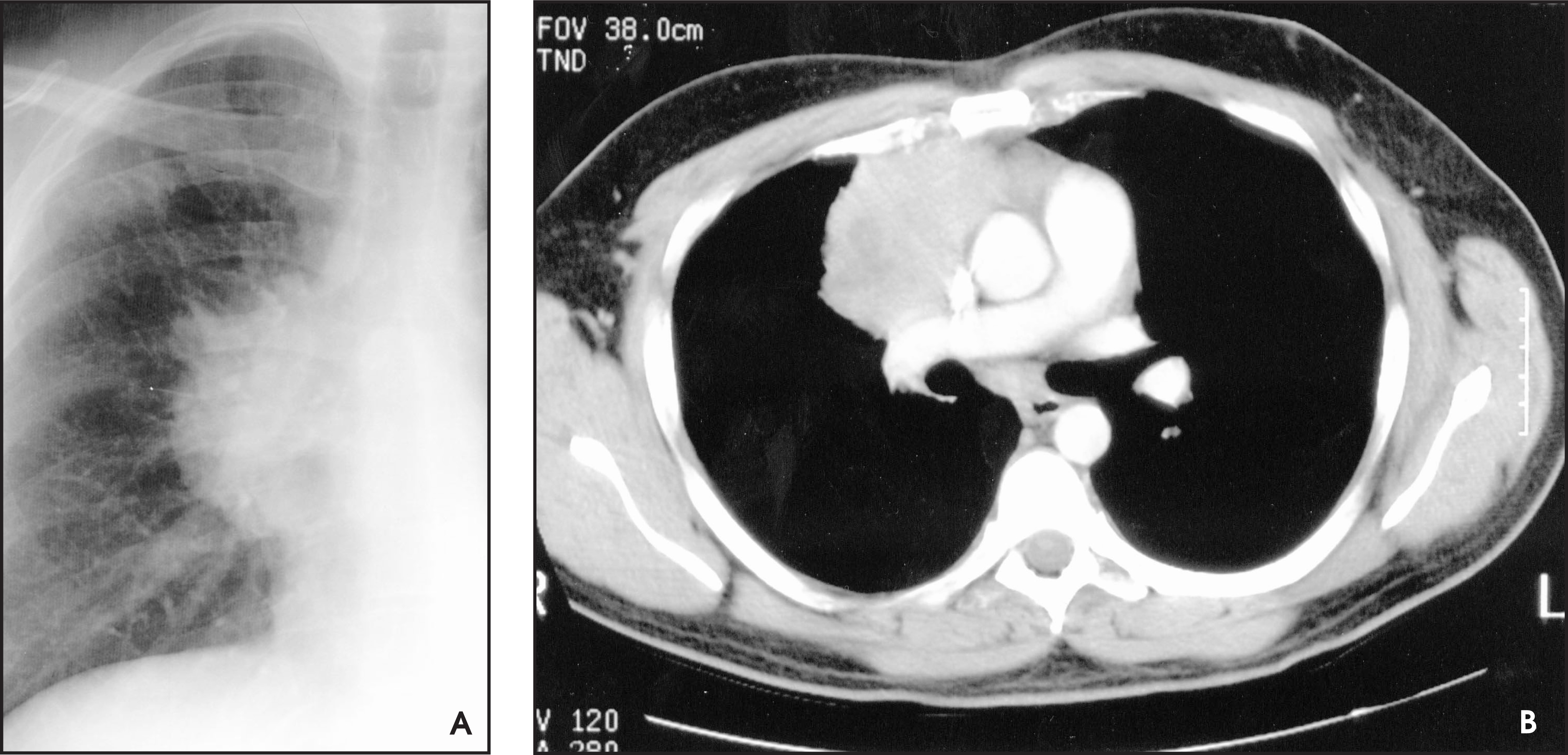
Thymoma and other thymic neoplasms
Thymomas are the most common primary tumor type in the anterior mediastinum.19-21 There appears to be no sex predilection for thymomas, but most cases occur in patients who are older than 20 years of age.22 There is a common association with myasthenia gravis (MG). Up to 50% of patients with thymoma have MG, but only 10% to 15% of patients with MG have thymomas,23 and some patients have been found to have attenuation of symptoms following resection of the thymoma.21 Half of patients with thymomas are asymptomatic.
Other parathymic manifestations associated with thymoma include a pure red-cell aplasia and hypogammaglobulinemia. Although most thymomas are benign and contained by fibrous capsule, a third of thymomas invade through their capsule even though they remain cytologically benign.21,24 These are termed invasive thymomas. There can be transdiaphragmatic extension, vascular invasion, or mediastinal fat invasion by these lesions and they may even involve the pleural surface mimicking a malignant mesothelioma.25 Approximately 30% to 35% of thymomas are malignant.
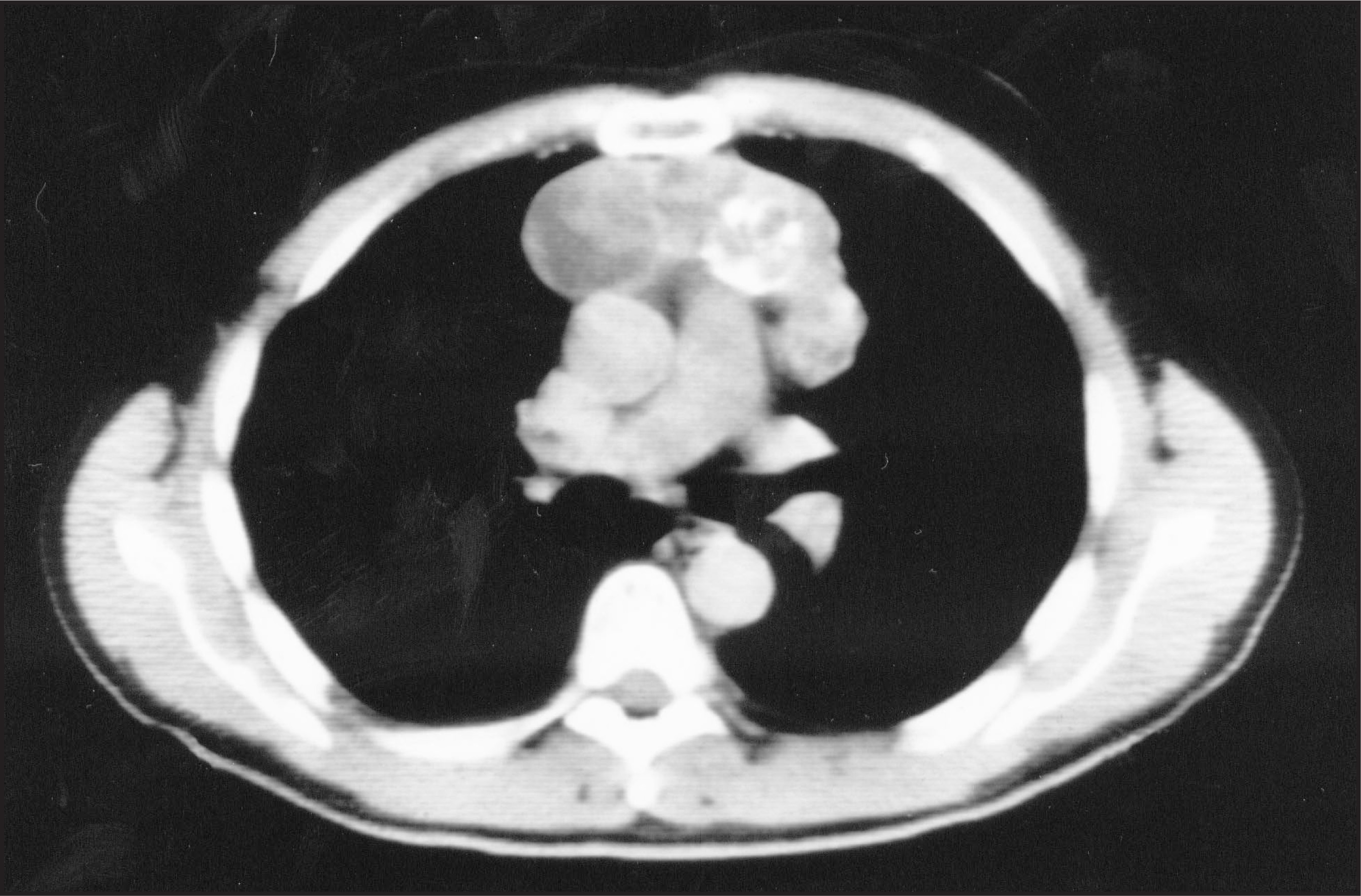
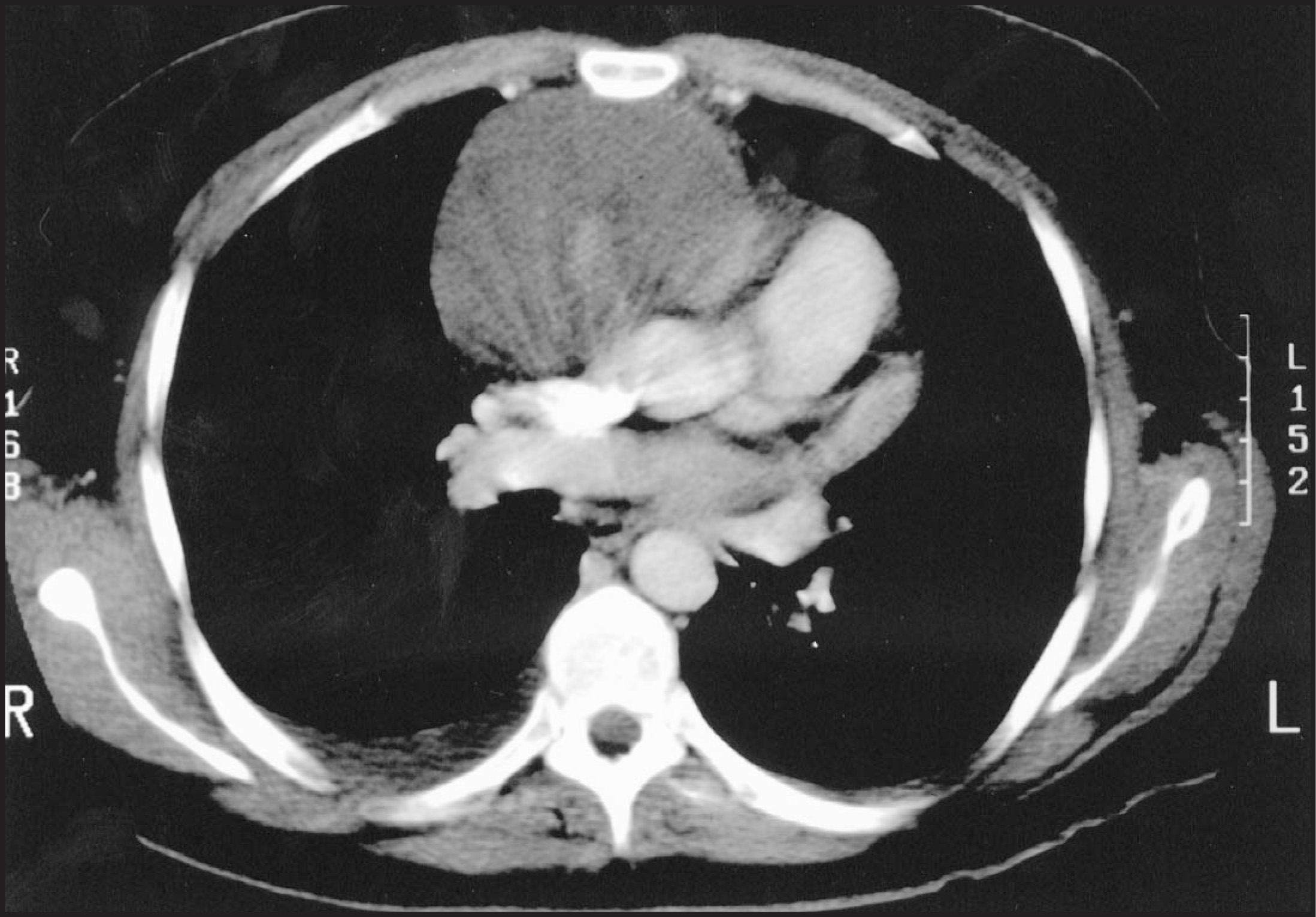
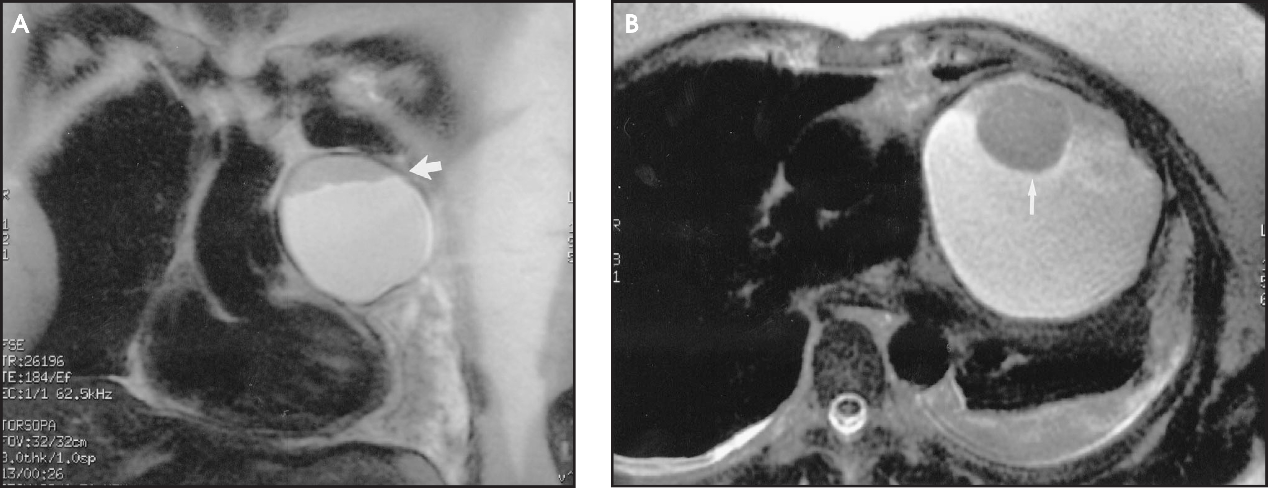
On CT, thymomas may demonstrate homogenous or heterogeneous attenuation depending on presence of hemorrhage, necrosis, or cystic components26 but are usually more solid in appearance than the normal thymus.22 They can appear as round, oval, or lobulated masses (figure 2). Calcification is seen in as many as 25% of cases and is usually coarse or irregular (figure 3). On MRI, thymomas can have intermediate-to high-signal intensity on T1-weighted images and high signal on T2-weighted images (figure 4). Contrast administration may help define vascular invasion; however, MRI does not offer a significant advantage over CT in the evaluation of thymomas.22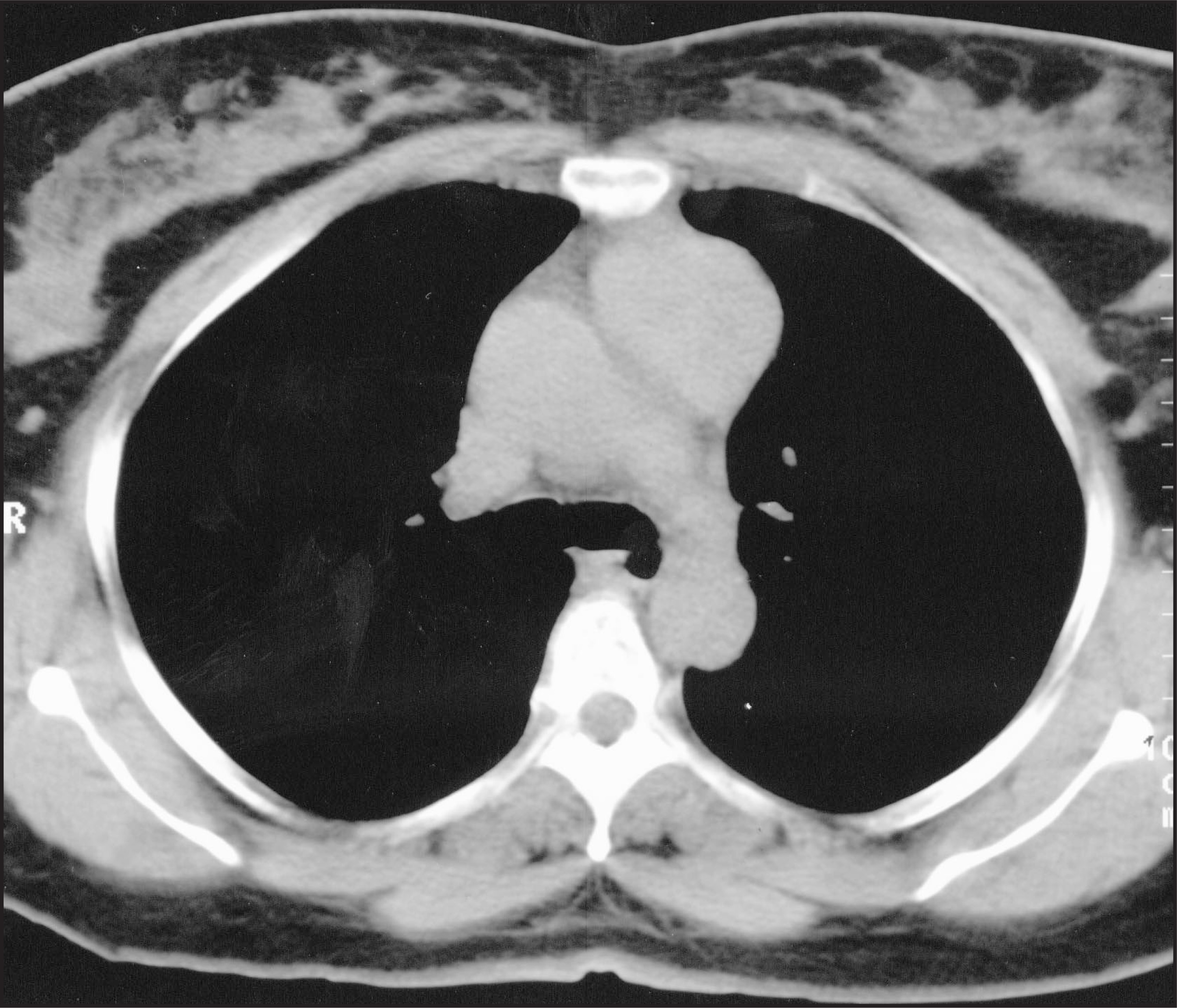
Cytologically malignant thymic neoplasms are termed thymic carcinoma and are distinctly separate from invasive thymoma. Although radiographically similar in their aggressive breaching of boundaries, the invasive thymoma does not demonstrate the cellular atypia of a carcinoma. The most common histologic subtypes of malignant thymic tumors are squamous cell and lymphoepithelioma-like carcinoma.27,28
Other thymic masses in the mediastinum include thymic cysts (figure 5), which may be congenital (due to a patent thymopharyngeal duct) or acquired due either to post inflammatory conditions or to association with a neoplasm.29-31 Thymolipomas are soft encapsulated masses containing mature adipose, thymic epithelial cells, and lymphoid tissue.20
Goiter
Substernal goiter accounts for 10% of all mediastinal masses. Approximately 75% of goiters extend into the anterior mediastinum while 20% to 25% involve the posterior mediastinum.32 CT shows these lesions to be contiguous with the thyroid in the neck, and demonstrates calcification and areas of cystic change33,34 (figure 6). Precontrast attenuation value may be high due to the presence of iodine,33,34 and there may be enhancement with intravenous contrast.35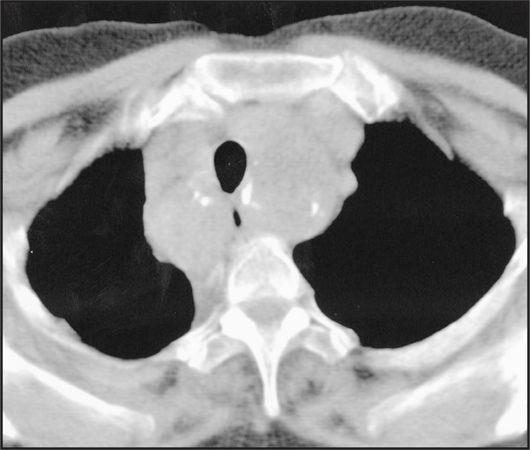
Germ-cell neoplasms
The mediastinum serves as the most common site of extragonadal involvement of germ-cell tumors in adult in the third decade.36 Neoplasms in this category include benign and malignant teratomas, seminomas, and non-seminomatous malignant germ-cell tumors (such as endodermal sinus tumors, embryonal carcinomas, mixed germ-cell tumors, and choriocarcinomas). Of these, teratomas are the most common neoplasms to present in the mediastinum.37,38 The characteristic pathologic and radiographic feature of a teratoma is the presence of one or more components from the three embryonic germ-cell layers, hair, calcium, or fat.
Plain radiography usually reveals a large, well-circumscribed mass causing displacement of adjacent structures. Calcification is seen in up to 80% of these lesions.39 On CT, a benign teratoma is usually cystic and may have fat up to 90% of the time whereas a malignant teratoma is usually more solid and contains fat only 40% of the time39 (figure 7). Fat-fluid levels, considered highly specific for the diagnosis of mature mediastinal teratoma, are uncommon40 (figure 8). CT findings of heterogeneity of the internal components of the mass and changes in the adjacent lung parenchyma, pleura, or pericardium may indicate tumor rupture.41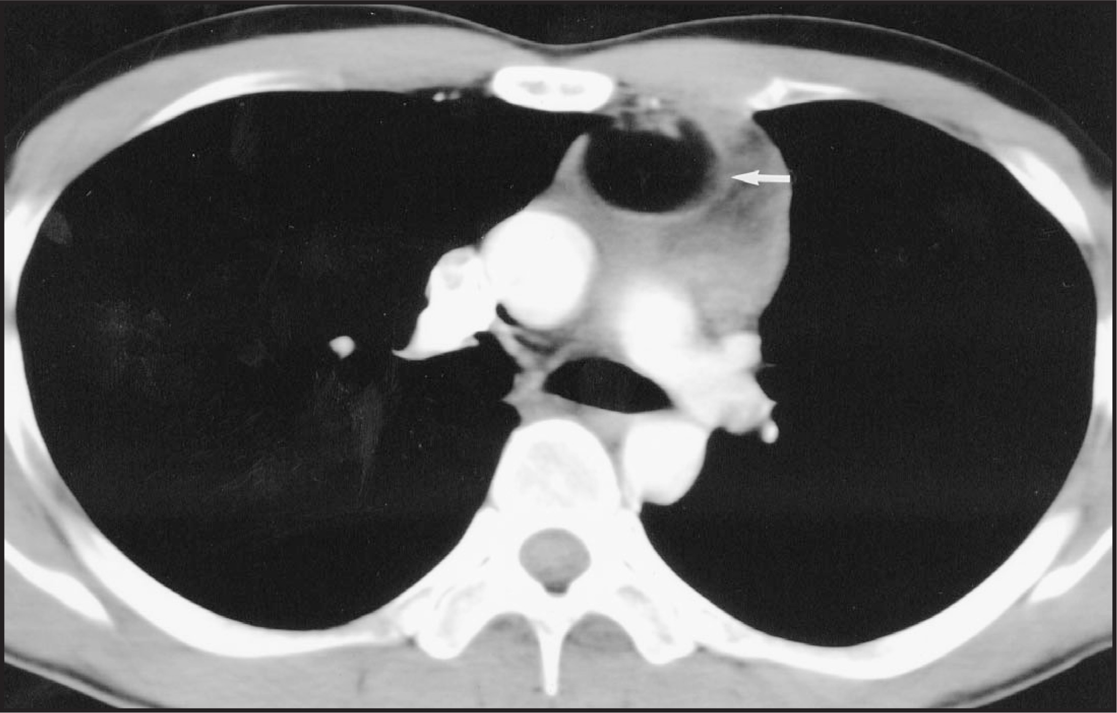
Seminomas are most common primary malignant tumor of the mediastinum and are often metastatic.42,43 They are associated with low levels of beta HCG in contrast to the non-seminomatous tumors that are associated with higher levels of beta HCG.44 They are usually large and lobular on CT and relatively homogenous in their attenuation.33 The non-seminomatous tumors are highly aggressive invasive tumors that are not usually resectable because of their local invasiveness and metastatic tendencies.45,46 On CT, they are usually large, lobulated, heterogeneous masses with areas of hemorrhage and necrosis.47
Middle mediastinum/vascular space
Foregut cysts
These are congenital malformations of the pulmonary and gastrointestinal tracts caused by abnormal budding of the bronchopulmonary foregut. These anomalies comprise 10% to 15% of primary mediastinal lesions. Bronchogenic cysts account for 54% to 63% of such anomalies.19,48 The cyst wall is usually lined by pseudostratified epithelium and they contain mucoid or serous material, hemorrhage, or pus.49,50 They typically present as masses in the paraesophageal and tracheobronchial axis and may compress adjacent structures.51 These cysts do not usually communicate with the airway. On CT, these lesions display water attenuation given their mostly fluid content (figures 9 and 10). On MR, they demonstrate correspondingly high T2 signal and possibly high T1 signal when high in proteinaceous material.52,53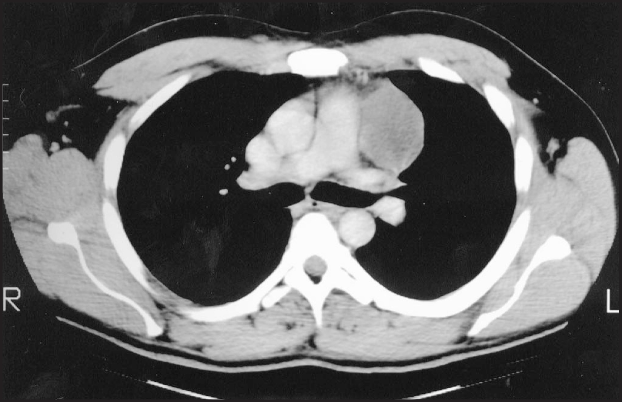
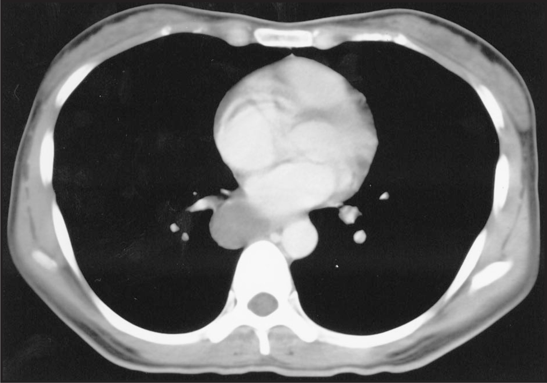
Lymph-node masses are other causes of middle mediastinal masses and include those from sarcoidosis, Castleman’s disease, bronchogenic carcinoma, and infectious causes such as tuberculosis.
Posterior mediastinum/post vascular space
Neurogenic tumors
Ninety percent of neurogenic tumors present as posterior mediastinal masses and represent 75% of posterior mediastinal masses.54,55 These tumors commonly arise either from peripheral nerves, or sympathetic or parasympathetic ganglia.
Schwannomas and neurofibromas are the most common of the posterior mediastinal masses and arise from peripheral nerves.56 They are benign masses that have similar appearances on CXR manifesting as lobulated paraspinal masses19,56 (figures 11 and 12). They may deform the adjacent bony structures,57 and characteristically grow into the spinal canal in a “dumbbell” or “hourglass” form.57 A plexiform neurofibroma is a subtype of neurofibroma that affects an entire nerve plexus.57 Multiple tumors of neural origin or a single plexiform tumor is considered pathognomonic for neurofibromatosis.58 On CT, schwannomas and neurofibromas, in addition to the characteristics described above, may enhance homogeneously, peripherally, or heterogeneously and may contain areas of low density corresponding to myelin, cystic change, or hemorrhage.59-61 MR is necessary in all cases of suspected neurogenic tumors in the mediastinum so intraspinal extension may be excluded.62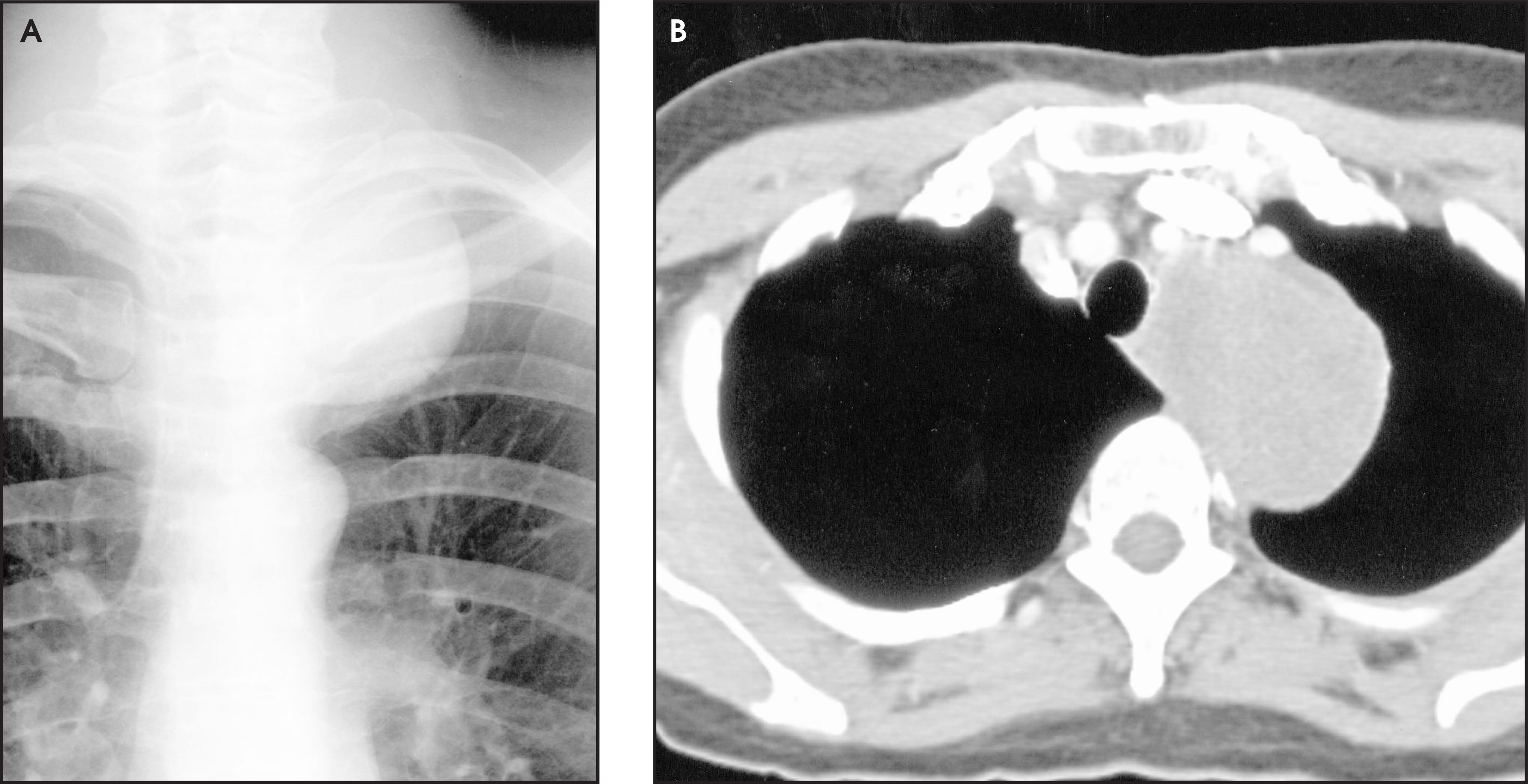
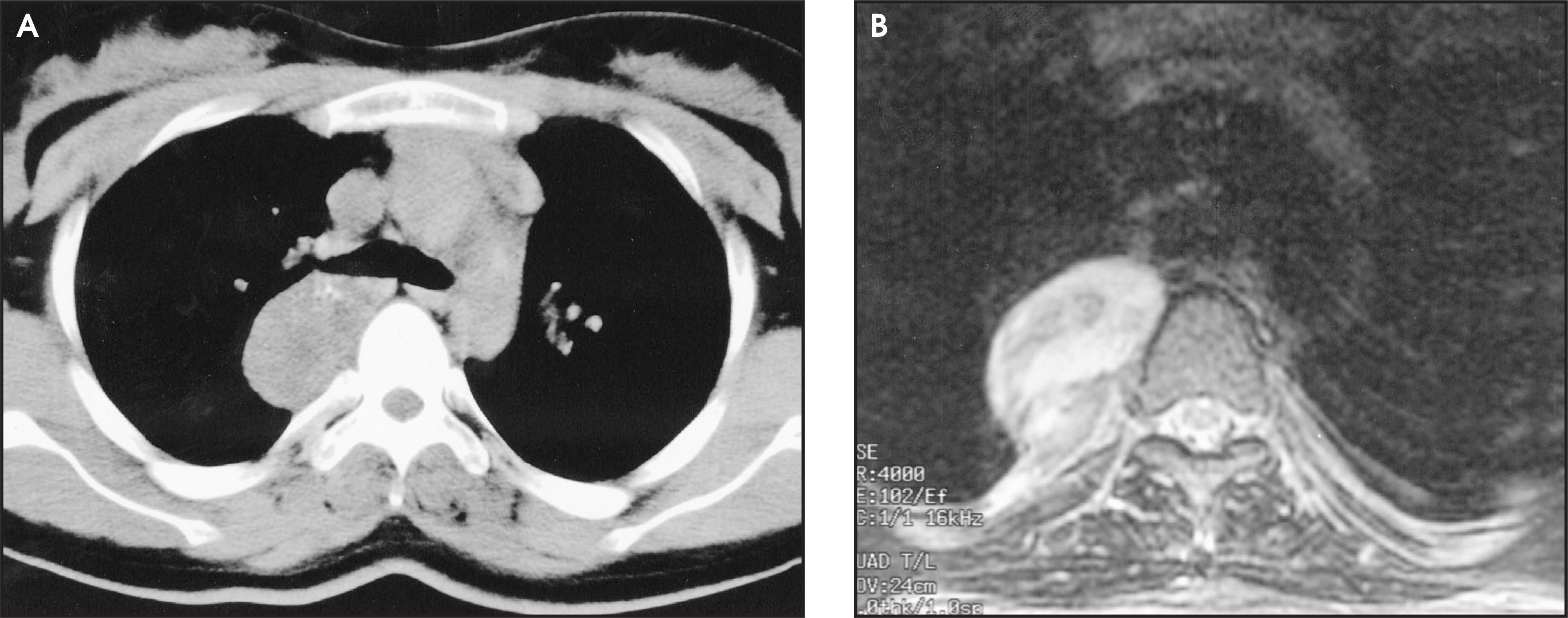
Malignant tumors of nerve sheath origin are the malignant equivalents of the schwannoma and neurofibroma. They do not, however, arise from preexisting schwannomas but may arise from a plexiform neurofibroma. Half of these malignant tumors occur in patients with neurofibromatosis.
Sympathetic ganglia tumors include ganglioneuroblastomas, ganglioneuromas, and neuroblastomas. These usually appear as broad-based, oblong masses adjacent to the spine.59
Lateral thoracic meningoceles
Lateral thoracic meningoceles are rare posterior mediastinal lesions that form as a result of herniation of the meninges through the intervertebral foramen. They are most common in patients with neurofibromatosis and are the most common cause of a posterior mediastinal mass in these patients.57,62 They radiologically manifest as a well-defined paraspinal mass and may be associated with bony erosion, widening of the neural foramina, and possibly kyphoscoliosis.58 MR can determine the presence of herniation of the subarachnoid space and signal intensity consistent with CSF58 (figure 13).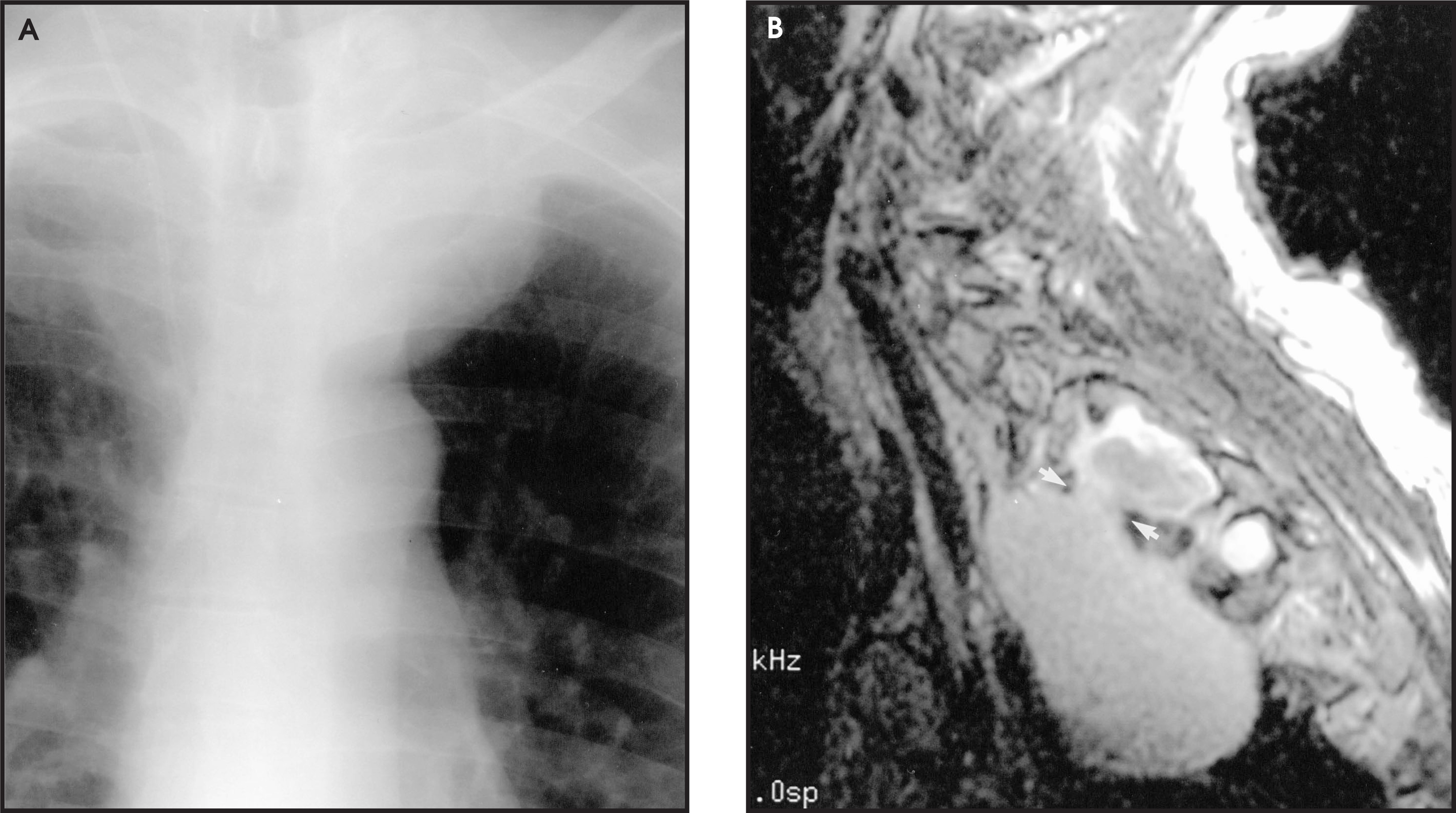
Esophageal lesions
Lesions within the mid- and distal esophagus can present as a posterior mediastinal mass. Esophageal carcinoma is an important part of the differential diagnosis, commonly occurring as the squamous cell type and less commonly as adenocarcinoma. On plain films, this may be recognized by an abnormal tracheoesophageal stripe, dilation of the esophagus proximal to the obstructing mass. Diagnosis is confirmed at endoscopy or by barium swallow. CT may demonstrate an intraluminal mass, thickening of the esophagus, and metastases. Benign lesions of the esophagus such as leiomyomas (figure 14), fibromas, and lipomas may also cause masses in this mediastinal compartment. They usually involve the lower third of the esophagus and do not commonly cause proximal esophageal dilation. Other anomalies that may present in this compartment include diverticula, hernias, or megaesophagus due to achalasia or scleroderma.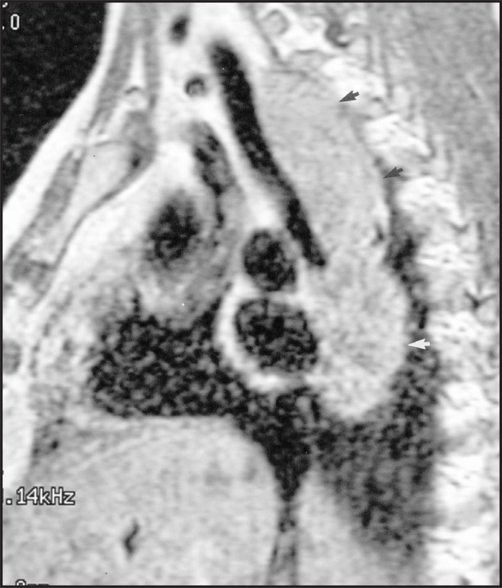
Paravertebral abnormalities
Extramedullary hematopoiesis may also present as a posterior mediastinal mass. Extramedullary hematopoiesis is seen most commonly in diseases of ineffective erythropoiesis such as thalassemia and spherocytosis and can also be seen in sickle cell anemia. Expansion of the medullary cavity is usually associated with multiple paraspinal masses58 (figure 15).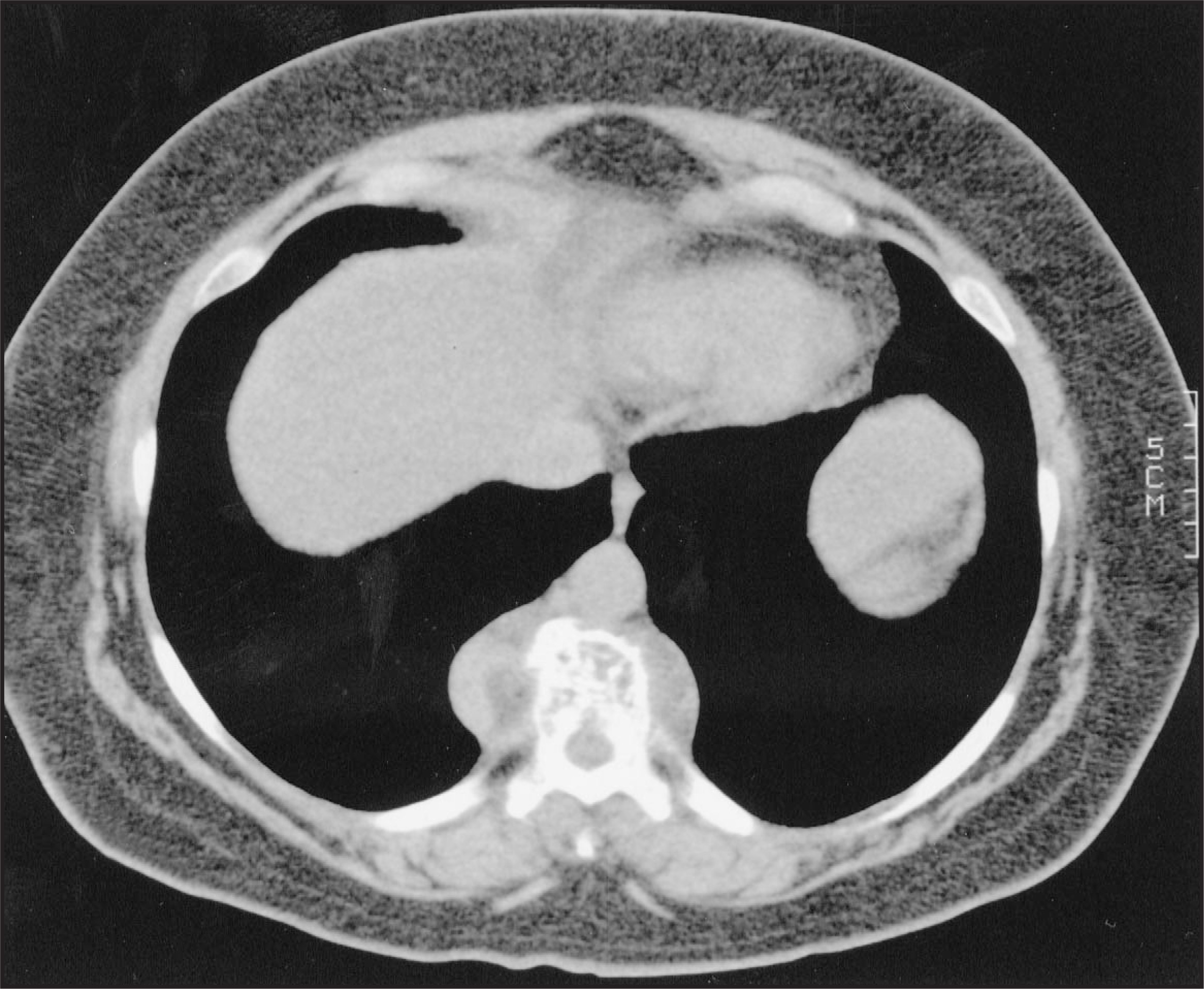
Conclusion
Mediastinal masses present a challenge, as they may include a vast number of diagnoses, each with complex imaging characteristics. Rigorous scrutiny of the normal contents of each compartment helps determine aberrancy. The final diagnosis depends as much on the clinical information (ie, patient demographics and symptoms), as the radiologic characteristics. Delineating the character and extent of a mass enough to guide proper diagnostic sampling and or therapeutic management remains an important radiologic role. AR
References
- Fraser R, Pare J, Fraser R, Fraser R, Pare J, Fraser R. Synopsis of Diseases of the Chest.. 1994:1-116.
- Heimburger I, Battersby J, Vellios F. Primary neoplasms of the mediastinum: A fifteen year experience.. Arch Surg.. 1963;86:120-126.
- Rubush J, Gardner I, Boyd W, Ehrenhaft J. Mediastinal tumors: A review of 186 cases.. J Thorac Cardiovasc Surg.. 1973;65:216-222.
- Silverman N, Sabiston D. Mediastinal masses.. Surg Clin North Am.. 1980;60:757-777.
- Wongsangiem M, Tangthangtham A. Primary tumors of the mediastinum: 190 case analysis (1975-1995).. J Med Assoc Thai.. 1996;79:689-697.
- Rebner M, Gross B, Robertson J. CT evaluation of mediastinal masses.. Comput Radiol.. 1987;11:103-110.
- Webb W. Magnetic resonance imaging of the chest.. Curr Opin Radiol.. 1989;1:40-43.
- Castellino R. The non-Hodgkins lymphomas: Practical concepts for the diagnostic radiologist.. Radiology.. 1991;178:315-321.
- Costello P, Jochelson M, Taveras J, Ferruci J. Radiology: Diagnosis–Imaging–Intervention.. 1996;Vol. 1:1-13.
- Klein J, Brant E, Helms A. Fundamentals of Diagnostic Radiology.. 1999:322-323.
- Vaeth J, Moskowitz S, Green J. Mediastinal Hodgkin’s disease.. Am J Roentgenol.. 1976;26:123-126.
- Lichtenstein A, Levine A, Taylor C. Primary mediastinal lymphoma in adults.. Am J Med.. 1980;68:509-514.
- Strickler J, Kurtin P. Mediastinal lymphoma.. Semin Diagn Pathol.. 1991;8:2-13.
- Kim H, Nosher J, Haas A. Cystic degeneration of thymic Hodgkins disease following radiation therapy.. Cancer.. 1985;55:354-356.
- Castellino R, Blank N, Hoppe R. Hodgkin disease: Contributions of chest CT in the initial staging evaluation.. Radiology.. 1986;160:603-605.
- Hopper K, Diehl L, Lesar M. Hodgkin disease: Clinical utility of CT in initial staging and treatment.. Radiology.. 1988;169:17-22.
- Kim H, Nosher J, Haas A. Cystic degeneration of thymic Hodgkin’s disease following radiation therapy.. Cancer.. 1985;55:354-356.
- Negendank W, Al-Katib A, Karanes C. Lymphomas: MR imaging contrast characteristics with clinical-pathologic correlations.. Radiology.. 1990;177:209-216.
- Wychulis A, Payne W, Clagett O. Surgical treatment of mediastinal tumors.. J Thorac Cardiovasc Surg.. 1972;62:379-391.
- Strollo D, Rosado de Christenson M, Jett J. Primary mediastinal tumors. Part I. Tumors of the anterior mediastinum.. Chest.. 1997;112:511-522.
- Lattes R. Thymoma and other tumors of the thymus: An analysis of 107 cases.. Cancer.. 1962;15:1224-1260.
- Morgenthaler T, Brown L, Colby T. Thymoma. (Symposium on intrathoracic neoplasm part ix.). Mayo Clin Proc. 1993;68:1110-1123.
- Osserman K, Genkins G. Studies in myaesthenia gravis: Review of 20-year experience in over 1200 patients.. Mt Sinai J Med.. 1971;38:497-537.
- Zerhouni E, Scott W, Baker R. Invasive thymomas: Diagnosis and evaluation by CT.. J Comput Assist Tomogr.. 1982;6:92-100.
- Moran C, Travis W, Rosado-de-Christenson M. Thymomas presenting as pleural tumors. Report of eight cases.. Am J Surg Pathol.. 1992;16:138-144.
- Rosado-de-Christenson M, Galobardes J, Moran C. Thymoma: Radiologic-pathologic correlation.. Radiographics.. 1992;12:151-168.
- Suster S, Rosai J. Thymic carcinoma: Clinicopathologic study of 60 cases.. Cancer.. 1991;67:1025-1032.
- Wick M, Weiland L, Scheithauer B. Primary thymic carcinomas.. Am J Surg Pathol.. 1982;6:613-630.
- Indeglia R, Shea M, Grage T. Congenital cysts of the thymus gland.. Arch Surg.. 1967;94:149-52.
- Suster S, Rosai J. Multilocular thymic cyst: An acquired reactive process.. Am J Surg Pathol.. 1991;15:388-398.
- Graeber G, Thompson L, Cohen D. Cystic lesions of the thymus.. J Thorac Cardiovasc Surg.. 1984;87:295-300.
- Tecce P, Fishman E, Kuhlman J. CT evaluation of the anterior mediastinum: Spectrum of disease.. Radiographics.. 1994;14:973-990.
- Brown L, Aughenbaugh G. Masses of the anterior mediastinum: CT and MR imaging.. AJR Am J Roentgenol.. 1991;157:1171-1180.
- Bashist B, Ellis K, Gold R. Computed tomography of intrathoracic goiters.. AJR Am J Roentgenol.. 1983;140:455-460.
- Glazer G, Axel L, Moss A. CT diagnosis of mediastinal thyroid.. AJR Am J Roentgenol.. 1982;138:495-498.
- Nichols C, Fox E. Extragonadal and pediatric germ cell tumors.. Hematol Oncol Clin North Am.. 1991;5:1189-1209.
- Davis R, Oldham H, Sabiston D. Primary cysts and neoplasms of the mediastinum: Recent changes in clinical presentation, methods of diagnosis, management, and results.. Ann Thorac Surg.. 1987;44:229-237.
- Cohen A, Thompson L, Edwards F, Bellamy R. Primary cysts and tumors of the mediastinum.. Ann Thorac Surg.. 1991;51:378-384.
- Quillin S, Siegel M. CT features of benign and malignant teratomas in children.. J Comput Assist Tomogr.. 1992;16:722-726.
- Moeller K, Rosado-de-Christenson M, Templeton P. Mediastinal mature teratoma: Imaging features.. AJR Am J Roentgenol.. 1997;169:985-990.
- Choi S, Lee J, Song K, Lim T. Mediastinal teratoma: CT differentiation of ruptured and unruptured tumors.. AJR Am J Roentgenol.. 1998;171:591-594.
- Hurt R, Bruckman J, Farrow G. Primary anterior mediastinal seminoma.. Cancer.. 1982;49:1658-1663.
- Bush S, Martinez A, Bagshaw M. Primary mediastinal seminoma.. Cancer.. 1981;48:1877-1882.
- Clamon G. Management of primary mediastinal seminoma.. Chest.. 1983;83:263-267.
- Knapp R, Hurt R, Payne W. Malignant germ cell tumors of mediastinum.. J Thorac Cardiovasc Surg.. 1985;89:82-89.
- Levitt R, Husband J, Glazer H. CT of primary germ cell tumors of the mediastinum.. Am J Roentgenol.. 1984;142:73-78.
- Blomlie V, Lien H, Fossa S. Computed tomography in primary non-seminomatous germ cell tumors of the mediastinum.. Acta Radiol.. 1988;29:289-292.
- Sirivella S, Ford W, Zikria E. Foregut cysts of the mediastinum. Results in 20 consecutive surgically treated patients.. J Thorac Cardiovasc Surg.. 1985;90:776-782.
- Kirwan W, Walbaum P, McCormack R. Cystic intrathoracic derivatives of the foregut and their complications.. Thorax.. 1973;28:424-428.
- Mendelson D, Rose J, Efremidis S. Bronchogenic cysts with high CT numbers.. AJR Am J Roentgenol.. 1983;140:463-465.
- LeBlanc J, Guttentag A, Shepard J, McLoud T. Imaging of mediastinal foregut cysts.. Can Assoc Radiol J.. 1994;45:381-386.
- Kuhlman J, Fishman E, Wang K. Mediastinal cysts: Diagnosis by CT and needle aspiration.. AJR Am J Roentgenol.. 1988;150:75-78.
- Nankata H, Egashira K, Wakanake H. MRI of bronchogenic cysts.. J Comput Assist Tomogr.. 1993;17:267-270.
- Benjamin S, McCormack L, Effler D. Primary tumors of the mediastinum.. Chest.. 1972;62:297-303.
- Davidson K, Walbaum P, McCormarck R. Intrathoracic neural tumors.. Thorax.. 1978;33:359-367.
- Reed J, Kagan-Hallett K, Feigin D. Neural tumors of the thorax: Subject review from the AFIP.. Radiology.. 1978;126:9-17.
- Aughenbaugh G. Thoracic manifestations of neurocutaneous diseases.. Radiol Clin North Am.. 1984;22:741-756.
- Strollo D, Rosado de Christenson M, Jett J. Primary mediastinal tumors. Part II. Tumors of the middle and posterior mediastinum.. Chest.. 1997;112:1344-1357.
- Kumar A, Kuhajda F, Martinez C. Computed tomography of the extracranial nerve sheath tumors with pathologic correlation.. J Comput Assist Tomogr.. 1983;7:857-865.
- Harkin R, Reed R. Atlas of Tumor Pathology: Tumors of the Peripheral Nervous System. 1982:25-150.
- Coleman B, Arger P, Dalinka M. CT of sarcomatous degeneration of neurofibromatosis.. AJR Am J Roentgenol.. 1983;140:383-387.
- Gefter W. Chest applications of magnetic resonance imaging: An update.. Radiol Clin North Am.. 1988;26:573-588.
Citation
. Nonvascular lesions of the mediastinum. Applied Radiology. 2002;31(5):33-41. doi:10.37549/AR1091.