Optimizing helical CT in children
Applied Radiology — Vol. 30 , Issue 12 , pp. 11 -17
DOI: 10.37549/AR1058
Published: December 1, 2001
Categories
Since its advent more than 25 years ago, computed tomography (CT) has become a well-established and increasingly utilized diagnostic tool. During the past decade, helical technology has accelerated this trend with a dramatic further reduction in time necessary for data acquisition of volume data sets. Familiarity with this pervasive technology is particularly important in children to optimize scan quality and minimize risk.
Although current literature abounds with information pertaining to current techniques and evolving applications for adult helical CT, information regarding the optimization of helical CT in infants and children is sparse.1-3 This is true despite the fact that many more variables must be addressed in the pediatric population. These include techniques for intravenous (IV) and oral contrast material administration, including variable delays in onset of scanning. Although successful helical CT data acquisition in children is complex and often requires attention to each individual child rather than adherence to protocol, diagnostic scanning can be achieved. To this end, the following material reviews contemporary pediatric body helical CT techniques.
Sedation
As CT scanning has become faster, the need for sedation has decreased. For example, the frequency of sedation dropped from 18% to 10% in young children as helical CT became available.4,5 Introduction of even faster multidetector scanners has further decreased the frequency of sedation in children <6 years of age to as low as 3%.6 Even with these reductions, the issue of safe and effective sedation remains an integral aspect of cross-sectional imaging in children. General guidelines and regulations concerning safe practice parameters for pediatric sedation have been published previously7-9 and support the basic principle of protecting the welfare of the child. Importantly, a minimal set of standards, including pre-sedation evaluation, selection of appropriate sedative agents, procedural monitoring, and implementation of specific discharge criteria, is essential.
Once the decision to sedate a child for the purpose of cross-sectional imaging has been made, there is a multitude of sedative agents from which to choose. Unfortunately, there is no singularly perfect agent. In general, successful, safe sedation stems from a limited group of drugs with which the radiologist is familiar (Table 1). Chloral hydrate and sodium pentobarbital are two of the most widely used sedative agents for pediatric diagnostic imaging purposes. Chloral hydrate, an oral medication given in doses of 50 to 100 mg/kg body weight (maximum dose of 2.0 grams) is most successful in children under about 15 kg. Intravenous administration of sodium pentobarbital in titrated doses of 1.0 to 3.0 mg/kg body weight with a total dose of 6.0 to 8.0 mg/kg (maximum dose of 200 mg) can also be safe and effective. When necessary, IV fentanyl citrate can be administered in conjunction with sodium pentobarbital in titrated doses of 1.0 μg/kg body weight (maximum dose of 4.0 μg/kg). This combination of drugs acts synergistically and is helpful when the child requires pain relief as well as sedation during a radiologic examination.
Oral contrast material
Although the use of oral contrast material in abdominal and pelvic CT examinations in children has become, for the most part, routine, its use has benefits and disadvantages. Oral contrast material is helpful in identification of the course and caliber of bowel, in distinction of bowel from adjacent solid structures or organs, and in determining luminal and mural abnormalities. The temporal information related to how far anterograde the contrast has passed can also be helpful in diagnosing bowel obstruction. However, certain clinical situations and diagnostic questions can be addressed in the absence of oral contrast material. In fact, the routine administration of gastrointestinal contrast material has been questioned10 and is not used routinely anymore at some institutions. Dense iodinated contrast may mask subtle mucosal enhancement abnormalities and may make detection of intraluminal or submucosal lesions difficult. Evaluation of the solid organs is usually not affected by the presence or absence of oral contrast material, particularly in a follow-up examination. In the setting of trauma, the use of oral contrast material is generally considered safe11,12; however, there is a risk of significant aspiration. The radiologist must take an active role in deciding the relative necessity of oral contrast material and must be prepared to handle any adverse outcome related to its use.
The issue of administration of oral contrast material to young children who require sedation prior to their examination is also controversial as oral intake of fluids increases the risk of aspiration.9,13 Oral contrast material is usually administered an hour or so prior to scanning, which interferes with most general nothing-by-mouth guidelines of 2 to 6 hours depending on the size of the child undergoing the examination and the liquid or solid nature of the ingested material. At our institution, oral contrast material may be administered up to the point the child is sedated for examinations for which contrast enhancement is believed necessary, such as cases of suspected bowel or mesenteric pathology (i.e., abscess) or in blunt abdominal trauma. We recently reviewed safety issues regarding our past 5 years of experience with administering oral contrast before sedation. In 337 consecutive cases, there were no instances of vomiting with aspiration (LF Donnelly, unpublished data, 1996–2000).
Agents used for standard opacification of the gastrointestinal tract usually contain water-soluble contrast diluted with liquids, such as fruit drink or soda. However, agents composed of dilute barium are also available. Table 2 presents appropriate amounts of oral contrast material given age- or weight-based considerations. Coaxing very young or sick children to drink enough oral contrast material is difficult. At our institution, adequate intraluminal contrast has been achieved by encouraging children to drink liquids (such as juice, water, or carbonated drinks) without contrast media. Figure 1 illustrates opacification of the gastrointestinal tract with juice in a child who refused to drink liquids containing iodinated contrast material.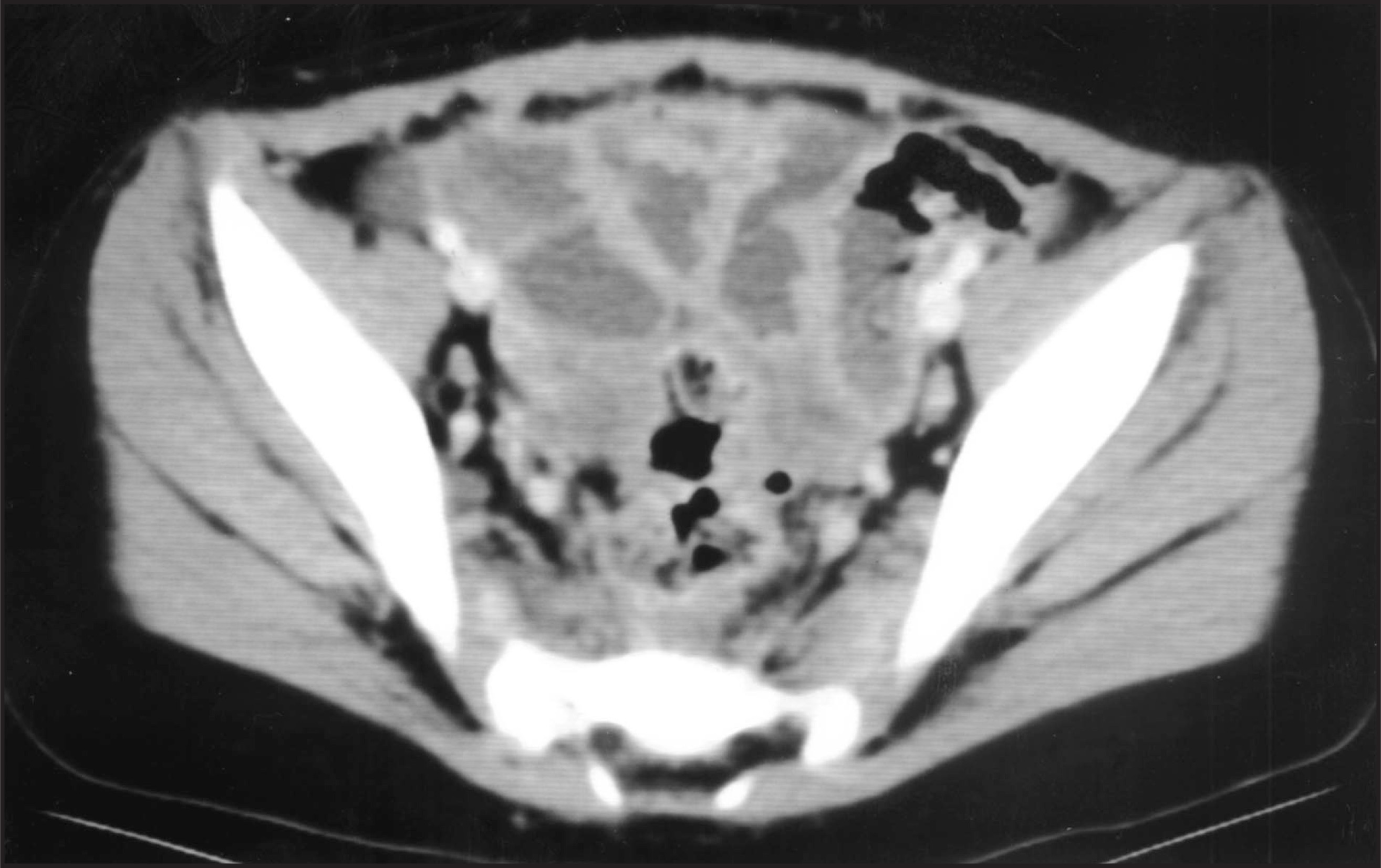
IV contrast material
The issue of appropriate administration of IV contrast is perhaps the most challenging and most important aspect of successful CT scanning in children. Multiple parameters that must be addressed include type of contrast, volume administered, location and caliber of intravenous access, method of contrast administration (manual injection versus power injection), and delay from time of injection to the initiation of scanning. Although the task may seem daunting, attention to these variables can lead to optimal contrast enhancement in most instances.
Generally, low-osmolar, nonionic IV contrast material is recommended in children.14 However, the use of specific contrast agents should be based on either individual or institutional standard of practice. The standard dose of IV contrast material for abdomen scanning is 2.0 mL/kg, not to exceed an adult dose of approximately 150 mL. When scanning the chest or performing CT angiography with a goal of vascular opacification instead of organ enhancement, 1.5 mL/kg is sufficient.15 With both single- and multidetector CT technology, CT angiography can be performed successfully, even in infants with relatively slow rates of administration and small volumes of IV contrast media15 (figure 2).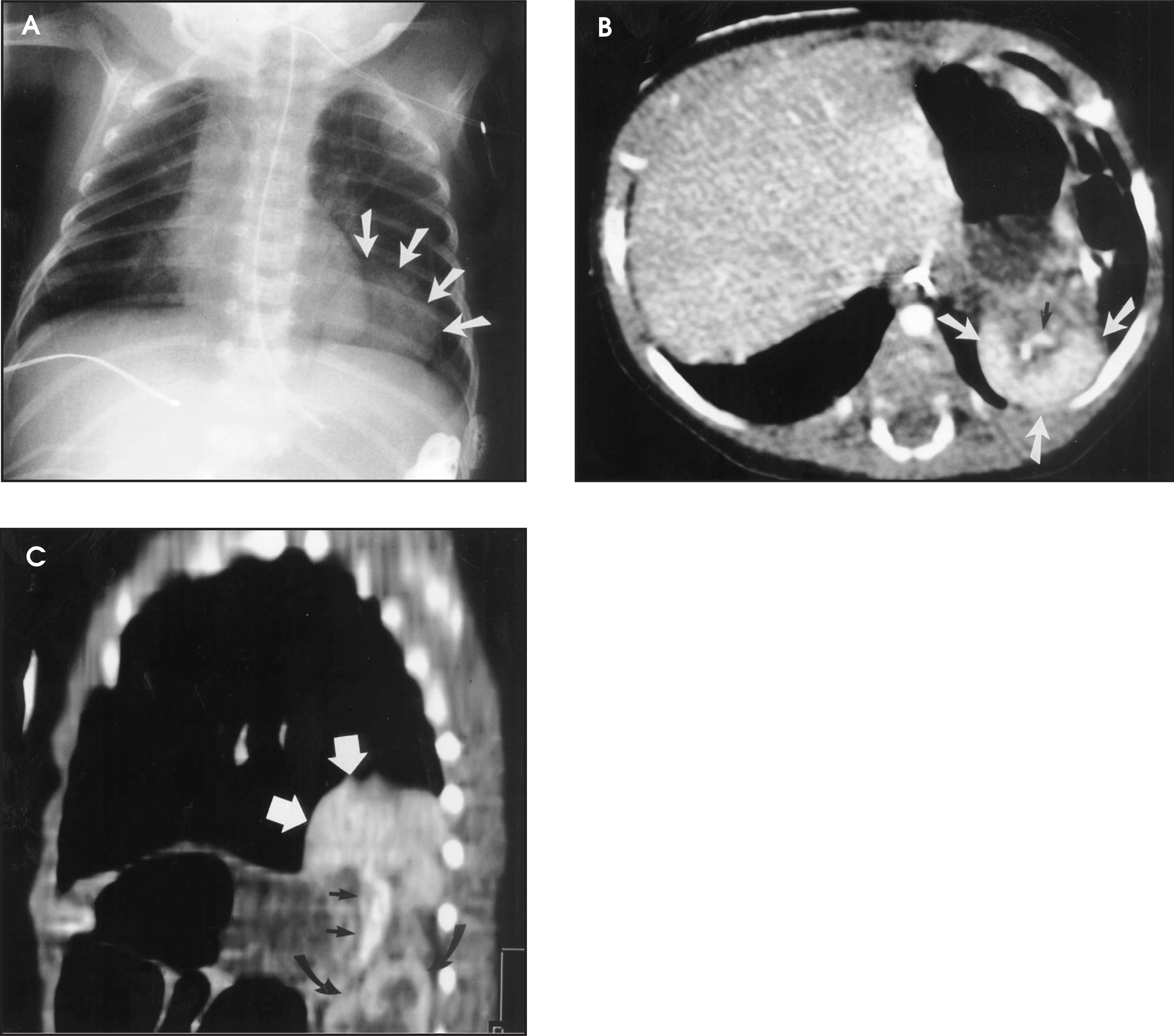
Administration of IV contrast may be through a peripheral catheter or central venous catheter. Methods of administration include manual (hand) bolus and power injector. Although for years manual injection has been standard practice in pediatric patients, power injectors have been used successfully with increasing frequency. This practice has been found to be safe.2,16,17 We use power injection through peripheral angiocatheters as small as 24 gauge, providing that the catheter functions well. Contraindications to the use of power injectors include absence of blood return through the catheter and inability to flush the catheter.16,18 Use of power injectors with central lines is controversial. Although this practice is probably more widespread than reports in the literature indicate,19 this practice is not sanctioned by the manufacturers.
Suggested rates of intravenous contrast administration are 1.0 to 1.5 mL/sec for 24-gauge angiocatheters and 1.5 to 2.0 mL/sec for 22-gauge angiocatheters.20 However, rates as low as 0.5 mL/sec have been used with acceptable enhancement.21 Generally speaking, target rates should be between 1.2 and 2.5 mL/sec to provide optimal vascular and solid organ enhancement. We use rates as high as 3.0 mL/sec for CT angiography despite the size of the child. Although manual injection of contrast material is regarded as safe and has provided quality scans for many years, the true rate of injection is imprecise. The rates vary between 1.0 and 3.0 mL/sec with an average rate of just under 2.0 mL/sec.16 Complications are no more common with power injection than with manual injection, provided that the catheter functions properly and is monitored during injection.16
The timing of scanning onset in relation to IV contrast administration (often referred to as scan delay) has changed with the advent of helical CT. In conventional slice-by-slice abdominal CT scanning, scanning was initiated after approximately one-half of the contrast bolus was administered. Recommendations for helical CT in children are to delay scanning of the abdomen until completion of IV contrast material administration. Reported delays from the end of contrast administration to the onset of diagnostic scanning vary from approximately 5 to 30 seconds.1 We have found that delays for abdominal scanning of 20 to 25 seconds, and 10 to 15 seconds for chest scanning, are reliable for both single and multislice helical CT. Chest scanning can also follow abdominal scanning with excellent enhancement of the cardiovascular structures.
With multidetector technology, neck, chest, abdomen, and pelvis scanning can be performed sequentially without the traditional splitting of IV contrast material.22 Multidetector helical technology allows even more rapid image acquisition, placing new demands on appropriate utilization and timing of IV contrast material administration. Using multislice equipment for abdominal imaging, we commonly use a 20-second delay in scanning initiation after completion of contrast administration. This allows for fairly consistent portal venous phase of contrast enhancement of the liver and parenchymal renal enhancement.23
In addition to these empiric delays, bolus tracking can also be used to determine scan onset, thus individualizing each CT. This is especially useful in pediatric body CT, where contrast material administration is complex. This technology confers only a slight increase in radiation dose to the patient. Although the usefulness of bolus tracking in adults is debatable,24 it has been found to be quite useful when scanning children17,21 (figure 3).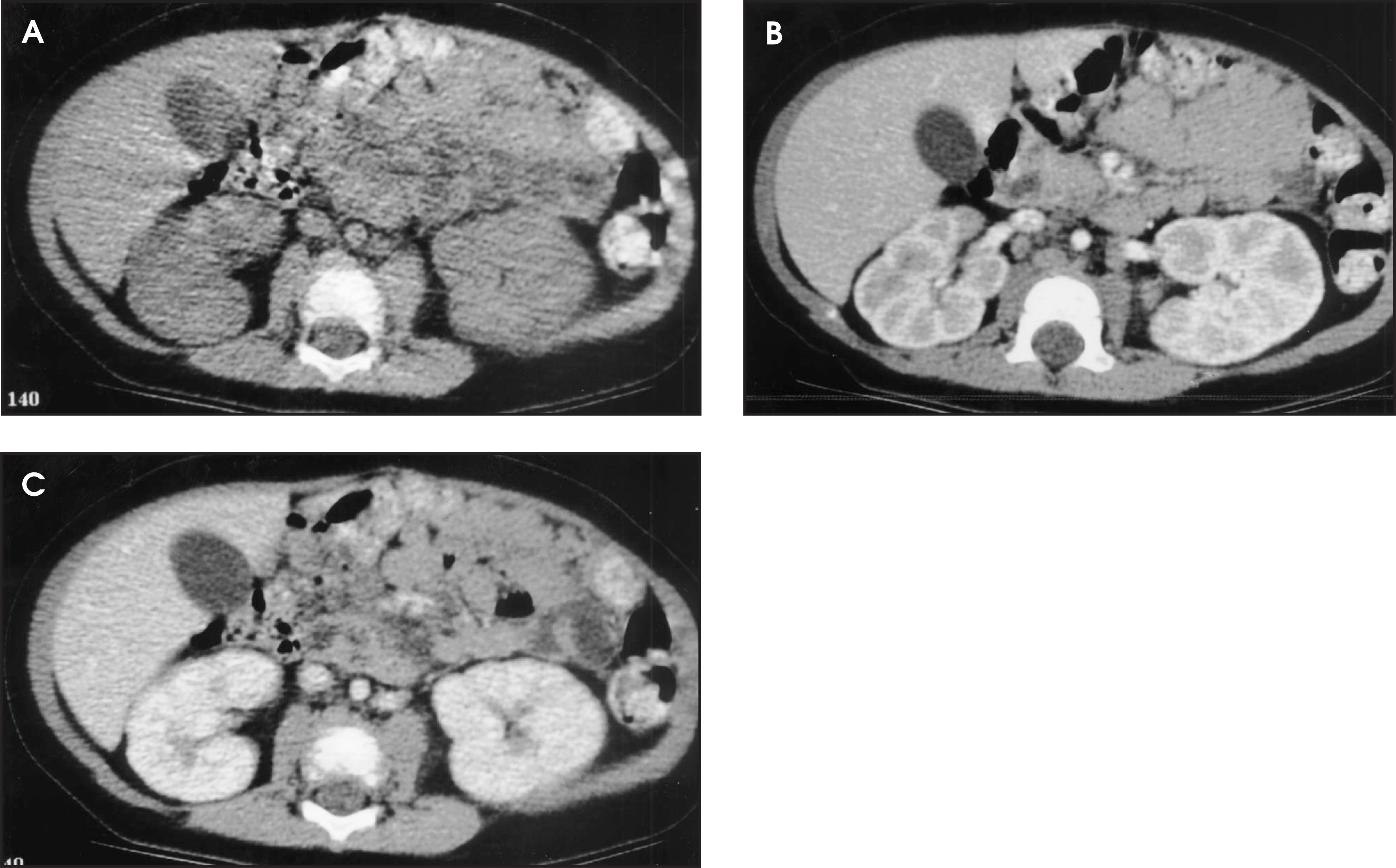
Scan parameters
In addition to the complexities surrounding issues of sedation and appropriate use of IV and oral contrast material in children, several other technical parameters deserve careful attention. These include tube current (milliampere [mA]), kilovoltage (kVp), table speed and slice thickness (“pitch”), and gantry rotation cycle time. These parameters are important since they determine image quality and radiation exposure. Despite this importance, specific recommendations and even general guidelines are lacking for pediatric helical CT. In general, investigators have recommended size-based adjustments in tube current (Table 3), and scanning at pitches of 1.5 to 2.0.1,25,26 These adjustments provide acceptable image quality while reducing radiation for general body indications. While lowering the kVp will also reduce radiation, there are no clinical guidelines for application of data that support sized-based kVp adjustments.27 In general, scanning should be performed using the fastest table speed and largest slice thickness indicated. This will, of course, depend on individual preferences, scan indication, and scanner manufacturer. A few studies support the use of low (or lower) tube current scanning in the chest28-31 and pelvis.32
The issue of radiation and CT is a timely topic.33 Computed tomography radiation can be considered a public health concern for the following reasons. First, CT accounts for up to 65% of medical radiation, and children account for approximately 11% of all CT examinations.34 Moreover, the rate of CT scans is increasing. Importantly, it has been shown that scanning is often obtained using adult technique (average pediatric mA was about 200), exposing the children to unnecessary radiation.35 This is important because children are more radiosensitive to the same organ dose than adults, and because a child’s longer life could allow radiation-induced malignancies to develop. We now realize that the relationship between low-level (CT) radiation and cancer is much closer than previously thought.36,37 Together, these facts mandate that radiation be minimized. Strategies include using other modalities that can provide sufficient diagnostic information, using sized-based scanning, and changing the paradigm of image quality from optimal (high dose) to acceptable (figures 2 and 3).
Conclusion
Helical CT scanning has become an invaluable tool for imaging children, although scanning children often presents unique challenges. Despite these challenges, diagnostic scanning can be achieved. Even in the most complex circumstances, attention to the individual child and addressing the diagnostic question with appropriate scan techniques can provide excellent results while minimizing risks. AR
References
- Frush D. RSNA Course Syllabus. 1999:17-31.
- Frush D, Donnelly L. Helical CT in children: Technical considerations and body applications. Radiology. 1998;209:37-48.
- Siegel M, Luker G. Pediatric applications of helical (spiral) CT. Radiol Clin North Am. 1995;33:997-1021.
- Kaste S, Young C, Holmes T, Baker D. Effect of helical CT on the frequency of sedation in pediatric patients. AJR Am J Roentgenol. 1997;168:1001-1003.
- White K. Reduced need for sedation in pediatric patients undergoing helical CT of the chest and abdomen. Pediatr Radiol. 1995;25:344-346.
- Pappas J, Donnelly L, Frush D. Marked reduction in the frequency of sedation of children using new multislice helical CT. Radiology. 2000;215:897-899.
- Guidelines for monitoring and management of pediatric patients during and after sedation for diagnostic and therapeutic procedures. Pediatrics. 1992;89:1110.
- 1995 Comprehensive Accreditation Manuals for Hospitals. 1995.
- Practice guidelines for sedation and analgesia by non-anesthesiologist: A report by the American Society of Anesthesiologists. Anesthesiology. 1996;84:459-471.
- Donnelly L. Commentary: Oral contrast material medium administration for abdominal CT: Reevaluating the benefits and disadvantages in the pediatric patient. Pediatr Radiol. 1997;27:770-772.
- Federle M, Yagan N, Peitzman A. Abdominal trauma: Use of oral contrast material for CT is safe. Radiology. 1997;205:91-93.
- Lim-Dunham J, Narra J, Benya E. Aspiration after administration of oral contrast material in children undergoing abdominal CT for trauma. AJR Am J Roentgenol. 1997;169:1015-1018.
- Frush D, Bisset G, Hall S. Pediatric sedation: The practice of safe sleep. AJR Am J Roentgenol. 1996;167:1381-1387.
- Cohen M, Smith J. Intravenous use of ionic and nonionic contrast agents in children. Radiology. 1994;191:793-794.
- Cohen R, Frush D, Donnelly L. Data acquisition for pediatric CT angiography: Problems and solutions. Pediatr Radiol. 2000;30:813-822.
- Kaste S, Young C. Safe use of power injectors with centeral and peripheral venous access devices for pediatric CT. Pediatr Radiol. 1996;26:499-501.
- Reuss L, Bulas D, Kushner D. Peak enhancement of the liver in children using power injection and helical CT. AJR Am J Roentgenol. 1998;170:677-681.
- Frush D, Siegel M, Bisset G. Challenges of pediatric spiral CT. RadioGraphics. 1997;17:939-959.
- Herts B, O’Malley C, Wirth S. Power injection of contrast media using central venous catheter feasibility, safety, and efficacy. AJR Am J Roentgenol. 2001;176:447-453.
- Frush D, Donnelly L. Helical CT: Technical considerations and body applications. Radiology. 1998;209:37-48.
- Frush D, Spencer E, Donnelly L. Optimizing contrast-enhanced abdominal CT in infants and children using bolus tracking. AJR Am J Roentgenol. 1999;172:1007-1013.
- Donnelly L, Frush D, Nelson R. Multislice helical CT: Facilitation of combined CT examination of the neck, chest, abdomen, and pelvis in children. AJR Am J Roentgenol. 2001;174:1620-1622.
- Frush D, Donnelly L, Bisset G. Technical innovation: Effect of scan delay on hepatic enhancement for pediatric abdominal multislice helical CT. AJR Am J Roentgenol. 2001;176:1559-1561.
- Paulson E, Fisher A, DeLong D. Helical liver CT with computer assisted bolus-tracking technology: Is it possible to predict which patient will not achieve a threshold of enhancement?. Radiology. 1998;209:787-792.
- Vade A, Demos T, Olson M. Evaluation of image quality using 1:1 pitch and 1.5:1 pitch helical CT in children: A comparative study. Pediatr Radiol. 1996;26:891-893.
- Donnelly L, Emery K, Brody A. Minimizing radiation dose for pediatric body applications of single-detector helical CT: Strategies at a large children’s hospital. AJR Am J Roentgenol. 2001;176:303-306.
- Huda W, Scalzetti E, Levin G. Technique factors and image quality as functions of patient weight at abdominal CT. Radiology. 2000;217:430-435.
- Ambrosino M, Roche K, Genieser N. Application of thin-section low-dose chest CT (TSCT) in the management of pediatric AIDS. Pediatr Radiol. 1995;25:393-400.
- Rogalla P, Stöver B, Scheer I. Low-dose spiral CT: Applicability to pediatric chest imaging. Pediatr Radiol. 1998;28:565-569.
- Ambrosino M, Genieser N, Roche K. Feasibility of high-resolution, low-dose chest CT in evaluating the pediatric chest. Pediatr Radiol. 1994;24:6-10.
- Lucaya J, Piqueras J, Garcia-Peña P. Low-dose high-resolution CT of the chest in children and young adults: Dose, cooperation, artifact incidence, and image quality. AJR Am J Roentgenol. 2000;175:985-992.
- Kamel I, Hernandez R, Martin J. Radiation dose reduction in CT of the pediatric pelvis. Radiology. 1994;190:683-687.
- Rogers L. Taking care of children: Check out the parameters used for helical CT. AJR Am J Roentgenol. 2001;176:287.
- Mettler F, Wiest P, Locken J. CT scanning: Patterns of use and dose. J Radiol Prot. 2000;20:353-359.
- Paterson A, Frush D, Donnelly L. Helical CT of the body: Are settings adjusted for pediatric patients?. AJR Am J Roentgenol. 2001;176:297-301.
- Brenner D, Elliston C, Hall E. Estimated risks of radiation-induced fatal cancer from pediatric CT. AJR Am J Roentgenol. 2001;176:289-296.
- Frush D. Pediatric helical CT: Techniques and applications. Med Imag Int. 1999;9:12-18.
Citation
. Optimizing helical CT in children. Applied Radiology. 2001;30(12):11-17. doi:10.37549/AR1058.