Osseous and multisystem involvement of tuberous sclerosis
Applied Radiology — Vol. 30 , Issue 6 , pp. 22 -24
DOI: 10.37549/AR1008
Published: June 1, 2001
Categories
Tuberous sclerosis (TS), or Bourneville's disease, is an autosomal dominant disease with a prevalence of 1:10,000 to 1:200,000 births1,2 showing variable penetrance. New mutations are responsible for a large number of these cases.3,4 The clinical triad includes epileptic seizures, mental retardation, and cutaneous lesions that are thought to be diagnostic.5 Multiple organ systems may be involved with this disease including the skin, central nervous system (CNS), ocular system, kidneys, heart, gastrointestinal tract, and skeletal system.
Osseous involvement in tuberous sclerosis
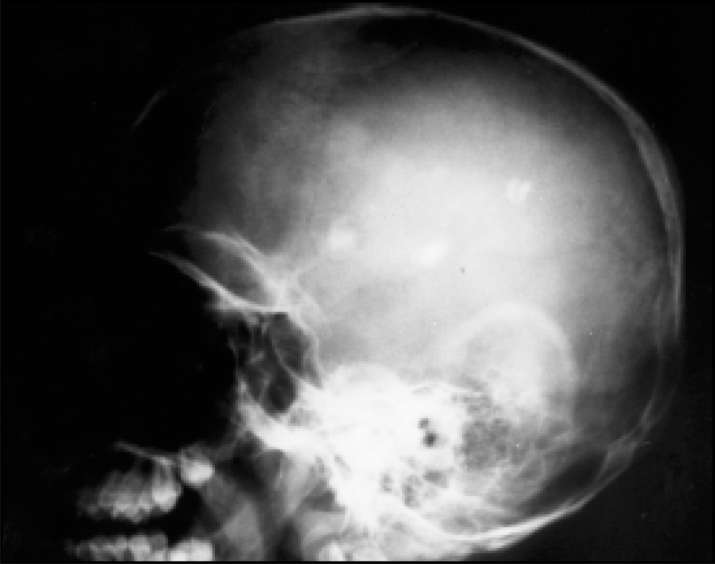
Cranial vault osseous abnormalities—These include patchy areas of calvarial sclerosis related to hyperostosis of the inner table, as well as prominence of the bony trabeculae in the diploic spaces (figure 1). The inner and outer tables may be thickened as well, with increased density seen on CT scans. The frontal and parietal regions are commonly involved. A common differential diagnosis for diploic thickening is long-term phenytoin therapy.
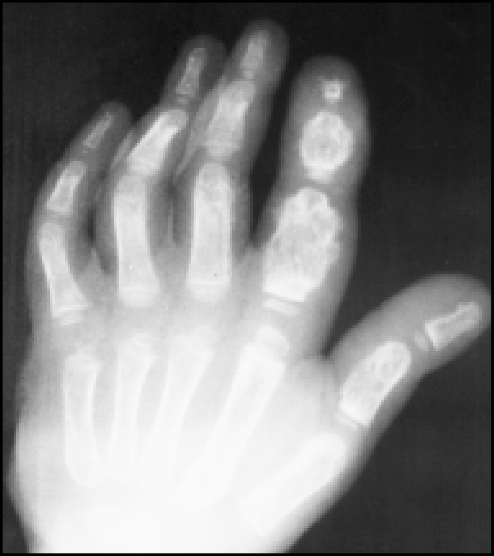
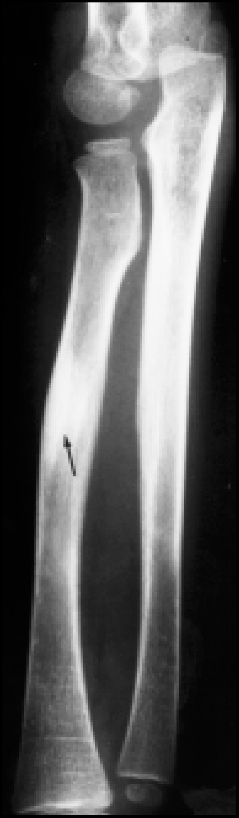
Extracranial osseous abnormalities—Osseous lesions in tuberous sclerosis can involve both the axial and appendicular skeleton and are fairly common in the disease. Cortical lesions include subperiosteal new bone deposition resulting in a thickened, irregular, often spiculated-appearing cortical contour, most often seen in the short tubular bones of the hands and feet but occasionally encountered in the long bones (figures 2 and 3). Cortical pits and depressions have also been described.6,7 Periungual fibromas of the skin often seen on the hands may lead to adjacent bony erosion.6,8
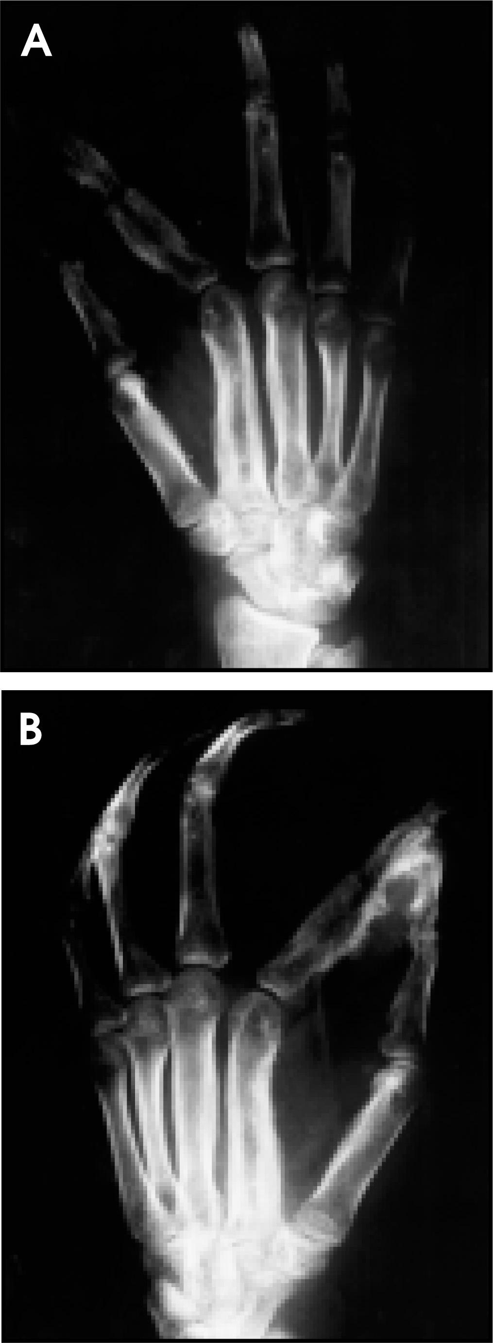
Varying size, round to oval cystic lesions with or without associated periosteal reaction may be seen in the phalanges, most commonly the distal phalanges (figure 4). Cystic areas may be encountered in the metacarpals and metatarsals as well and represent islands of fibrous tissue. Differential diagnosis includes neurofibromatosis and sarcoidosis.
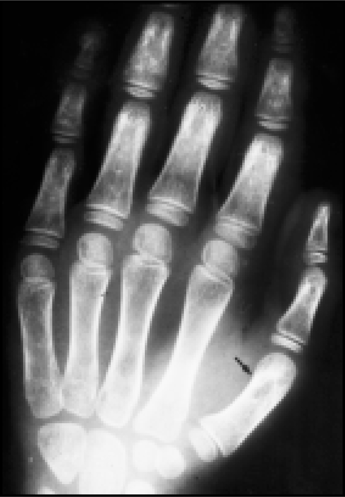
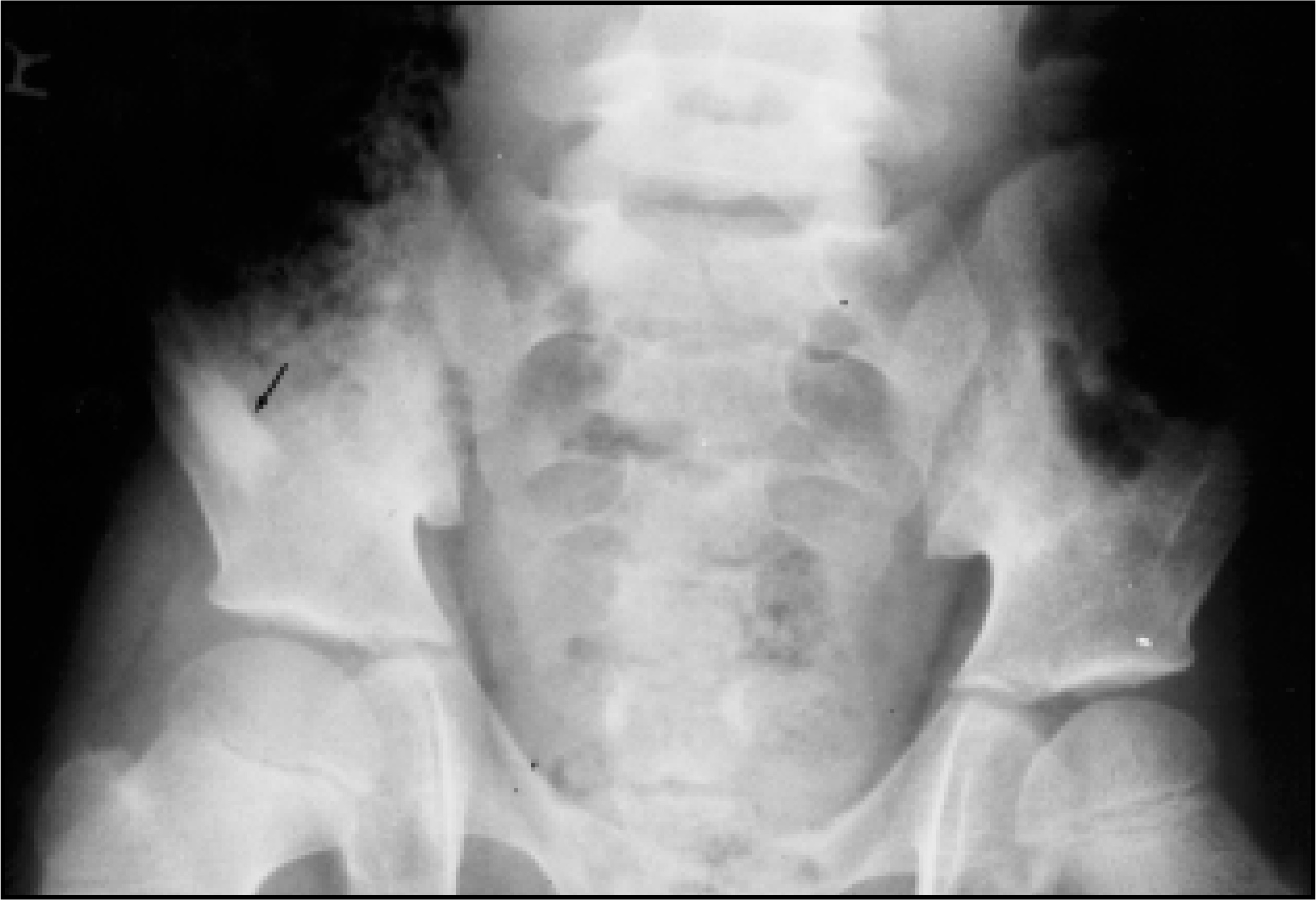
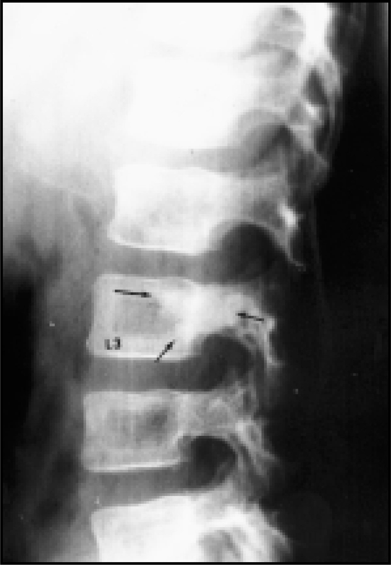
Medullary involvement in tuberous sclerosis presents as abnormal areas of osteoblastic activity, the so-called “bone islands.” The lesions are usually oval or flame-shaped, and may be found in the long bones, spine, or pelvis (figures 5, 6 and 7). These lesions range from a few millimeters to several centimeters in size and may enlarge slowly over several years, but characteristically do not expand the bone.
Extraosseous multisystem involvement
Cutaneous—Adenoma sebaceum occurs in 80% to 90% of cases but may not be present till after puberty. Shagreen patches occur in 20% to 50% of cases, characterized by dull red or tan firm plaques with a wrinkled surface protruding slightly above the surrounding skin. These lesions frequently are seen in the lumbar region and occasionally are painful. Hypopigmented macules may be noted at birth or in the neonatal period.3,7,9,10
Central nervous system—The CNS lesions include subependymal hamartomas, small nodular lesions in the subependymal region of the ventricles, a common CNS finding. These lesions may calcify11-13 and may be seen on plain skull radiographs. Subependymal nodules with prominent contrast enhancement suggest the presence of a giant cell astrocytoma, most commonly located at the foramen of Monro and seen in 5% to 10% of affected individuals.
Ocular lesions—The most frequently encountered ocular finding is a retinal hamartoma, located at the optic disc. These lesions are usually multiple and may be detected with the opthalmoscope. Like hamartomas of the brain, calcification may occur.14At birth, 52% of patients will have this lesion, making this the most common abnormality detected at birth.5
Renal lesions—Approximately 55% of patients will develop a renal lesion including cysts, angiomyolipomas, renal cell carcinomas, and aneurysms. At least 50% of all renal angiomyolipomas occur in patients with TS. On CT, detection of fat within these lesions is diagnostic.
Lungs—Approximately 1% of patients with TS will develop pulmonary symptoms. These usually occur late in the disease. Radiographically, these lesions are identical to lymphangioleiomyomatosis (LAM). Almost all of the affected patients are female. A common complication is spontaneous pneumothorax. On rare occasions, a patient may develop a chylothorax.
Cardiac lesions—Rhabdomyoma is the most common cardiac tumor in infants and children, and approximately 50% to 70% of these tumors are associated with TS.
Summary
Osseous involvement in tuberous sclerosis can have a varied radiographic appearance with involvement of both the axial and appendicular skeleton. While many of the lesions have a characteristic radiological appearance, some are unusual, and other differential diagnoses need to be considered. AR
References
- Critchley M, Earl C. Tuberous sclerosis and allied conditions. Brain. 1932;55:311.
- Thibault J, Manuelidis E. Tuberous sclerosis in a premature infant. Report of a case and review of the literature. Neurology. 1970;20:139-146.
- Bundey S. The signifance of a white macule on the skin of a child. Dev Med Child Neurol. 1970;12:805-806.
- Medley B, McLeod R, Houser O. Tuberous sclerosis. Semin Roentgenol. 1976;11:35-54.
- Feldman F. Diagnosis of Bone and Joint Disorders. 1995:4353-4393.
- Green G. The radiology of tuberous sclerosis. Clin Radiol. 1968;19:135-147.
- Aughenbaugh G. Thoracic manifestations of neurocutaneous diseases. Radiol Clin North Am. 1984;22:741-756.
- Bender B, Yunis E. The pathology of tuberous sclerosis. Pathol Annu. 1982;17:339-382.
- Fitzpatrick T, Szabo G, Hori Y. White leaf-shaped macules: Earliest visible signs of tuberous sclerosis. Arch Dermatol. 1968;98(1):1-6.
- Gold A, Freeman J. Depigmented nevi: The earliest signs of tuberous sclerosis. Pediatrics. 1965;35:1003.
- Starshak R, Wells R, Sty J, Gregg D. Diagnostic Imaging of Infants and Children. 1992.
- Terada T, Nakai E, Moriwaki H. Tuberous sclerosis with an atypical skull change: Case report. Neurosurgery. 1985;16:804-807.
- Brown B. The radiologic features of bone changes in tuberous sclerosis with a case report. J Can Assoc Radiol. 1961;12:1.
- Slone R, Gutierrez F, Fisher A. Thoracic Imaging: A Practical Approach. 1999:111-112.
Citation
. Osseous and multisystem involvement of tuberous sclerosis. Applied Radiology. 2001;30(6):22-24. doi:10.37549/AR1008.