Overlap Syndrome
Journal of Pediatric Case Reports — Vol. 1 , Issue 7
Published: October 1, 2026
1 University of Arizona College of Medicine–Phoenix, Phoenix, Arizona
2 Department of Radiology, Phoenix Children’s Hospital, Phoenix, Arizona
3 Department of Radiology, Cincinnati Children’s Hospital, University of Cincinnati College of Medicine, Cincinnati, Ohio
Abstract
Autoimmune hepatitis with primary sclerosing cholangitis, also known as overlap syndrome, is a rare occurrence in the pediatric population. Children with overlap syndrome present with findings related to each disease. In addition, there is a high association with coexisting inflammatory bowel disease. Imaging, particularly MRI with MR cholangiopancreatography, pre- and post-hepatobiliary contrast administration imaging, and MR elastography, allows for a complete assessment of the liver and can be used in the diagnosis and surveillance of each component of liver disease.
Keywords
gastrointestinal, intestine, autoimmune
Categories
Case Summary
A teenager with a history of ulcerative colitis, autoimmune hepatitis (AIH), and sclerosing cholangitis presented with jaundice and hyperbilirubinemia. He had previously had symptoms related to liver failure and portal hypertension with low platelet levels, esophageal varices, and variceal bleeding.
Imaging Findings
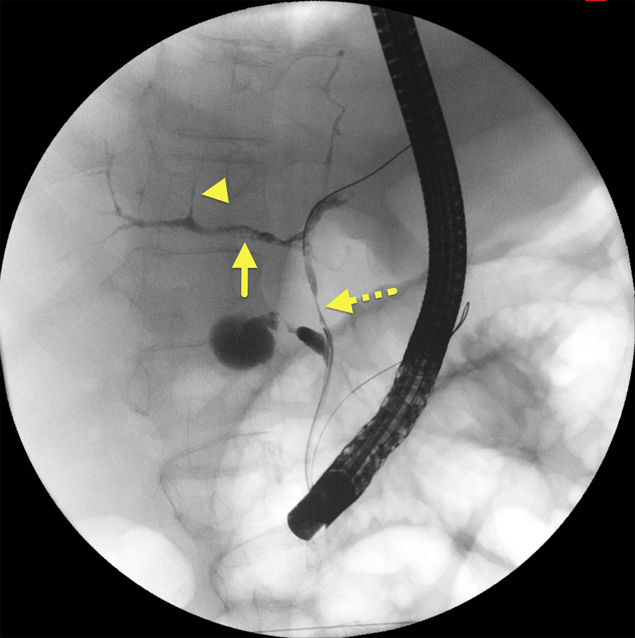
An endoscopic retrograde cholangiopancreatography (ERCP) (Figure 1) demonstrated diffuse irregularity of intra- and extrahepatic bile ducts, pruning of intrahepatic bile ducts, narrowing of the distal common bile duct, and filling defects within the right central intrahepatic ducts consistent with primary sclerosing cholangitis (PSC).
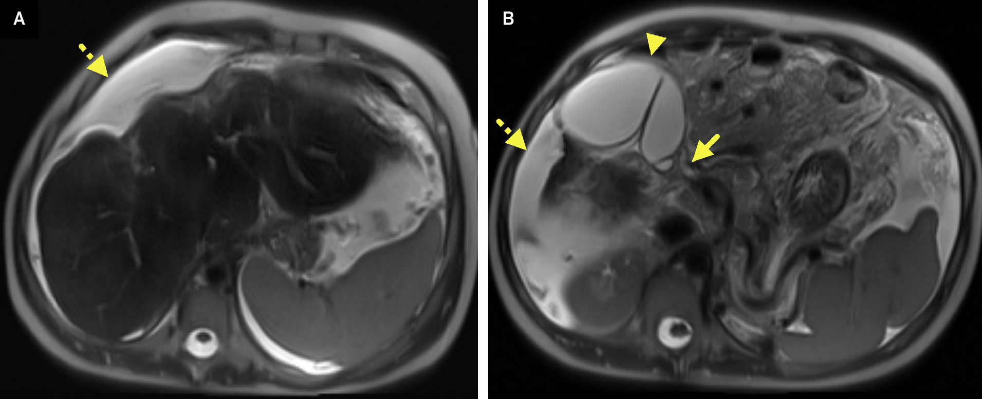
MR cholangiopancreatography (MRCP) (Figure 2) showed the liver to be small, with a macronodular contour and wispy increased T2 signal consistent with fibrosis. There was a large volume of ascites. Intrahepatic bile ducts were not well visualized. The common bile duct was relatively enlarged, and the gallbladder was dilated.
Diagnosis
Overlap syndrome.
The differential diagnosis includes viral hepatitis, choledocholithiasis, ascending cholangitis, and primary biliary cirrhosis.
Discussion
AIH is a liver disease characterized by lymphoplasmacytic infiltration, lobular inflammation, and damage.1 It occurs in 1 in 200,000 people in the general United States population and is strongly associated with HLA DR3 and DR4 alleles.1,2 Patients present with findings of liver failure: thrombocytopenia, coagulopathy, jaundice, abdominal pain, and elevated serum transaminases.2 AIH can be categorized into AIH type 1 (positive for ANA and smooth muscle antibody) or type 2 (positive for liver kidney microsomal antibody) disease. Immunosuppression is the mainstay of treatment. Those with acute liver failure and encephalopathy will need a liver transplant. The risk for recurrence of AIH in transplanted patients is 16-43%.1,2
Over the past decade, there have been significant advances in the imaging and quantification of liver fibrosis. US and MR elastography have been used to measure liver stiffness, a value used as a surrogate for liver fibrosis. In patients with known hepatic fibrosis, routine screening with MRI using a hepatobiliary agent is recommended.3 Studies in children have shown that hepatobiliary agents help to improve lesion detection and improve the diagnostic confidence when a liver lesion is identified.4
PSC occurs in 0.2-1.5 per 100,000 children.2 Its presentation is insidious, with signs and symptoms of growth delay, fat-soluble vitamin deficiencies, jaundice, and pruritus.2 PSC can be diagnosed via cholangiogram. MRCP is currently the test of choice due to its noninvasive nature. All types of cholangiograms can show multiple dilations and strictures, giving the bile ducts a beaded appearance.2,5 ERCP allows for biopsy and the therapeutic dilation of strictures with stenting to slow the development of end stage liver disease. On biopsy, periductal onion skin fibrosis is seen in 40% of cases.2 Treatment of PSC is usually supportive.
Overlap syndrome occurs in 6-11% of patients with AIH.1 These patients have characteristics of both AIH and PSC. Additionally, inflammatory bowel disease is present in 41% of patients with overlap syndrome.1 Immunosuppression combined with ursodeoxycholic acid has demonstrated improvement in serum transaminase levels but has not been associated with improved patient outcomes.1,2 AIH patients with elevated GGT or poor response to immunosuppression warrant screening with MRCP to evaluate for PSC and overlap syndrome.2 Characteristic imaging findings of overlap syndrome include macro-regenerative nodules, peripheral liver atrophy, and biliary ductal irregularity.1
Patients with sclerosing cholangitis are at risk for developing cholangiocarcinoma at some time in their life. This risk increases from the time of diagnosis and is said to occur in 9% at 10 years and 19% of patients at 20 years from diagnosis.2 While this risk is high, cholangiocarcinoma is still rare in the pediatric population, occurring in 1% of pediatric patients with PSC. Because of the elevated risk, patients require frequent monitoring with MRCP, especially as they age.2,5,6
Conclusion
AIH with PSC, also known as overlap syndrome, is a rare occurrence in the pediatric population. Children with overlap syndrome present with findings related to each disease. In addition, there is a high association with coexisting inflammatory bowel disease. Imaging, particularly MRI with MRCP, pre- and post-hepatobiliary contrast administration imaging, and MR elastography, allows for a complete assessment of the liver and can be used in the diagnosis and surveillance of each component of the liver disease.
References
- Malik N, Venkatesh S. Imaging of autoimmune hepatitis and overlap syndromes. Abdom Radiol (NY). 2017;42(1):19-27. doi:10.1007/s00261-016-1019-x.
- Kerkar N, Chan A. Autoimmune hepatitis, sclerosing cholangitis, and autoimmune sclerosing cholangitis or overlap syndrome. Clin Liver Dis. 2018;22(4):689-702. doi:10.1016/j.cld.2018.06.005.
- Kolbe A, Podberesky D, Zhang B, Towbin A. The impact of hepatocyte phase imaging from infancy to young adulthood in patients with a known or suspected liver lesion. Pediatr Radiol. 2015;45(3):354-365. doi:10.1007/s00247-014-3160-4.
- Schooler G, Squires J, Alazraki A. Pediatric hepatoblastoma, hepatocellular carcinoma, and other hepatic neoplasms: consensus imaging recommendations from american college of radiology pediatric liver reporting and data system (LI-RADS) working group. Radiology. 2020;296(3):493-497. doi:10.1148/radiol.2020200751.
- Khoshpouri P, Habibabadi R, Hazhirkarzar B. Imaging features of primary sclerosing cholangitis: from diagnosis to liver transplant follow-up. Radiographics. 2019;39(7):1938-1964. doi:10.1148/rg.2019180213.
- Culver E, Chapman R. Systematic review: management options for primary sclerosing cholangitis and its variant forms - igg4-associated cholangitis and overlap with autoimmune hepatitis. Aliment Pharmacol Ther. 2011;33(12):1273-1291. doi:10.1111/j.1365-2036.2011.04658.x.
Disclosures
The authors have no conflicts of interest to disclose. None of the authors received outside funding for the production of this original manuscript and no part of this article has been previously published elsewhere.
Citation
. Overlap Syndrome. Journal of Pediatric Case Reports. 2026;1(7). doi:10.37549/JPCR-26-0113.