Pediatric body MR imaging: Our approach
Applied Radiology — Vol. 39 , Issue 4 , pp. 8 -19
DOI: 10.37549/AR1748
Published: April 1, 2010
Categories
Pediatric body magnetic resonance imaging (MRI) has made many advances since its inception. It offers exquisite soft-tissue contrast without exposure to ionizing radiation, which is important in the pediatric population. The advancement to 3.0 Tesla (T) magnets, new sequences, multichannel coils and parallel imaging allows for increased resolution and decreased image acquisition times.1,2
MRI is quickly becoming the modality of choice for pediatric abdominal masses. It plays an important role in the diagnosis of disease, staging and monitoring response to therapy.3-6 Therefore, good quality images that can demonstrate the lesion consistently are needed.
Body MRI in children is challenging due to the small size of the individuals and the variable extent of cooperation with prolonged image acquisition times. MRI is sensitive to motion artifact, which can significantly degrade image quality, and requires multiple sequences. These factors demonstrate the need for faster scanning techniques and steps for improving patient cooperation.7-9 A brief discussion of MR sequences will be followed by useful techniques including our protocols for pediatric sedation and the sequences employed at our institution to obtain consistent diagnostic quality images, while minimizing the need for full patient cooperation.
T1-weighted sequences
T1-weighted (T1W) images are fundamental to body MRI. The primary information provided by T1W imaging is the following:
presence of abnormally increased fluid content or fibrous tissue (low signal intensity on T1W),
presence of subacute blood or concentrated protein (high signal intensity on T1W), and
presence of fat content (high signal intensity on T1W).
The routine use of fat suppression sequences may provide additional information.7,10
T1W spin echo
This is a breathing-averaged data acquisition. We usually incorporate fat suppression to improve the dynamic range of intra-abdominal tissue signal intensities. On spin echo sequences, a longer time of repetition (TR) is used to compensate for the prolonged T1 relaxation, and a shorter time to echo (TE) is used to compensate for accelerated decay if a 3T field is being used.11
Spoiled gradient echo (SGE)
SGE sequences with the use of phased-array multicoil imaging may be used to replace longer duration sequences such as T1W spin-echo sequences.
Limitations: SGE is very sensitive to motion and requires patient cooperation.
3-dimensional gradient echo
Three-dimensional gradient echo (3D-GE) has been used extensively for MR angiography (MRA) and has evolved into a useful technique for body MRI.7 The imaging requires reduction of flip angle from 70 degrees to 90 degrees in MRA to 15 degrees to 20 degrees for body imaging. This shorter flip angle predominantly determines the contrast of the images. The advantage over 2-dimensional imaging is that it provides thinner, 2.5 to 3.0 mm sections which can then be reconstructed into other imaging planes. The fat suppression is also of superior quality and uniformity.
Limitation: The disadvantage is that there is a diminished contrast-to-noise ratio. This limitation has caused many to question the use of this technique. Gadolinium-enhanced fat-suppressed post-contrast imaging helps to boost the contrast-to-noise ratio and is useful for detecting flowing blood compared with areas of flow void or decreased signal in spin-echo and fast spin-echo sequences.10
Motion insensitive MPRAGE
In patients who are unable to cooperate with lying still for an examination, SGE sequences may be modified into a single-shot technique using the minimum TR to achieve diagnostic images. Such sequences include Water Excitation T1W Magnetization Prepared Rapid Acquisition Gradient Echo (WE-MPRAGE) and Turbo Fast Low Angle Shot (TurboFLASH). These sequences display image contrast by suppression of transverse magnetization by a gradient pulse. Therefore, the only magnetization that remains is that of longitudinal magnetization, which reflects T1 contrast. An inversion time of 0.5 sec provides optimal T1W image contrast, which leads to slice-to-slice TR of 1.5 sec.12
Limitations: Using this technique, it is not possible to obtain high-resolution, or predictable, T1W imaging. The WE-MPRAGE slice-by-slice technique cannot be used for dynamic gadolinium-enhanced imaging, particularly during the most important phase of hepatic arterial imaging, i.e. the hepatic-arterial dominant phase. As each slice requires approximately 1.5 sec to acquire, the time duration between the top and bottom liver slices is too great to “snapshot” the entire liver during this phase.
T2-weighted (T2W) sequences
T2-weighted images are important in abdominal MR especially for use in hepatic imaging. They help in lesion characterization and help in the assessment of diffuse liver disease. Presence of perfusion abnormalities, edema, fibrosis and abnormal iron deposition can be easily characterized on T2W images.7,10
Echo train spin echo (ETSE)
This sequence is based on a series of 180 degree refocusing pulses instead of a single 180 degree pulse. It requires a shorter time to acquire T2W images with almost no artifact from magnetic susceptibility.13,14 Acquiring this sequence as an echo train permits shortening the sequence from 15 min to 4 to 5 min. Fat suppression is often used in conjunction with this sequence to improve the dynamic range of intra-abdominal signal intensities.
Limitation: Since fat is very high in signal intensity on these sequences, usually fat suppression is required to optimize the contrast between fat and water. Fat suppression should generally be applied for at least one set of images of the abdomen to ensure optimal contrast between high-signal abnormalities, such as cystic masses, and fluid collections adjacent to intra-abdominal fat.
Single-shot ETSE
This sequence uses very long echo train lengths and half Fourier imaging to provide high-quality images in a short duration. Image acquisition is sufficiently short to essentially “freeze” motion. We routinely employ a single-shot ETSE technique termed HASTE (Half Fourier Acquisition Single Shot Turbo Spin Echo) or single-shot fast spin-echo.7,13,15 This is a slice-by-slice technique where a single slice-selective excitation pulse is followed by a series of echoes, typically using a train of 80 to 180 pulses, each separated by 3 msec, to fill in the k-space of the entire slice.7 This sequence requires 1.2 to 1.5 sec before continuing to the next slice. Since the motion-sensitive component represents such a small fraction of the total acquisition period, this sequence is relatively insensitive to breathing motion artifact. In contrast, conventional spin-echo requires longer acquisition times and is therefore motion sensitive.16
ETSE is the sequence of choice for MR cholangiopancreatography (MRCP),7 MR urography17 and fetal MRI. MRCP is based on a modified ETSE sequence, which uses a longer time to echo on the order of 250 to 500 msec. The longer time to echo causes the fluid in the gallbladder, pancreatic duct and bile duct to appear bright. There are various methods for obtaining these sequences, most commonly thin sections of 3 mm to 4 mm for higher resolution, or a thick slab of 3 cm to 4 cm sections with the goal of including the pancreatic and bile ducts in a single image.
Limitations: There is less contrast between tissues due to decreased T2 differences. In the case of hepatic imaging, the T2 difference between diseased and background normal liver tissue may be subtle; and because of the T2 averaging effects of summated multiple T2 echoes, this contrast may be blurred. The lesions with similar T2 values may differ on T1 values, which emphasize the importance of acquiring T1W images.10
Data acquisition parameters
During the last 5 years, one of the advances in MRI has been the advent of more sophisticated 3T systems. Compared with standard 1.5T systems, a higher field strength provides a higher signal-to-noise ratio (SNR), higher spatial resolution, faster speed and better fat suppression. The high SNR is crucial in younger children because of their smaller size.1,2,18,19 Both thinner and multiple images can be obtained in a single data acquisition with 3T.
Limitations: Artifacts are more common with 3T as compared with 1.5T.1,2,18 The recognition of these artifacts is important in order to optimize image quality. Magnetic susceptibility artifacts result from microscopic gradients or variations of magnetic field that occur near the interfaces of various tissues. There is more signal dropout and increase in the number of localized regions of low and high signal intensities when a 3T system is used. The chemical shift artifact is also another impediment. Due to the increase in main field strength, the frequency difference between fat and water increases almost twofold from 220 Hz at 1.5T to 440 Hz at 3T.1,2,18,20 These artifacts can be minimized by using increased bandwidth. However, this technique can lead to a decrease in SNR, which degrades by the square root of bandwidth. Standing-wave artifacts are also of concern in 3T imaging and result in production of artifactual bright and dark signals in the image. The presence of large volumes of fluid, such as ascites, makes this artifact more conspicuous.21-23
Receiver coil
Because high spatial resolution and SNR are crucial, the use of receiver coils minimizes the noise from nonimaged body parts. Phased-array coils, which contain multiple elements in contrast to the single-channel coil, are now commonly used.1,2 The coils used for 1.5T and3T are different and may not be substituted for one another. High-quality coils improve pediatric imaging.
MR examination techniques
It is important to devise strategies for sedation and protocols based on age, developmental level and the type of study ordered prior to the MRI procedure. For the purpose of categorizing appropriate strategies, we have broadly classified children into 3 age groups based upon their general level of cooperativeness: older children (6 to 18 years of age), small children (1.5 to 6 years of age) and infants (<1.5years of age). Sedation techniques and the specific protocols for each age group will be discussed.
Sedation
For any sedation procedure, children should be NPO for at least 4 hours prior to the examination. Prior to moderately sedating a pediatric patient, it is critical to do a thorough assessment including patient history, baseline vital signs, weight, airway examination, oropharyngeal classification, and a cardiopulmonary examination in order to recognize potential factors that may place the patient at increased risk for complications. General anesthesia is chosen when a pediatric patient has co-morbidities that are contraindications to conscious or moderate sedation or when timed breathing suspension would assist in improving the diagnostic quality of the images.24-26
Monitoring of vital signs is essential for moderately sedated patients. A nurse remains in the procedure area throughout the study and periodically records vital signs (pulse oxygenation, ventilation, circulation and temperature). When conscious sedation is selected, the person sedating the patient should be trained in advanced life support. At our institution we use Versed®(midazolam, Ben Venue Laboratories Inc., Bedford, OH) for minimal sedation and anxiolysis. For moderate sedation, we use chloral hydrate (Pharmaceutical Associates Inc., Greenville, SC) in children <2 years of age, and Nembutal (pentobarbital sodium, Ovation Pharmaceuticals, Deerfield, IL) for those >2years old. Versed can also be used in conjunction if needed. Monitored, conscious sedation is most appropriate for healthy children and short diagnostic examinations. Titration of medications is based upon our sedation continuum described in Table 1.
When the MRI is completed, the patient should be taken to a quiet area to recover and be given ample time to awaken before they are discharged. Recovery time varies depending on the type and amount of drugs used. We ensure that the patient returns to his/her baseline status with intact protective reflexes prior to discharge. The caregivers should be given verbal and written discharge instructions prior to leaving the facility with a contact phone number for concerns or questions after discharge.
General considerations
In the pediatric population, we generally aim for scanning times of approximately 20 min for upper abdominal studies and 35 min for abdomen and pelvis studies.
We prefer to image neonates at 3T (Table 2) rather than 1.5T (Table 3) due to the small size of the patients. The higher SNR of 3T allows for thinner sections and higher image quality of WE-MPRAGE imaging.11,12 A compelling feature of WE-MPRAGE is that it is a single-shot technique and therefore does not require patient cooperation.12 For identical reasons, the single-shot T2W ETSE sequence is used in all MR protocols.
Due to their higher intrinsic SNR, breathing-averaged T1W and T2W sequences form an important part of an MRI strategy. Image quality and soft-tissue contrast resolution are often improved with the use of fat suppression.11 As a time-saving device, T2W sequences should be performed as echo train sequences, which may also be considered on T1W sequences. It may be essential to use fat suppression on T2W ETSE sequences for liver imaging.7,27 Fat suppression ensures optimal contrast between high-signal abnormalities, such as fluid collections and cystic masses, and adjacent intra-abdominal or pelvic fat.28
MR protocols
Older children (6-18 years of age)
Older children are generally sufficiently cooperative to be imaged with standard body MR protocols at either 1.5T or 3T. On modernMRI equipment our basic protocol includes (Figure 1):
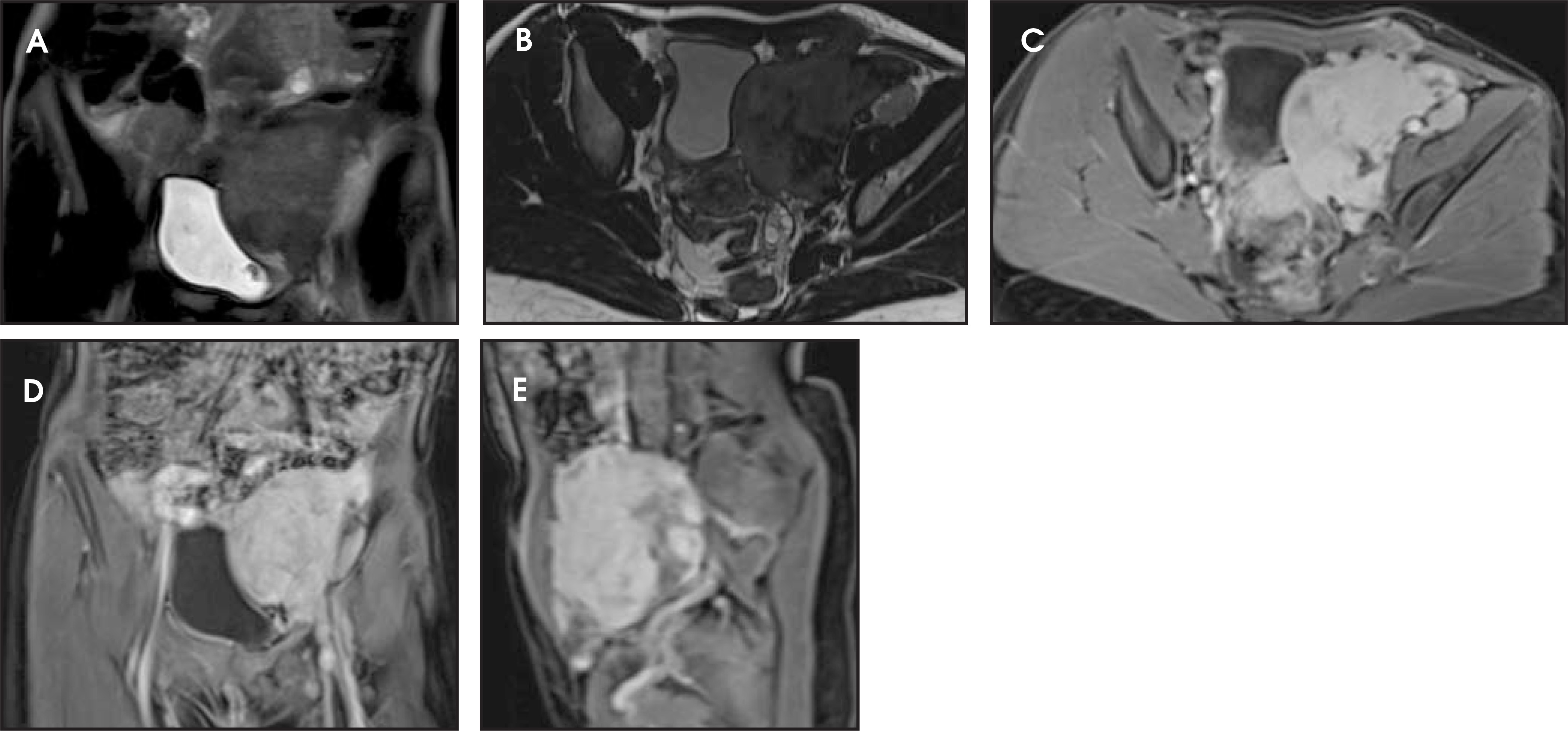
SS-T2-ETSE (standard and with fat suppression)
T1W in-phase/out-of-phase GE, fat suppression 3D-T1-GE
Gadolinium administration
Three passes using transverse 3D-T1-GE
Properly prepared and coached to cooperate with breath-hold techniques, the best quality of body MRI studies can be anticipated in children between 12 to 18 years of age. Conscious sedation is routine in this age group.
Small children (1.5-6 years of age)
This age group is generally more alert and anxious, and therefore more likely to require pharmaceutical sedation and restraints. Pillows, adhesive straps, foam sponges and tape can be used to immobilize the child. Moderate sedation is routinely used in this age group.
Although it is not imperative to image this age group at 3T, we routinely do so, since the higher SNR will provide the best anatomic information. Our protocol includes (Figure 2):
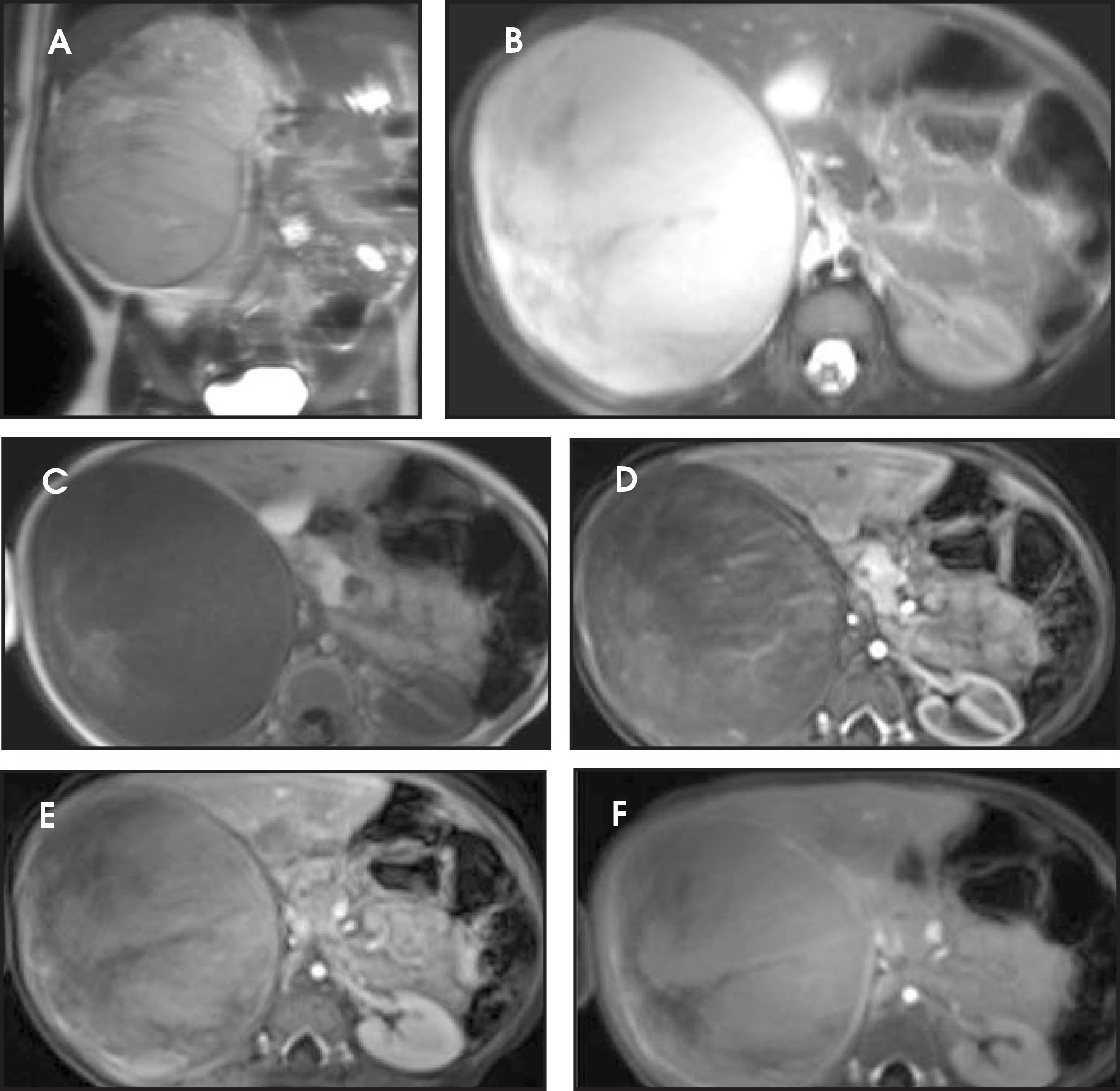
Multiplanar noncontrasted fat-suppressed T1W spin echo
Fat-suppressed T2W ETSE sequences
Gadolinium administration
WE-MPRAGE images in both the transverse and coronal planes
Suspended respiration imaging may also be indicated in selected individuals. Older children within this age group may be sufficiently cooperative to undergo standard body MR protocols without sedation.
Infants (<1.5 years of age)
At our institution, imaging of infants can often be performed without sedation by organizing their feeding and sleep schedules according to the timing of the MRI study. Multiple protocols for sedation and general anesthesia for the pediatric population are available and the choice of protocol strongly depends upon the radiologist’s training and the availability of trained nursing staff and anesthesiologists.
Because the anatomy is smaller in infants, we generally prefer to image them at 3T due to the higher SNR for better contrast and spatial resolution. Our basic MRI protocol in infants is the same as that for small children (Figure 3).
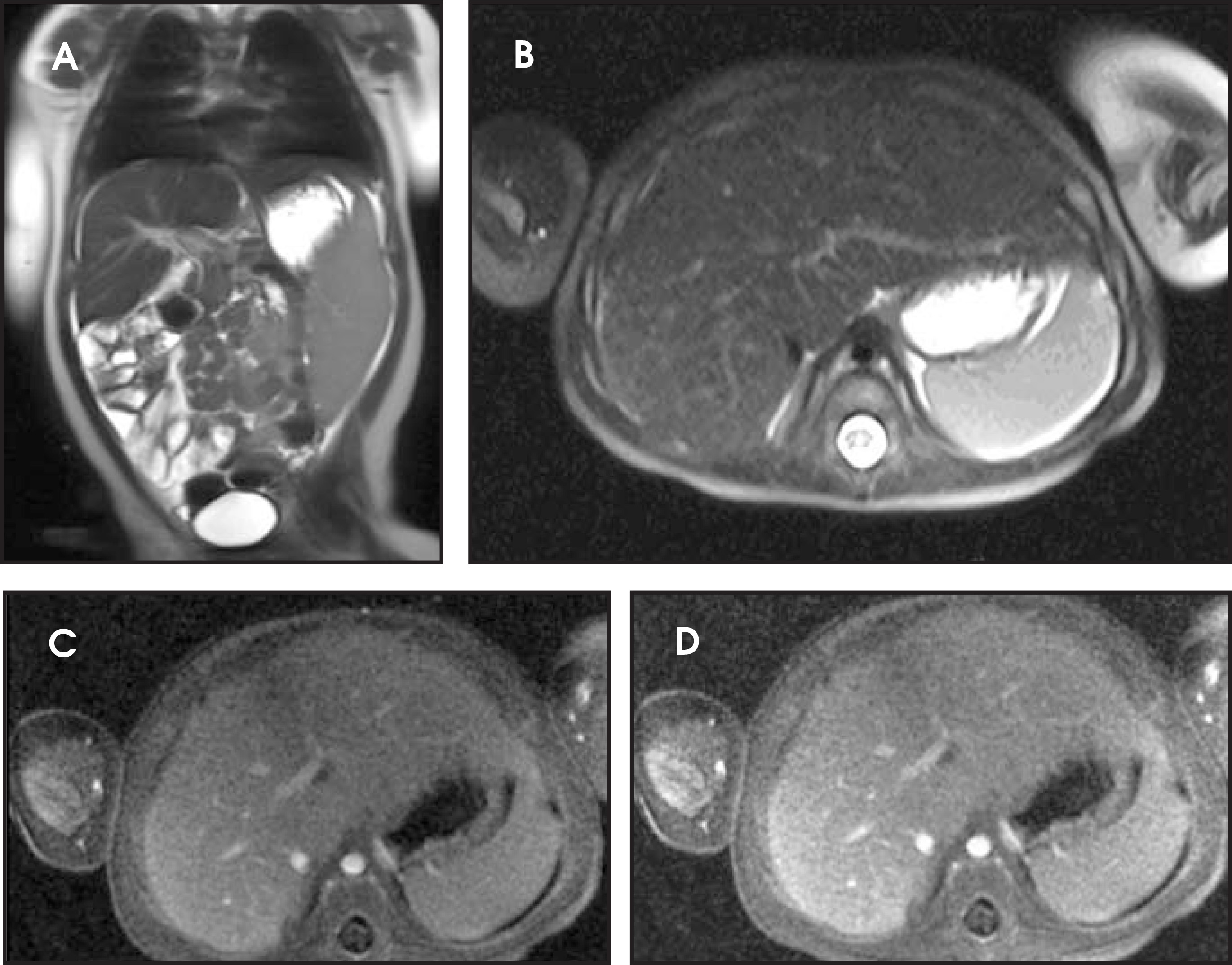
Multiplanar noncontrasted fat-suppressed T1W spin echo
Fat-suppressed T2W ETSE sequences
Gadolinium administration
WE-MPRAGE images in both the transverse and coronal planes
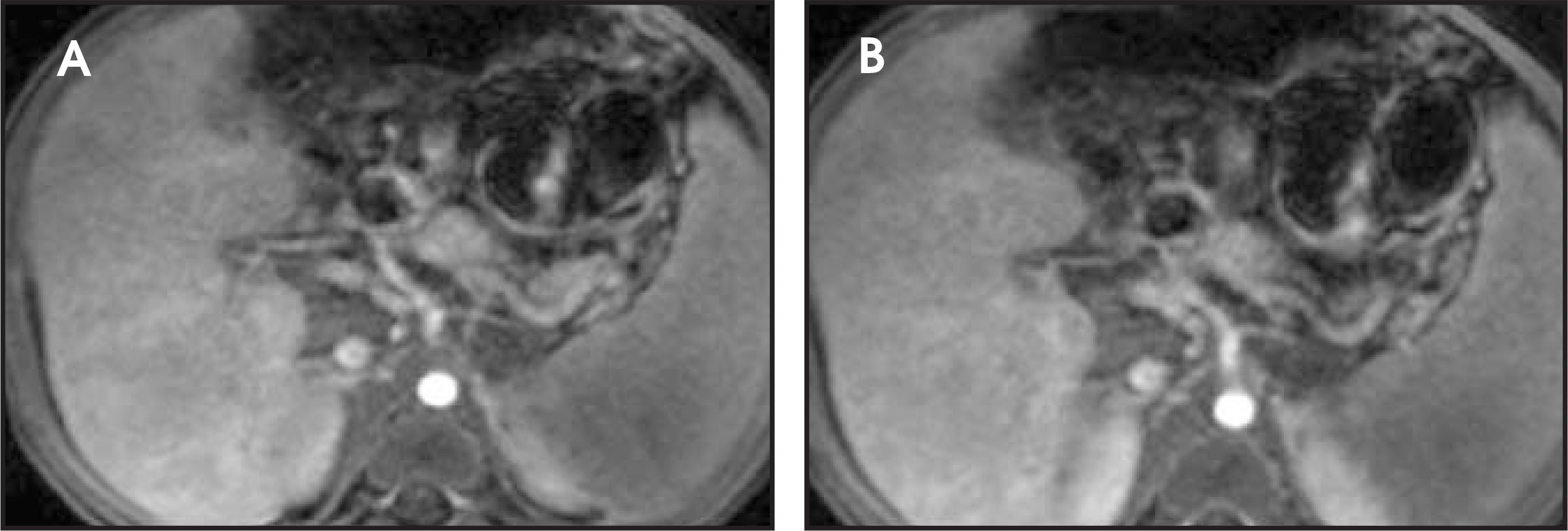
In selected patients in which thin-section and more dynamic imaging is required following gadolinium administration, patients will undergo general anesthesia and may be imaged with respiration-suspended 3D gradient-echo imaging. This approach must be carefully choreographed with the anesthesiology team in order to accurately time the suspension of respiration with the data acquisition (Figure 4).
Gadolinium-based contrast agents (GBCA)
In children more so than adults, the physician should pay special attention to the choice of appropriate gadolinium-based contrast agents (GBCAs) because of the potential for osseous deposition of gadolinium. As a result, we do not recommend the use of nonionic linear GBCAs, which exhibit lower conditional stability.29,30 Although the incidence of renal failure, and hence the risk of nephrogenic systemic fibrosis, is considerably lower in the pediatric population, it is imperative to follow guidelines at least as stringent as those used for adults.
Conclusion
MRI has the potential to become the best imaging modality for a wide range of pediatric illnesses, especially focal liver disease. Abdominal MRI is undoubtedly a powerful tool for diagnosis and surveillance of many pediatric illnesses. It provides excellent anatomic information without the use of ionizing radiation, which is very important in the pediatric population. The advancement to 3T magnets increases soft-tissue contrast and provides faster image acquisition times. The protocols and sedation techniques described above have successfully and consistently produced high-quality images at our institution.
References
- MacKenzie J, Vasanawala S. Advances in pediatric MR imaging. Magn Reson Imaging Clin N Am. 2008;16:385-402.
- Siegel M, Chung E, Conran R. Pediatric liver: Focal masses. Magn Reson Imaging Clin N Am. 2008;16:437-452.
- Semelka R, Worawattanakul S, Kelekis N. Liver lesion detection, characterization, and effect on patient management: Comparison of single phase spiral CT andcurrent MRI techniques. J Magn Reson Imaging. 1997;7:1040-1047.
- Ichikawa T, Haradome H, Hachiya J. Pancreatic ductal adenocarcinoma: Preoperative assessment with helical CT versus dynamic MR imaging. Radiology. 1997;202:655-662.
- Semelka R, Shoenut J, Kroeker M. Renal lesions: Controlled comparisons between CT and 1.5-T MR imaging with non-enhanced and gadolinium-enhanced fat-suppressed spin-echo and breath-hold FLASH techniques. Radiology. 1992;182:425-430.
- Semelka R, Kelekis N, Molina P. Pancreatic masses with inconclusive findings on spiral CT: Is there a role for MRI?. J Magn Reson Imaging. 1996;6:585-588.
- Semelka R, Martin D, Balci N. Magnetic resonance imaging of the liver: How I do it. J Gastroenterol Hepatol. 2006;21:632-637.
- Semelka R, Simm F, Recht M. T1-weighted sequence for MR imaging of liver: Comparison of three techniques for single breath, whole volume acquisition at 1.0and 1.5 T. Radiology. 1991;180:629-635.
- Semelka R, Balci N, Op de Beeck B, Reinhold C. Evaluation of a 10-minute comprehensive MR imaging examination of the upper abdomen. Radiology. 1999;211:189-195.
- Kelekis N, Semelka R, Worawattanakul S. Hepatocellular carcinoma in North America: A multi-institutional study of appearance on T1-weighted, T2-weighted, andserial gadolinium enhanced gradient-echo images. Am J Roentgenol. 1998;170:1005-1013.
- Ramalho M, Altun E, Herédia V. Liver MR imaging: 1.5T versus 3T. Magn Reson Imaging Clin N Am. 2007;15:321-347.
- Altun E, Semelka R, Dale B, Elias J. Water excitation MPRAGE: An alternative sequence for postcontrast imaging of the abdomen in noncooperative patients at 1.5 Teslaand 3.0 Tesla MRI. J Magn Reson Imaging. 2008;27:1146-1154.
- Huang J, Raman S, Vuong N, Sayre J, Lu D. Utility of breath-hold fast-recovery fast spin-echo T2 versus respiratory-triggered fast spin-echo T2 in clinical hepatic imaging. AJR Am J Roentgenol. 2005;184:842-846.
- Huang I, Emery K, Laor T. Fast-recovery fast spin-echo T2-weighted MR imaging: A free-breathing alternative to fast spin-echo in the pediatric abdomen. Pediatr Radiol. 2008;38:675-679.
- Semelka R, Kelekis N, Thomasson D. HASTE MR imaging: Description of technique and preliminary results in the abdomen. J. Magn Reson Imaging. 1996;6:698-699.
- Gaa J, Hatabu H, Jenkins R. Liver masses: Replacement of conventional T2-weighted spin-echo MR imaging with breath-hold MR imaging. Radiology. 1996;200:459-464.
- Grattan-Smith J, Little S, Jones R. MR urography in children: How we do it. Pediatr Radiol. 2008;38(Suppl):S3-S17.
- Anupindi S, Jaramillo D. Pediatric magnetic resonance imaging techniques. Magn Reson Imaging Clin N Am. 2002;10:189-207.
- Schenk J, Friebe B, Ley S. Visualization of intrarenal vessels by 3.0-T MR angiography in comparison with digital subtraction angiography using renal specimens. Pediatr Radiol. 2006;36:1075-1081.
- Hecht E, Lee R, Taouli B, Sodickson D. Perspectives on body MR imaging at ultrahigh field. Magn Reson Imaging Clin N Am. 2007;15:449-465.
- Soher B, Dale B, Merkle E. A review of MR physics: 3T versus 1.5T. Magn Reson Imaging Clin N Am. 2007;15:277-290.
- Merkle E, Dale B, Paulson E. Abdominal MR Imaging at 3T. Magn Reson Imaging Clin N Am. 2006;14:17-26.
- Kuhl C, Träber F, Schild H. Whole-body high-field-strength (3.0-T) MR Imaging in clinical practice. Part I. Technical considerations and clinical applications. Radiology. 2008;246:675-696.
- Mason K, Zurakowski D, Connor L. Infant sedation for MR imaging and CT: Oral versus intravenous pentobarbital. Radiology. 2004;233:723-728.
- Mason K, Sanborn P, Zurakowski D. Superiority of pentobarbital versus chloral hydrate for sedation in infants during imaging. Radiology. 2004;230:537-542.
- Weisenbach J, Vadon G, Storcz J. Sedation and sleep induction instead of anesthesia in infants and young children during CT and MRI examinations. [Article in Hungarian.] Orv Hetil. 1996;137:629-632.
- Zapparoli M, Semelka R, Altun E. 3.0-T MRI evaluation of patients with chronic liver diseases:Initial observations. J Magn Reson Imaging. 2008;26:650-660.
- Tsurusaki M, Semelka R, Zapparoli M. Quantitative and qualitative comparison of 3.0T and 1.5T MR imaging of the liver in patients with diffuse parenchymal liverdisease. Eur J Radiol.
- Wertman R, Altun E, Martin D. Risk of nephrogenic systemic fibrosis: Evaluation of gadolinium chelate contrast agents at four American universities. Radiology. 2008;248:799-806.
- White G, Gibby W, Tweedle M. Comparison of Gd (DTPA-BMA) (Omniscan) versus Gd (HP-DO3A) (ProHance) relative to gadolinium retention in human bone tissueby inductively coupled plasma mass spectroscopy. Invest Radiol. 2006;41:272-278.
Citation
. Pediatric body MR imaging: Our approach. Applied Radiology. 2010;39(4):8-19. doi:10.37549/AR1748.