Pediatric neuroradiology, part 1: Embryologic basis for brain malformation
Applied Radiology — Vol. 38 , Issue 7 , pp. 29 -40
DOI: 10.37549/AR1703
Published: July 1, 2009
Categories
Knowledge of basic brain embryology provides the foundation for making diagnoses of brain malformations, heritable diseases, congenital neoplasms and even disorders of postnatal development.
Malformation is defined as defective or abnormal formation, especially when acquired during development. An anomaly is a marked deviation from normal, especially as a result of congenital or hereditary defects. The term syndrome is defined as a set of symptoms occurring together. A syndrome may be due to malformation or hereditary defects.1
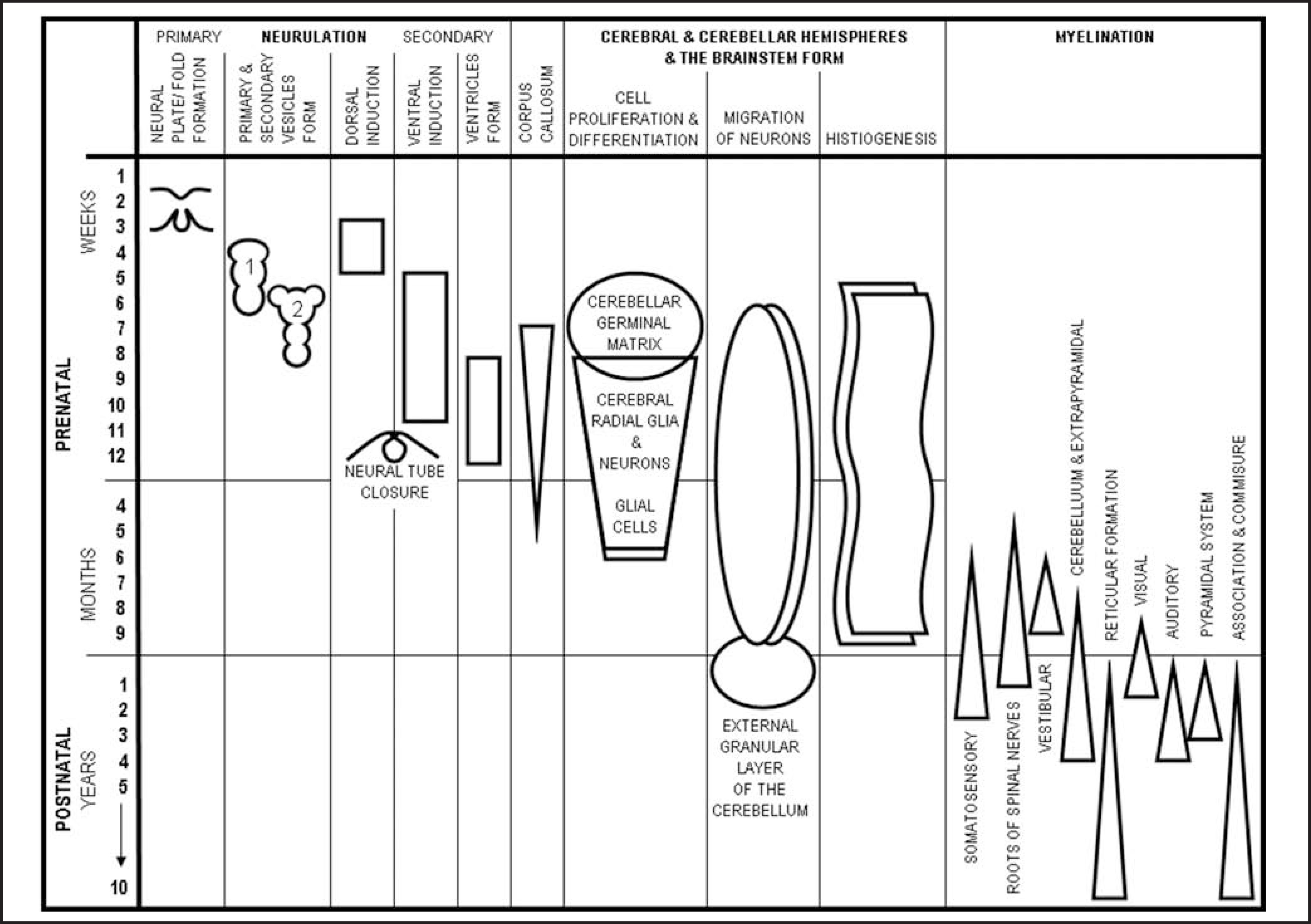
Timing of brain malformations and anomalies can be estimated through critical assessment of absent or malformed structures.2 (Figure 1) Heritable neurological diseases are caused by genetic errors that cause defects in the normal processes of brain formation and typically have imaging stigmata that, when learned, are easily recognizable. Congenital brain neoplasms, malformations and other neurological diseases may be associated with hydrocephalus and can develop at almost any time of brain development. Recognition of the imaging milestones in postnatal brain maturation, primarily the process of myelination, is important in differentiating dysmyelination from degenerative processes.
Brain embryology
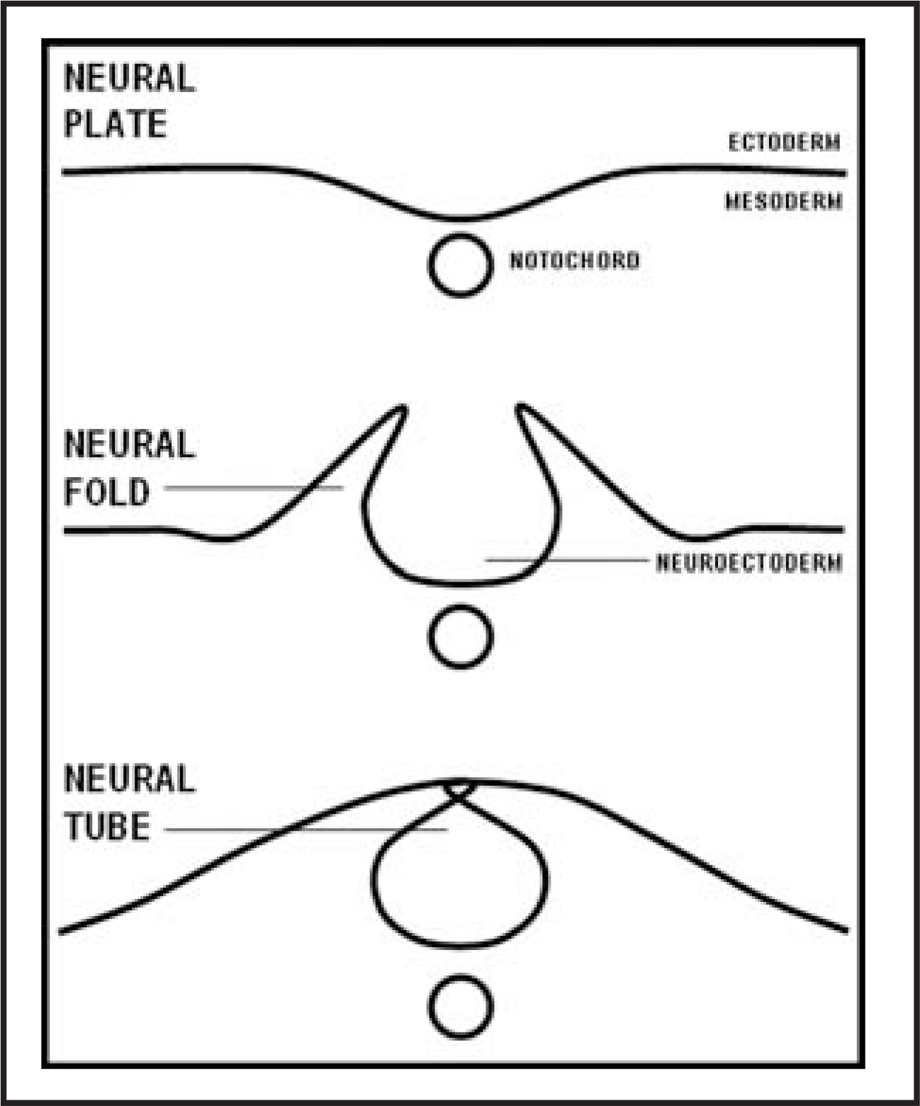
The earliest steps in the development of the brain occur at about 17 days of gestation when the neural plate, a thickening of the ectoderm, forms in the dorsal midline of the embryo and begins to differentiate into neurons. By the 20th day of gestation, the neural tube is formed and begins to close in the early stages of neurulation (Figure 2). Neurulation continues through stages of vesicle formation and the dorsal and ventral stages of induction.
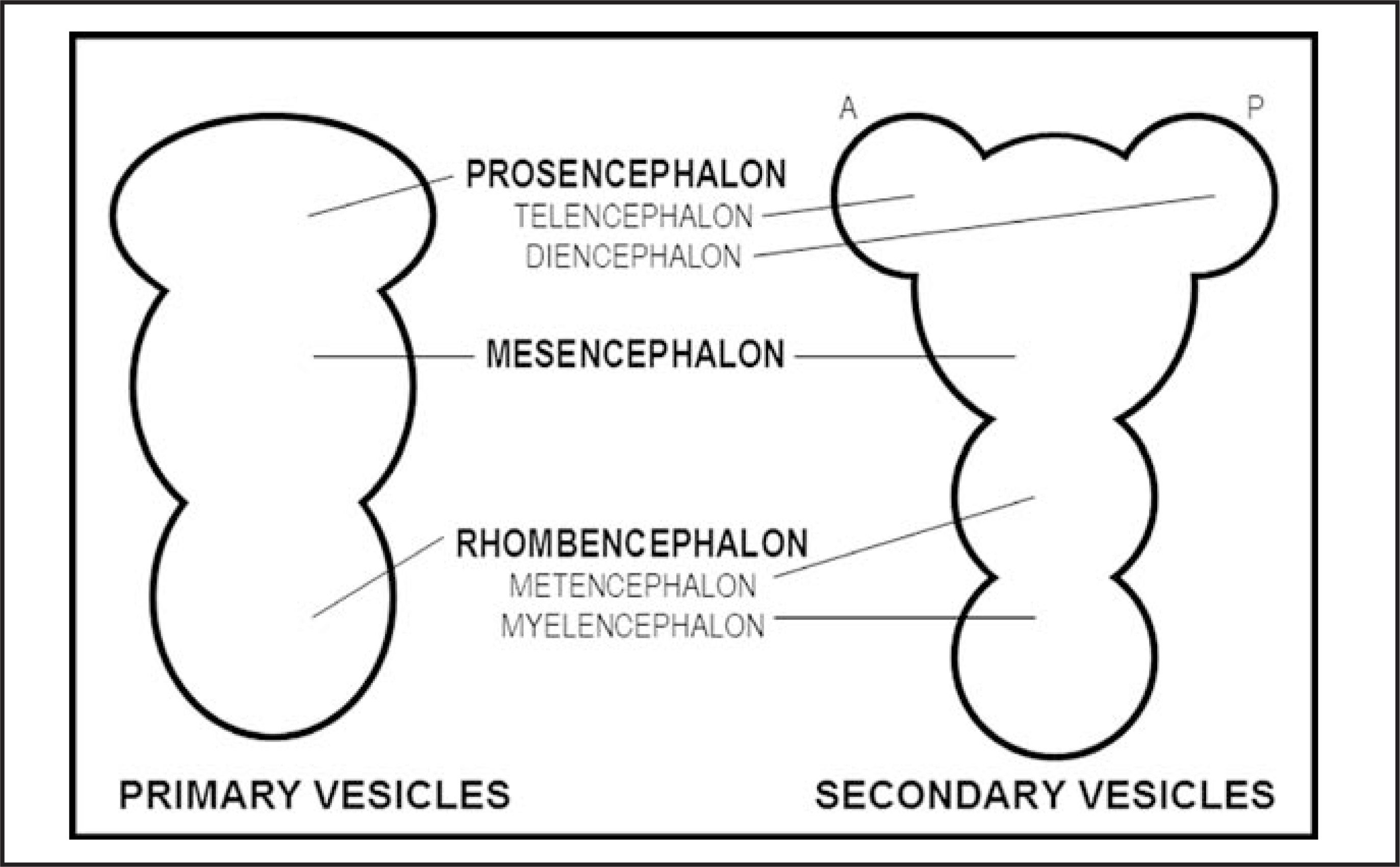
The brain forms at the rostral end of the neural tube. By the middle of the fourth week of gestation, 3 distinct primary vesicles have developed. As the primary vesicles mature and are folded, they differentiate into secondary vesicles during the fifth week of gestation.3,4
The 3 primary vesicles are the forebrain (prosencephalon), midbrain (mesencephalon) and hindbrain (rhombencephalon). The secondary vesicles arise from the primary vesicles: the prosencephalon divides into the telencephalon anteriorly and the diencephalon posteriorly; the rhombencephalon divides into the anterior metencephalon and the posterior myelencephalon. The mesencephalon remains a single vesicle and retains the name mesencephalon (Figure 3).
At the same time, cavities that will become the ventricular system form within each vesicle. The lateral ventricles develop in the forebrain (prosencephalon). The third ventricle develops from the cavity in the midbrain (mesencephalon) and the fourth ventricle from the cavity in the hindbrain (rhombencephalon). The foramina of Monro connect the lateral and third ventricles; the third ventricle drains to the fourth ventricle via the aqueduct of Sylvius. As this process occurs, the choroid plexus develops from blood vessels that invade the ventricles from the diencephalon and the myelencephalon.5
Differentiation of the secondary vesicles occurs rapidly. The telencephalon expands to commence formation of the cerebral hemispheres by week 11 of gestation. Importantly, each cerebral hemisphere is formed individually through the process of neuronal proliferation. During this time the cerebral cortex, basal ganglia and anterior commissure are formed. Cortical cells continue to migrate throughout gestation until about the 35th week. The insular cortex and early formation of the Sylvian fissure occur during weeks 11 to 28 of gestation through a process termed operculization. Definition of the sulci and gyri, which define the lobes of the cerebral hemispheres, is not complete until the 35th week. The diencephalon develops into the epithalamus, thalamus, hypothalamus, globi pallidi, the pineal gland and the neurohypophysis of the pituitary gland.5–9
The cerebral commissures of the telencephalon begin to form during the seventh week of gestation when a thickening of the lamina terminalis arises at the rostral end of the neural tube, becoming the lamina reuniens and the massa commissuralis.9 These cells are the site of origin of the anterior commissure and the corpus callosum, respectively. The corpus callosum is the largest of the decussating white matter tracts. Its progression of development is reported to be in sequence, beginning with the posterior aspect of the genu, followed by the body, splenium, anterior genu and the rostrum during weeks 10 to 12 of gestation.10 This sequence of events has been challenged, raising controversy.6,7
Structures arising from the mesencephalon are the superior and inferior colliculi of the tectum, cerebral peduncles, optic lobes, optic tectum, tegmentum and somatic motor neurons of cranial nerves III and IV. The cerebellum and pons arise from the metencephalon portion of the rhombencephalon. Like the cerebral hemispheres, the cerebellar hemispheres are formed by paired dorsal swellings that grow individually and are aligned at the midline. The myelencepahlon portion of the rhombencephalon develops into nerve fibers that form the medulla oblongata. Somatic motor nerves of cranial nerves VI and XII and the visceral motor neurons of cranial nerves V, VII, IX, X and XI are developed from the myelencephalon. The rostral neural tube is contiguous with the myelencephalon and forms the spinal cord.5
As each malformation is described, timing and the basis of the abnormal embryological process will be referenced. I will not discuss spinal pathology in this article.
Brain malformations
Neurulation
Dorsal induction and ventral induction are 2 processes of neurulation in brain embryology that occur subsequent to the early formation of the primaryand secondary vesicles.
Neurulation (3 to 4 weeks)
Dorsal induction occurs at 3 to 4 weeks gestation and is the process by which the neural tube closes, forming the spinal cord. There are 3 phases of dorsal induction; neurulation, canalization and retrogressive differentiation. Failed closure of the rostral end of the neural tube can result in anencephaly, a defect in which brain tissue is completely absent, a malformation that is incompatible with postnatal life. Other major malformations of abnormal dorsal induction are cephalocele and the Chiari II malformation.11
Cephalocele
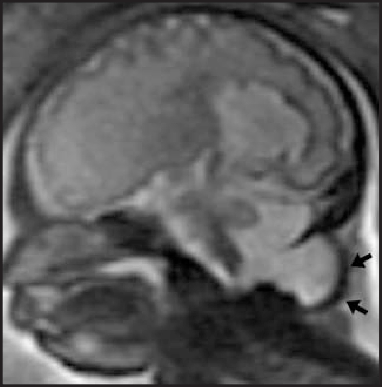
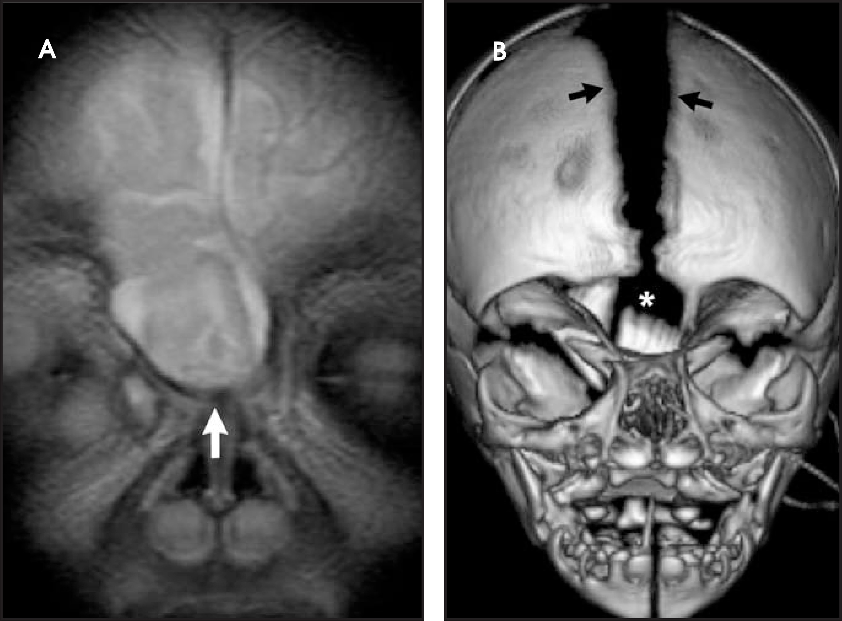
Cephalocele is an extension of intracranial contents (e.g. meninges, CSF and/or brain) through a dural and calvarial defect. The type of malformation is named for its anatomic location and the contents included in the herniated tissue. The defect is usually midline and is typically occipital in those of European descent (Figure 4) and frontoethmoidal in those with Asian heritage (Figure 5). Chiari III malformation is an occipital, C1-to-C2 encephalocele that may contain cerebellar tissue and CSF. 12
Chiari malformations
The Chiari malformations I through IV are not a continuum. The number designations I, II, III or IV do not imply a progression of severity of a single brain malformation. They are numerous malformations that can occur during neurulation of the hindbrain and commonly are associated with hydrocephalus. Some of the Chiari malformations are controversial, such as Chiari IV: hypoplasia of the cerebellum alone or in association with Chiari II.13 Chiari zero is also a controversial designation: indicating normal position of the cerebellar tonsils on imaging studies, but clinical presentation of headache, which is reminiscent of the experiences of patients with Chiari I (personal communication, David M. Frim, MD, Chairman of Neurosurgery, The University of Chicago, Chicago, IL).
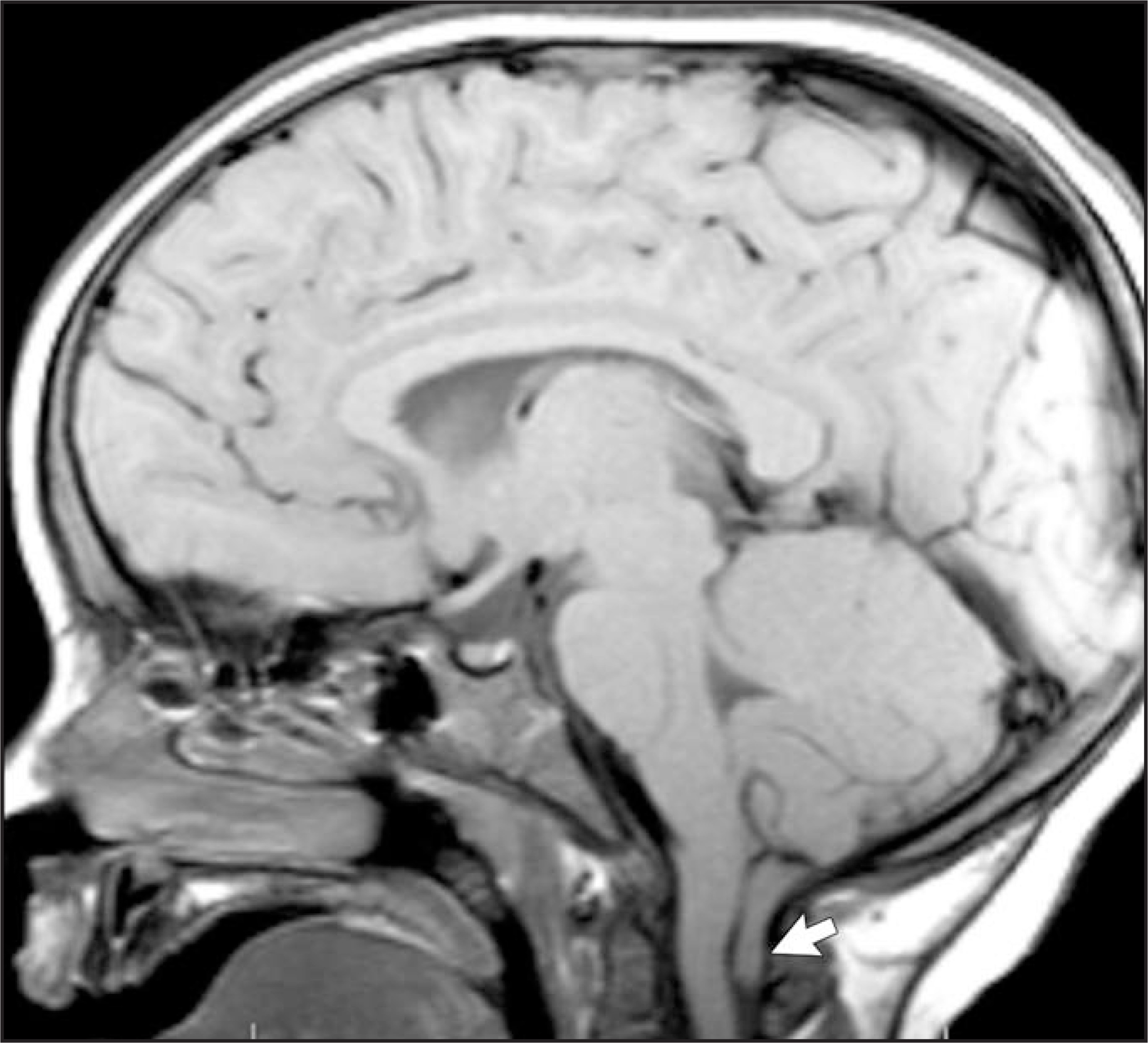
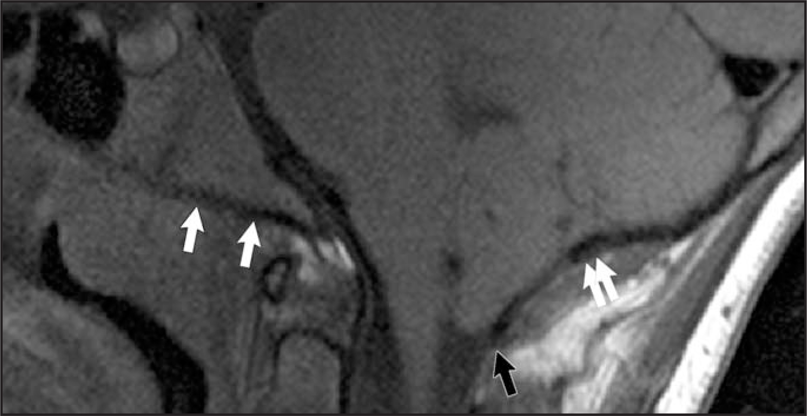
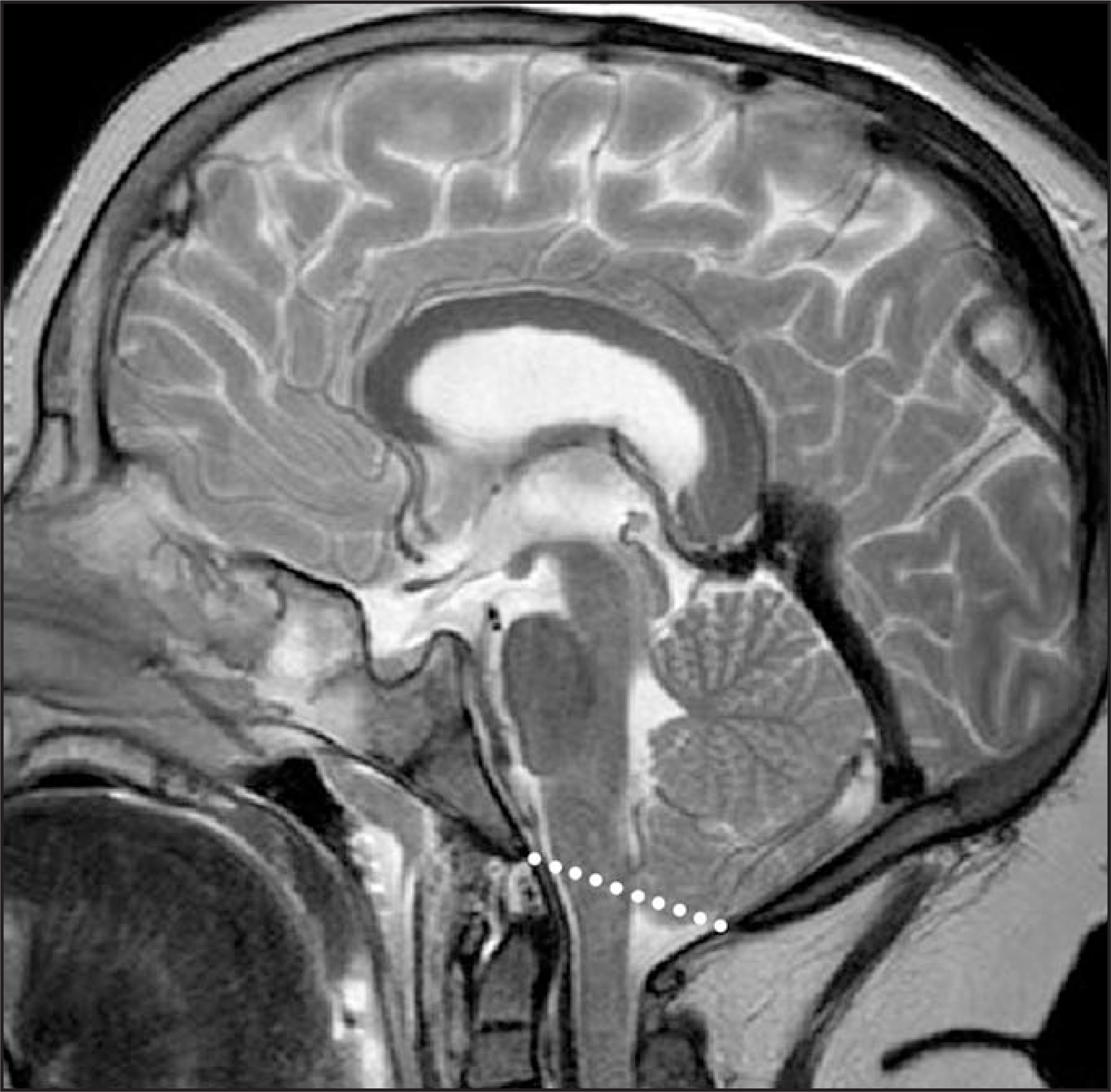
Chiari I malformation is characterized by low-lying cerebellar tonsils (Figure 6). The posterior fossa may be small because of shortening of the clivus. The foramen magnum is defined on sagittal magnetic resonance (MR) images by the ventral and dorsal margins of the occipital bone, i.e. the clivus (basion) and occiput (opisthion). Horizontal orientation of the clivus and cupping of the occiput is seen in many patients, contributing to smallness of the posterior fossa (Figure 7). Normal cerebellar tonsils are oval in shape and should lie <5 mm below a line drawn between the ventral and dorsal borders of the foramen magnum (Figure 8).14
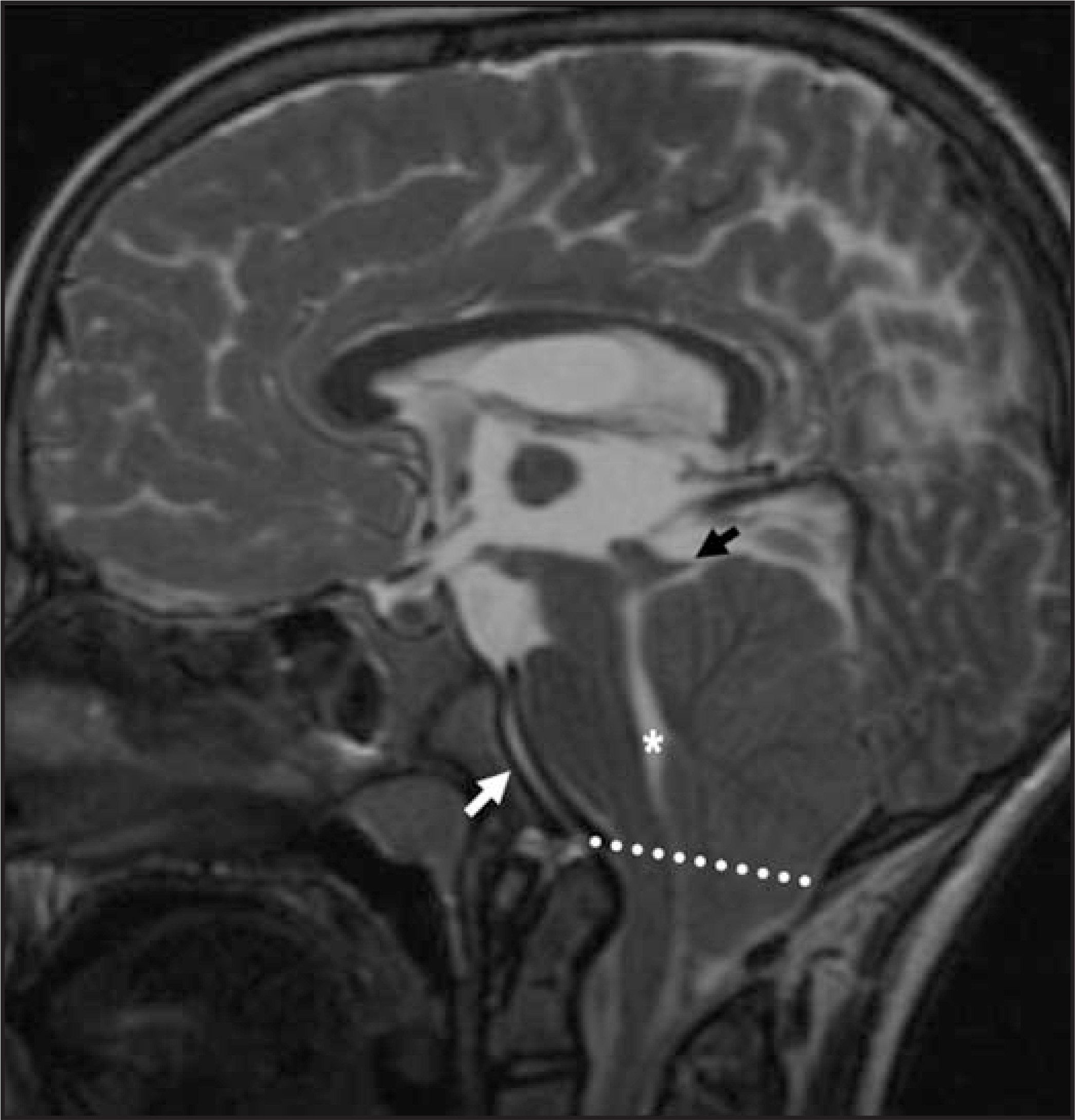
Chiari II malformation and meningomyelocele are nearly always associated. The posterior fossa is small and the tentorium is low lying, resulting in crowding of the cerebellum and brainstem into the cervical medullary junction and upper cervical spinal canal. This crowding results in kinking of the medullary cervical junction and elongation of the fourth ventricle (Figure 9). Although many patients are developmentally normal, agenesis of the corpus callosum and cortical migration anomalies may accompany Chiari II malformation.15
Neurulation (5 to 10 weeks)
Ventral induction takes place during weeks 5 to 10 of neurulation. The brain segments, neuronal proliferation occurs and the face is formed. The primary and secondary vesicles (prosencephalon, mesencephalon and rhombencephalon) form the cerebrum, midbrain, cerebellum and lower brainstem. The cerebrum and cerebellum each form 2 distinct hemispheres.
Failure of these neural proliferation processes results in midline supratentorial anomalies such as holoprosencephaly, agenesis of the corpus callosum, pituitary maldevelopment and posterior fossa malformations such as Dandy-Walker malformation, cerebellar hypoplasia and rhombencephalosynapsis. Hydrocephalus due to aqueductal stenosis can also occur during this time.11
Hydropcephalus
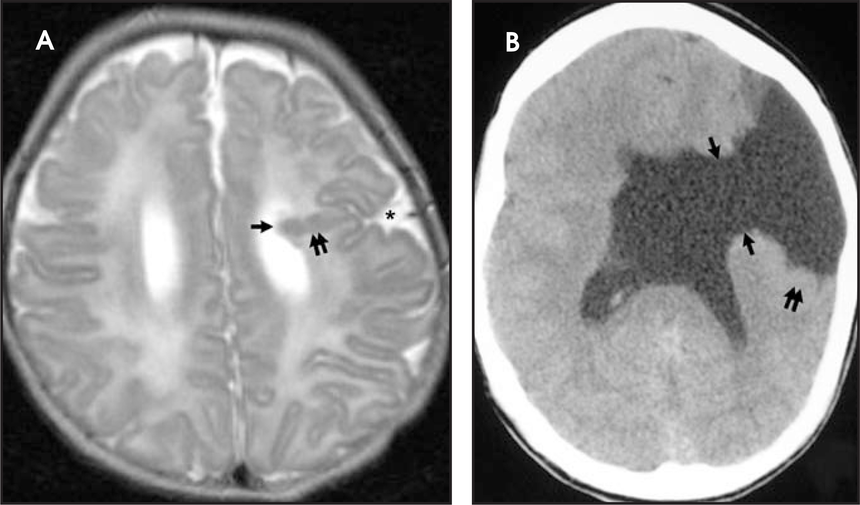
Hydrocephalus is an enlargement of the ventricular system in the brain and implies there is elevated intracranial pressure. Almost all of the malformations, diseases and syndromes mentioned in this article can be associated with hydrocephalus.
The cavities that become the ventricles, and the foramina and aqueducts connecting them, form during weeks 4 to 12 of gestation. Obstruction at any point of the ventricular system due to failure in the formation of these cavities can occur at any time. Other sources of ventricular obstruction are canalization of the foramina and aqueducts, overproduction of CSF by the choroid plexus, or diminished reabsorption through the arachnoid villi. Obstruction can occur prenatally, from the time of formation, to birth, or postnatally and result in hydrocephalus.
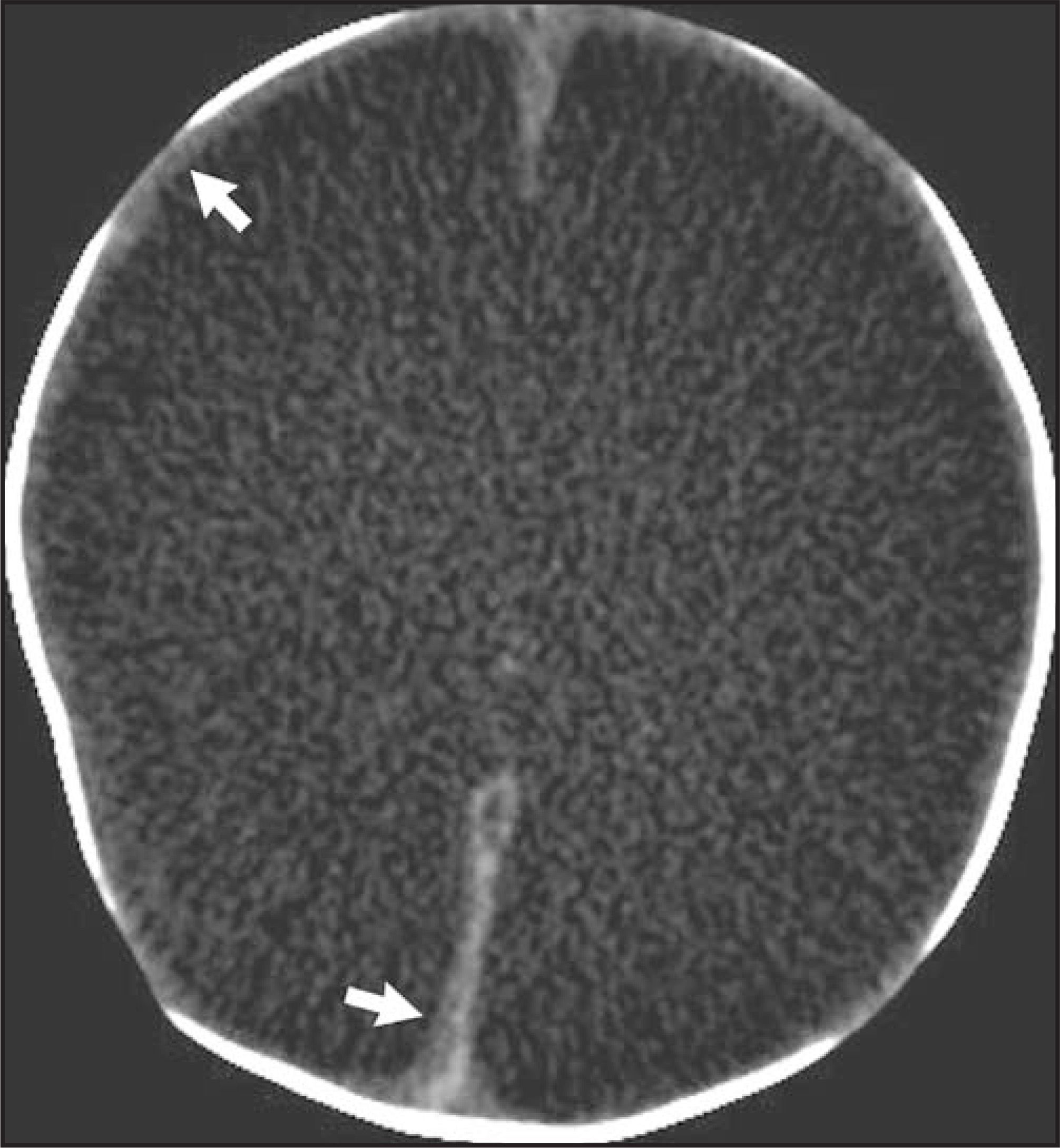
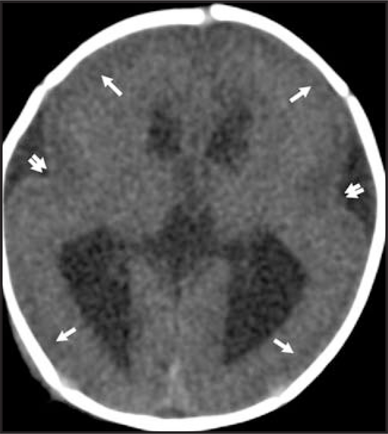
When severe, hydrocephalus may be difficult to differentiate from hydranencephaly, which is an extreme form of cerebral encephalomalacia, probably the result of occlusion of both internal carotid arteries and infarction of all cerebral tissue (Figure 10).9,16
Aqueductal stenosis
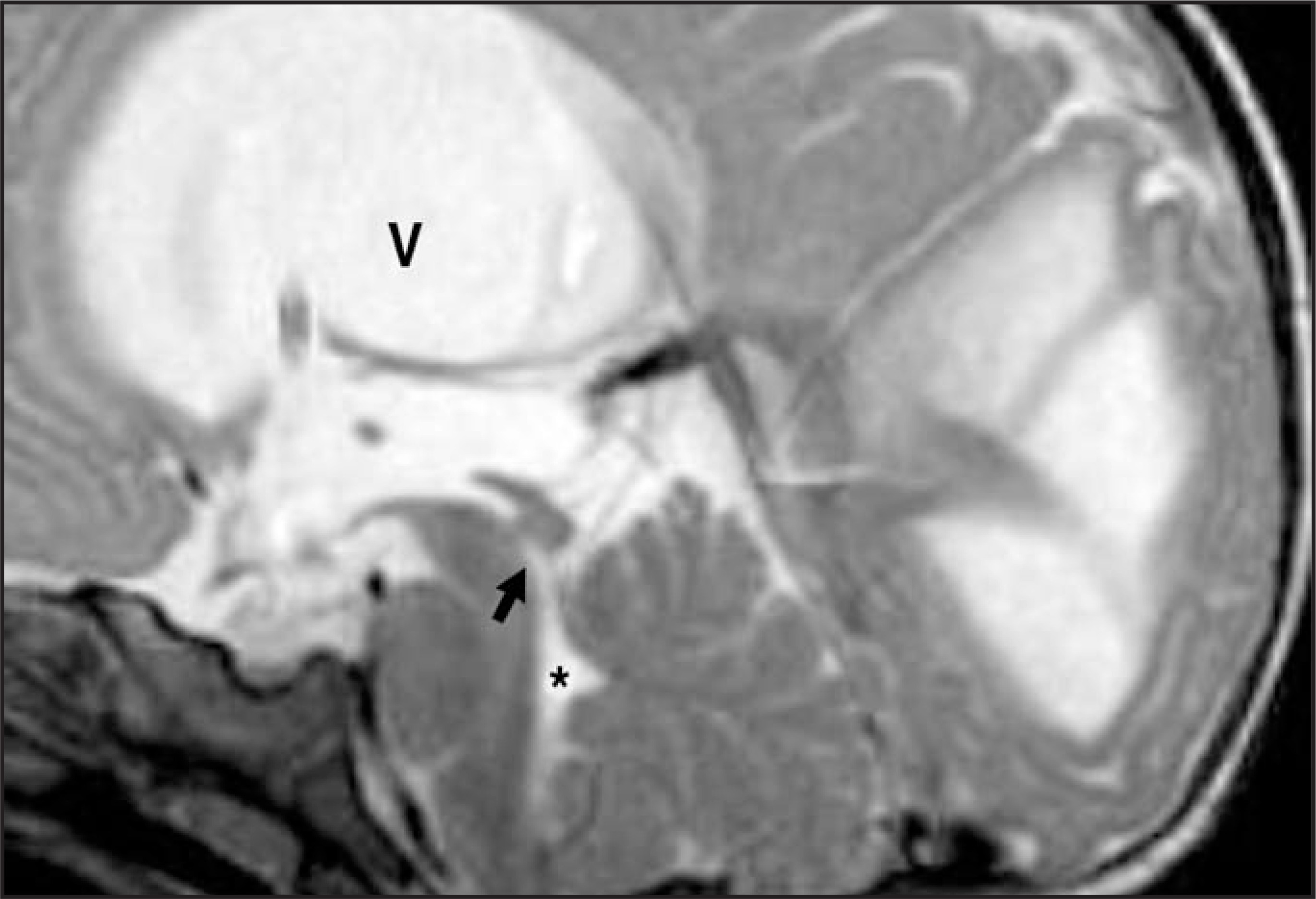
Congenital stenosis of the aqueduct of Sylvius can be due to intrinsic or extrinsic narrowing or malformation of the aqueduct. Abnormal histiogenesis and proliferation of periaqueductal grey matter in the midbrain can result in primary stenosis or formation of numerous minute channels through the aqueduct. Mass effect on the quadrigeminal plate from supratentorial hydrocephalus, or a mass, can cause secondary narrowing of the aqueduct. Pre- and postnatal infection, inflammatory disease or intraventricular hemorrhage can lead to acquired aqueductal stenosis due to fibrosis or gliosis, leading to stenosis. X-linked forms of aqueductal stenosis are also described. Stenosis results in lateral and third ventricle hydrocephalus; the fourth ventricle remains normal in volume (Figure 11).17
Holoprosencephaly
Holoprosencephaly occurs due to failure of proliferation of cerebral tissue to form 2 separate cerebral hemispheres. Normally, the right and left cerebral hemispheres form independently in a unified process of neuronal proliferation. Although prosencephalon formation abnormalities are programmed for failure earlier in gestation, even before the neural tube closes, it is during the proliferative phase of ventral induction that holoprosencephaly manifests. When the hemispheres fail to develop into 2 separate hemispheres but rather form a single, midline mass of cerebral tissue, the result is holoprosencephaly.
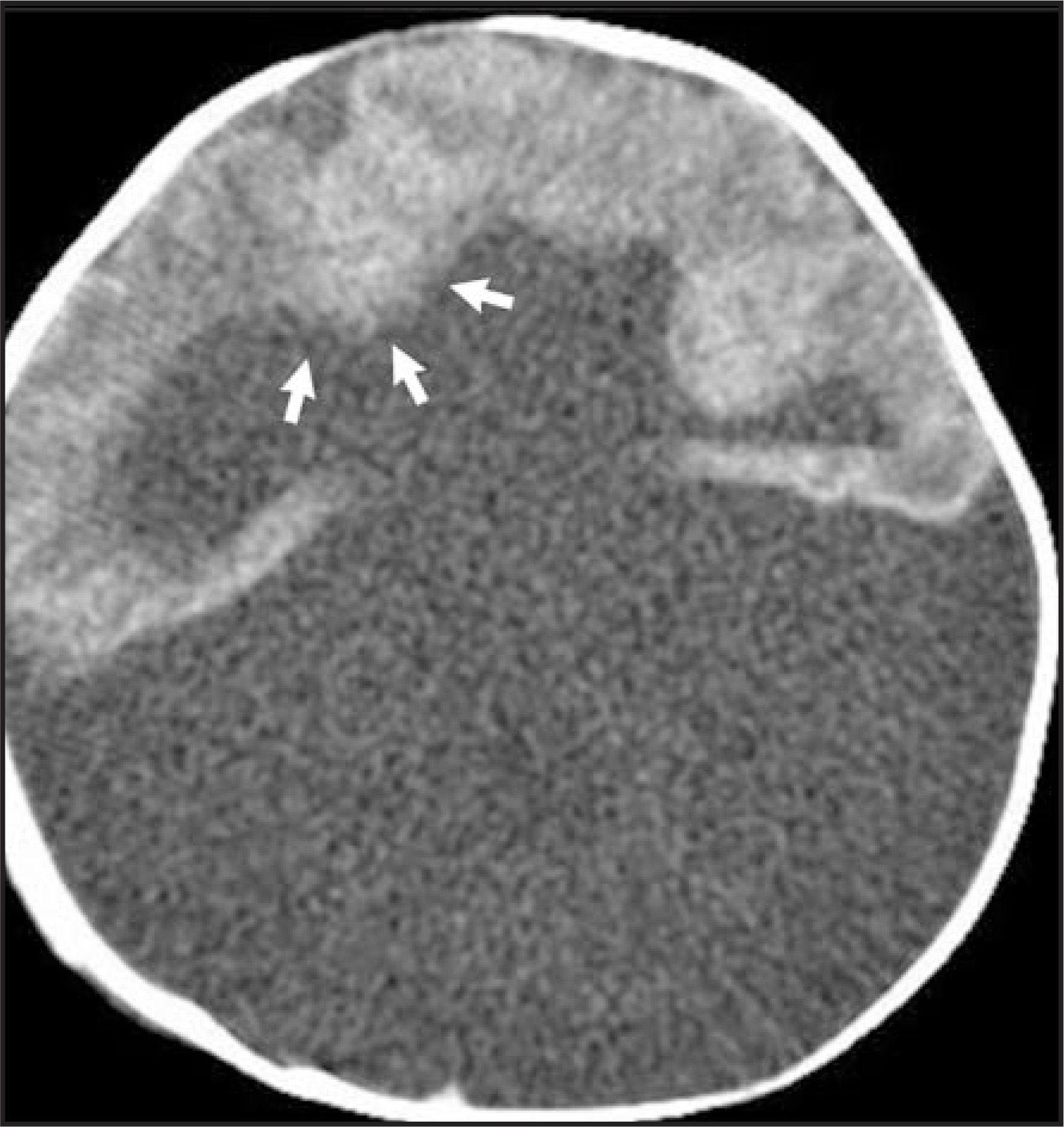
The most severe form of holoprosencephaly is termed alobar, because the cerebral tissue bears no resemblance to normally defined cerebral lobes (Figure 12). The lateral ventricles are also abnormal, forming a midline monoventricle. Septo-optic dysplasia is the mildest form of the holoprosencephaly spectrum: the septum pellucidum and the optic nerves are atrophic (Figure 13). Pituitary gland malfunction is part of the syndrome of septo-optic dysplasia (Figure 14). Semilobar and lobar forms of holoprosencephaly describe the degree to which the frontal, temporal, parietal and occipital lobes are defined. The degree of cerebral malformation is less severe than in the alobar form. Other midline structures, the falx and septum pellucidum are dysplastic. Schizencephaly is associated in 50% of cases.9,17,18
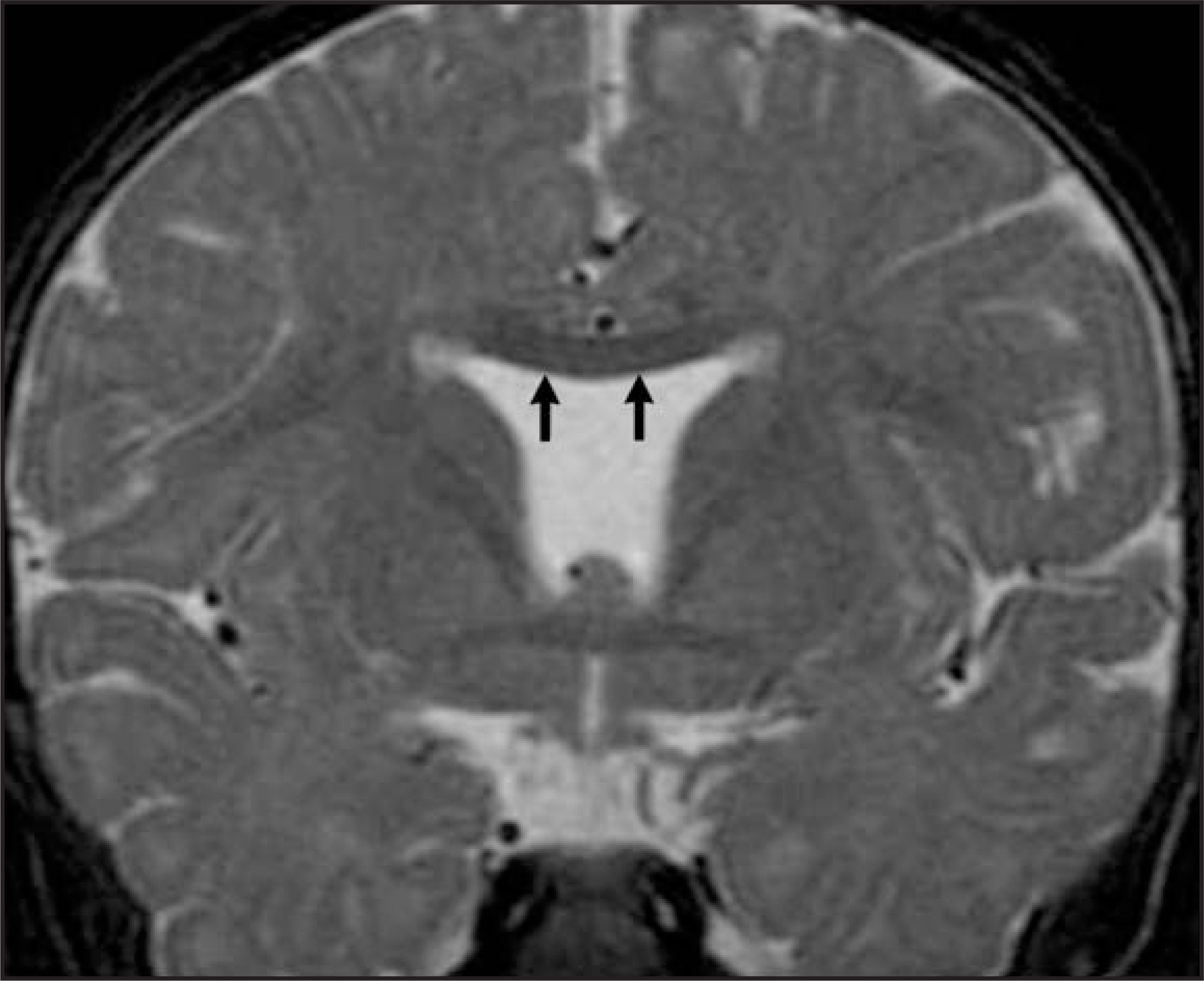
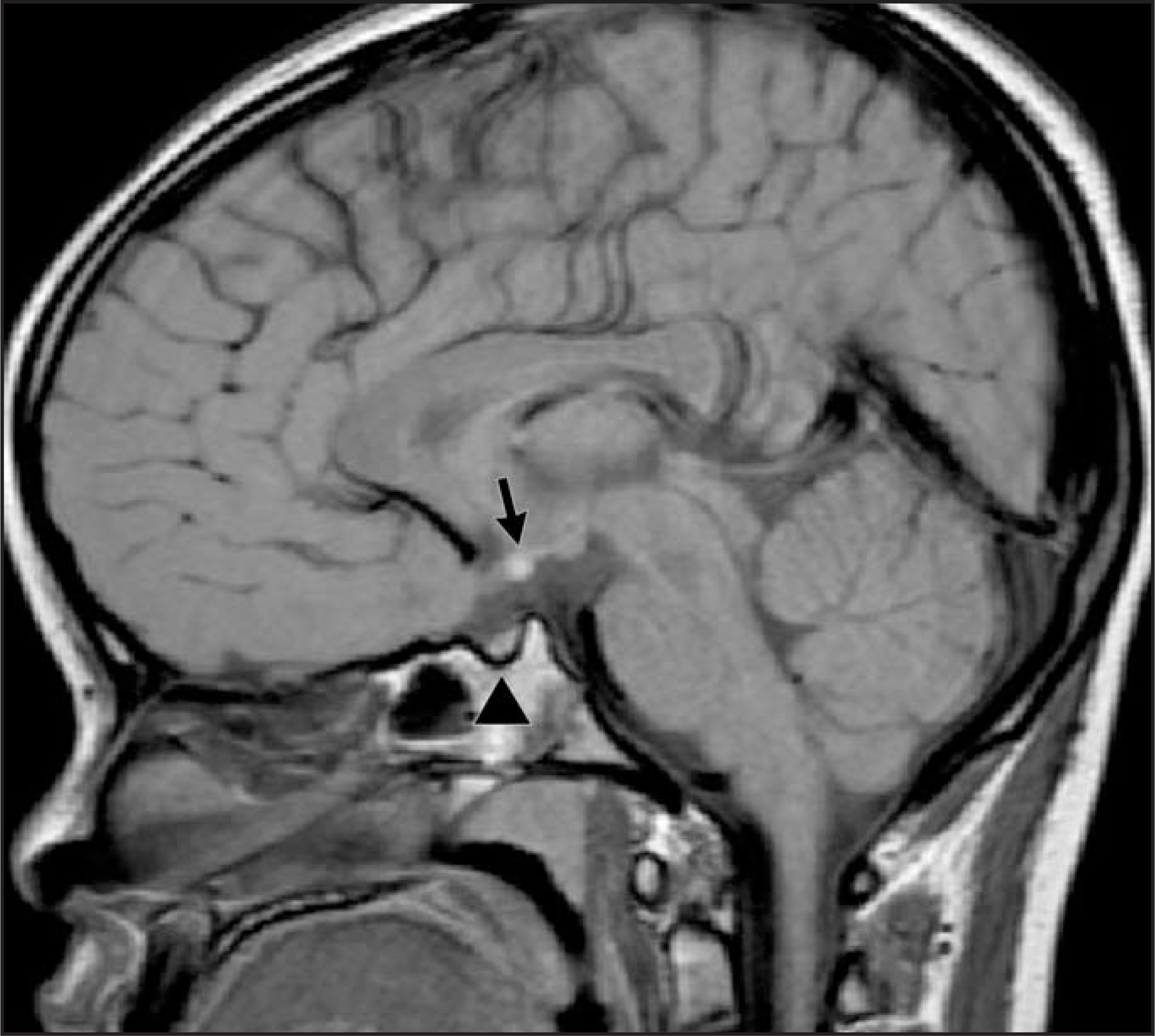
Because there is a temporal relationship between facial formation and neuronal proliferation, facial malformation is usually seen in patients with holoprosencephaly. Facial malformations are due to abnormal development of the premaxillary segments of the face and result in arrhinia and midline facial clefts.19
Agenesis of the corpus callosum
Agenesis of the corpus callosum is one of the most common malformations of the brain.20 The corpus callosum begins to form in the seventh week of gestation and is complete by 18 to 20 weeks. There has been controversy regarding the definitive order in which segments of the corpus callosum are formed, but its absence is known to be associated with a range of findings including normal development, Dandy-Walker complex, Chiari II malformation, numerous syndromes, and the absence may be accompanied by seizure disorders and mental retardation.6, 7
Radiographic findings of dysgenesis or agenesis of the corpus callosum include absence of, or a malformed corpus callosum (Figure 15), and parallel orientation of the lateral ventricles; normally the frontal horns lie closer together than the occipital horns of the lateral ventricles. The occipital horns and atria of the lateral ventricles may be dilated, a finding termed colpocephaly (Figure 15). When the corpus callosum is completely or partially absent the cingulate gyrus does not form normally, allowing interhemispheric gyri to radiate toward the roof of the lateral ventricles. The neurons that normally cross the midline to form the corpus callosum course along the interhemispheric fissure, in groups of white matter called Probst bundles (Figure 15). These bundles lie along the superior medial surface of the lateral ventricles, indenting the ventricle which causes a bull’s horn configuration.20
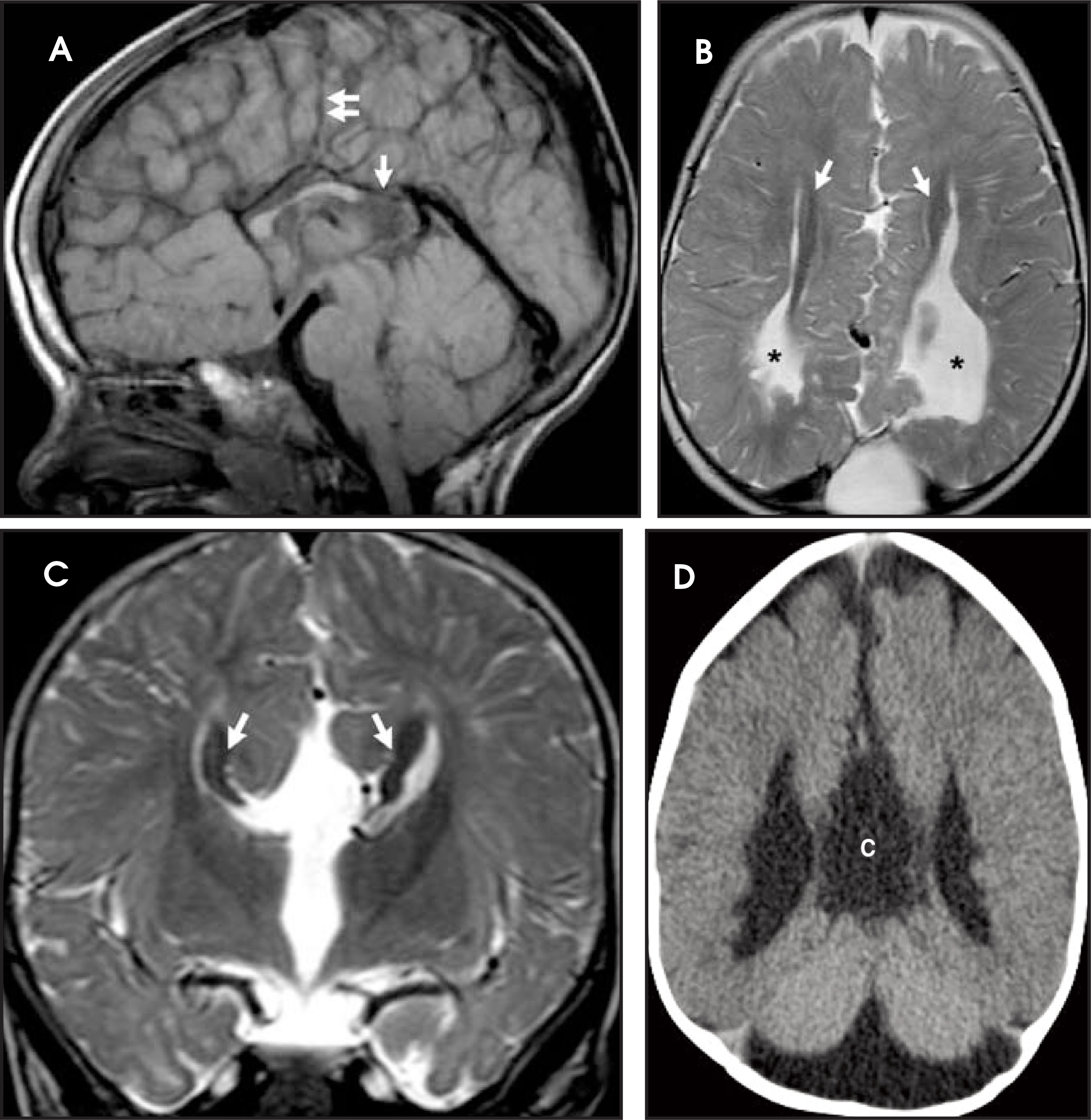
The roof of the third ventricle can be displaced upward because the corpus callosum is not limiting its superior expansion; an interhemispheric cyst or lipoma may be associated (Figure 15).20
The Dandy-Walker complex
The Dandy-Walker complex is the result of malformation of the metencephalon portion of the rhombencephalon leading to atresia of the cerebellar outlet foramina. As a result, the roof of the fourth ventricle does not develop normally and there is hypogenesis or agenesis of the cerebellar vermis. The fourth ventricle therefore communicates freely with extra-axial fluid in the posterior fossa (Figure 16). The tentorium and position of the torcular Herophili are elevated; i.e. the posterior fossa is enlarged (Figure 16). The Dandy-Walker complex encompasses a range of hypoplasia or dysplasia of the cerebellar hemispheres and/or vermis which can be found in patients with numerous diagnoses of chromosomal anomalies and syndromes.11,21
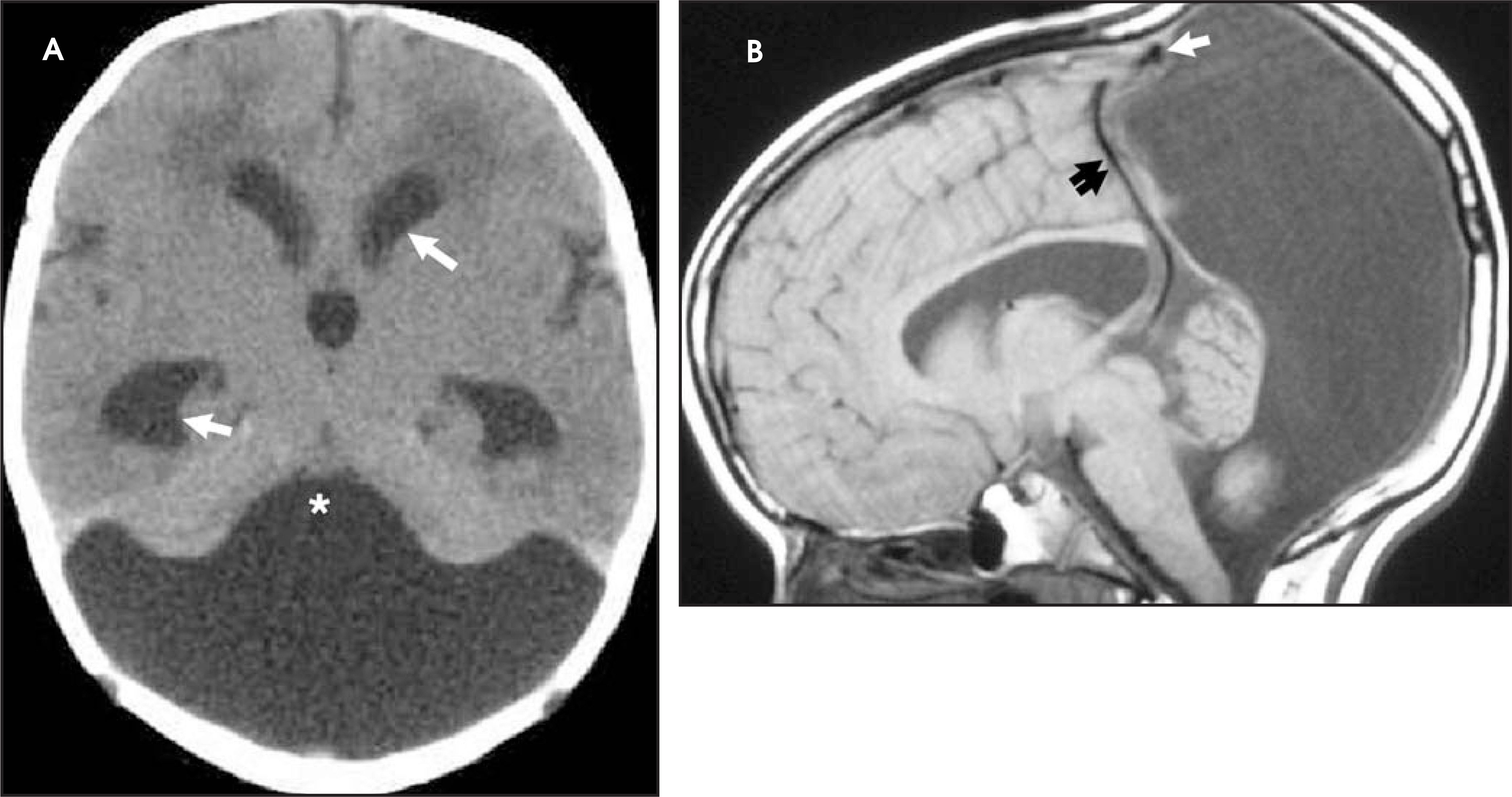
Cerebellar hypoplasia
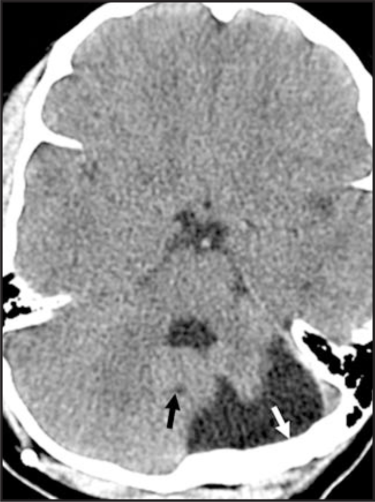
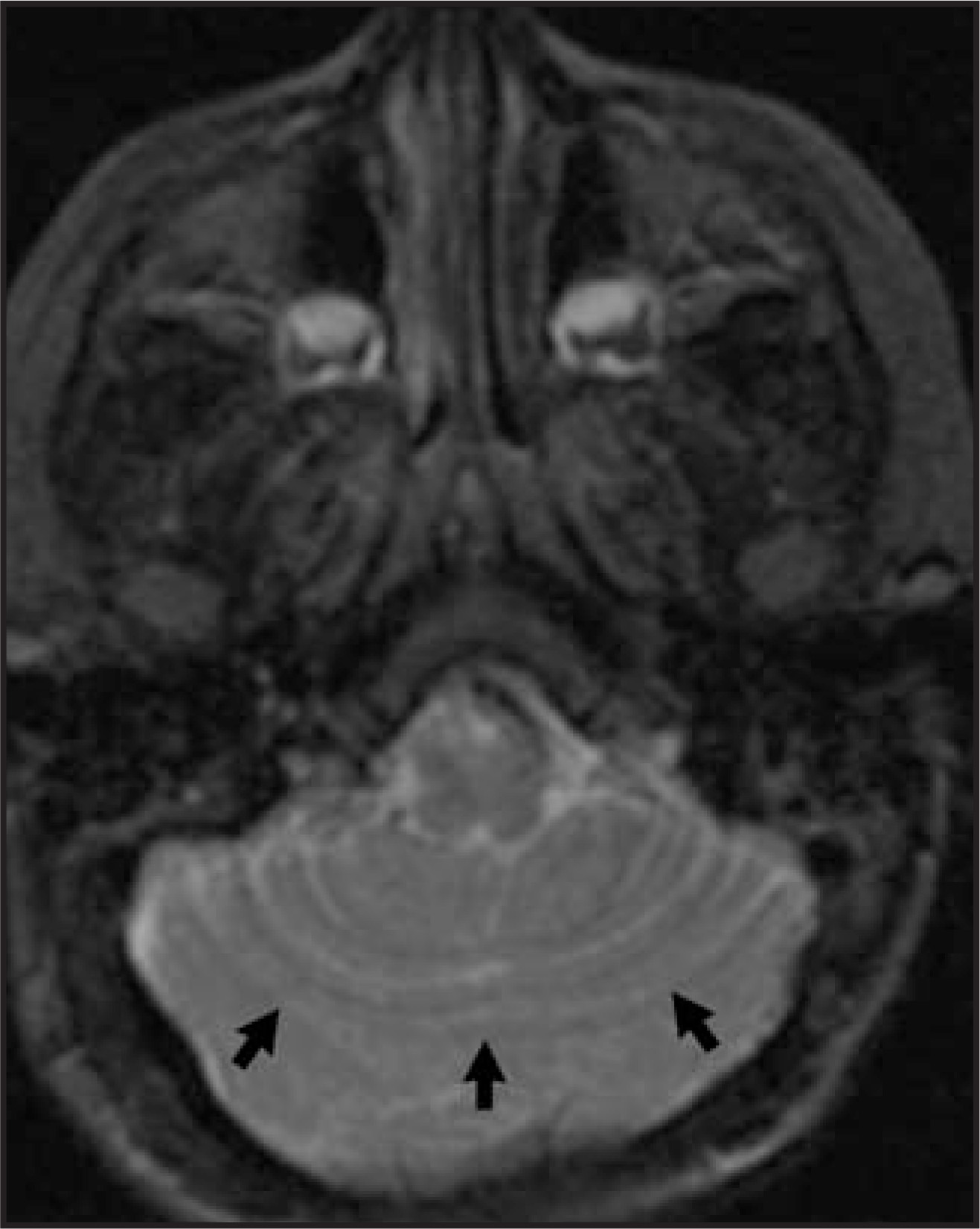
Cerebellar hypoplasia may be diffuse or can be limited to a single hemisphere and may involve the vermis. Differentiating hypoplasia from other cerebellar malformations, dysplasia and atrophy requires determining that the posterior fossa is normal volume and determining the absence of an associated cyst in communication with the fourth ventrice (Figure 17).21
Rhombencephalosynapsis
Although rhombencephalosynapsis is usually described as a “fusion” anomaly, or dysplasia of the cerebellar hemispheres and vermis, and has been described in a patient with holoprosencephaly,9,21 I believe this malformation is probably the result of failed neuronal proliferation of the cerebellar hemispheres, much like holoprosencephaly of the cerebral hemispheres.
Imaging studies reveal an absence of the normal formation of midline structures of the posterior fossa including the vermis and mesencephalic structures. The cerebellar hemispheres are continuous across the midline (Figure 18).9
Neuronal proliferation, migration and histiogenesis (8 to 21 weeks)
During this phase of development neuronal cells undergo proliferation, differentiation and histiogenesis. Neuronal stem cells migrate from the germinal matrix to the cerebral cortex with the goal of producing organized cortical layering. Failure of this process results in microcephaly, megalencephaly, heterotopia, focal cortical dysplasia, polymicrogyria, lissencephaly, hemimegalencephaly, schizencephaly, anomalies of operculization, and phakomatoses. Phakomatoses and other inheritable neurologic diseases will be discussed in part 2 of this article. Regulators of cortical malformation have been identified and associated with specific malformations of the cerebral cortex through molecular genetic studies.22 Vascular malformations are thought to be formed during this time; indeed, many malformations of the cerebral cortices are accompanied by abnormal vasculature.11 Vascular anomalies will not be discussed further in this article.
Microcephaly and megalencephaly
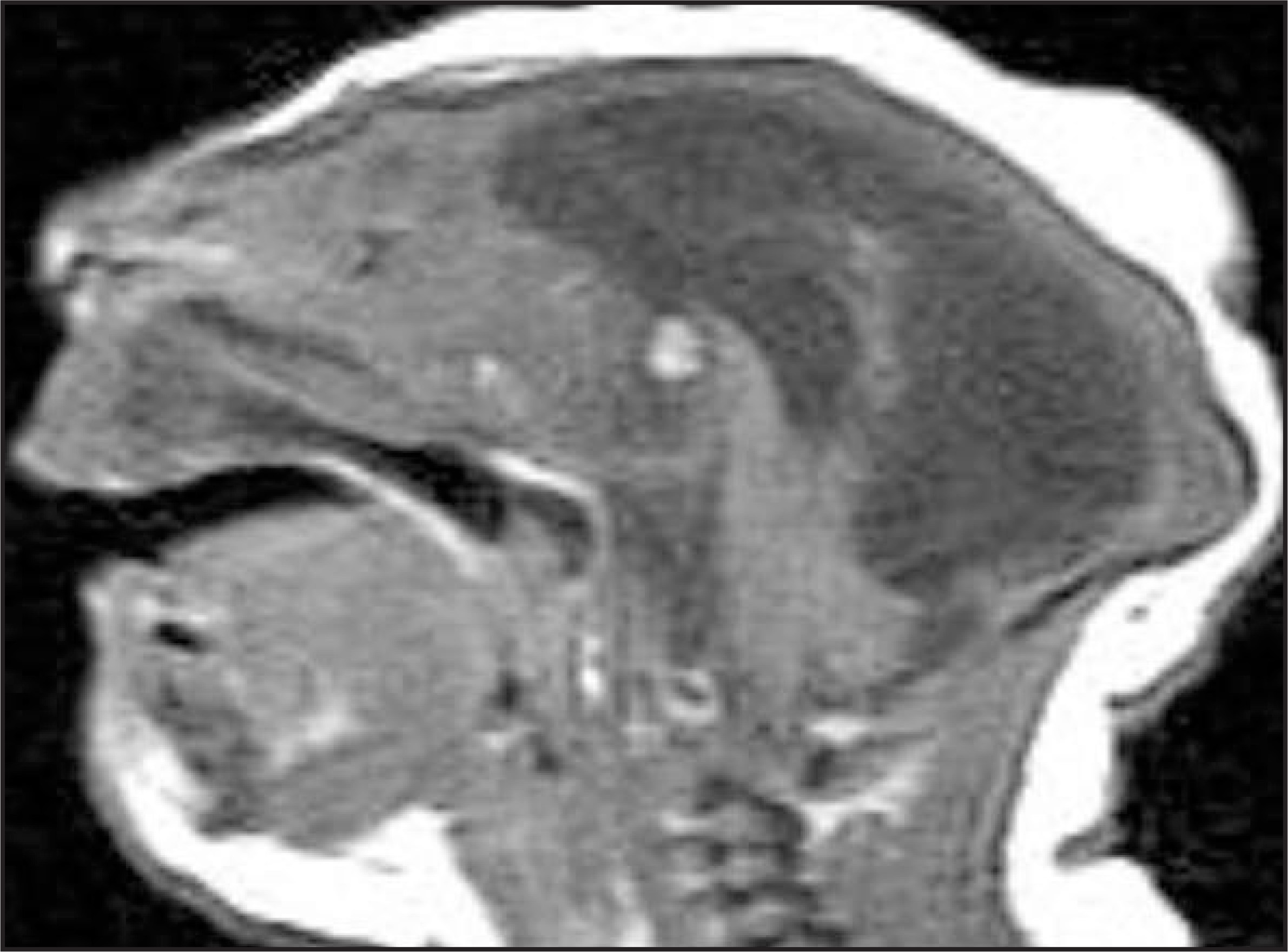
Microcephaly and megalencephaly are due to disorders of neuronal and glial proliferation or excess or reduced apoptosis. Microcephaly is a malformation secondary to abnormal stem-cell proliferation or apoptosis after normal proliferation of stem cells. By definition, the head circumference in these children is ≥3 standard deviations below the norm. There are fewer gyri, the depth of the sulci is shallow and the volume of white matter is diminished (Figure 19).
Megalencephaly is the result of a generalized increase in neuronal and glial proliferation or diminished apoptosis.9
Focal cortical dysplasia
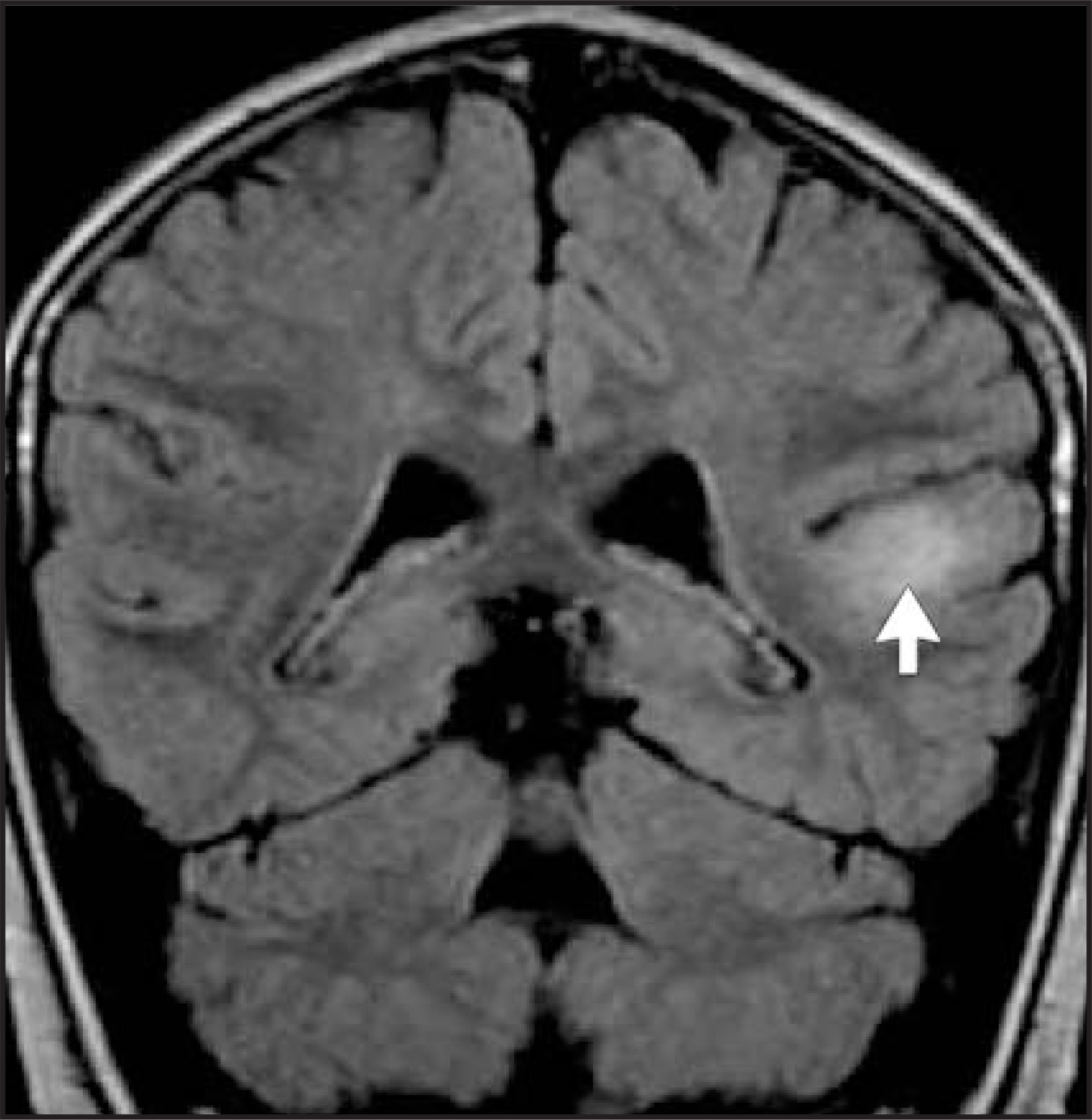
Focal cortical dysplasia is the result of abnormal migration of neurons to the cerebral cortical cell layers. Histologically, the cortical cells are also abnormal. Some forms of cortical dysplasia contain balloon cells, and may show abnormal signal and architecture extending from the germinal matrix through the deep and subcortical white matter. Imaging findings are variable, depending on the involvement of white matter and may show focal blurring of the grey-white junction, or thinning or thickening of the affected cerebral cortex, which usually has high T2 signal on MR (Figure 20).9
Heterotopia
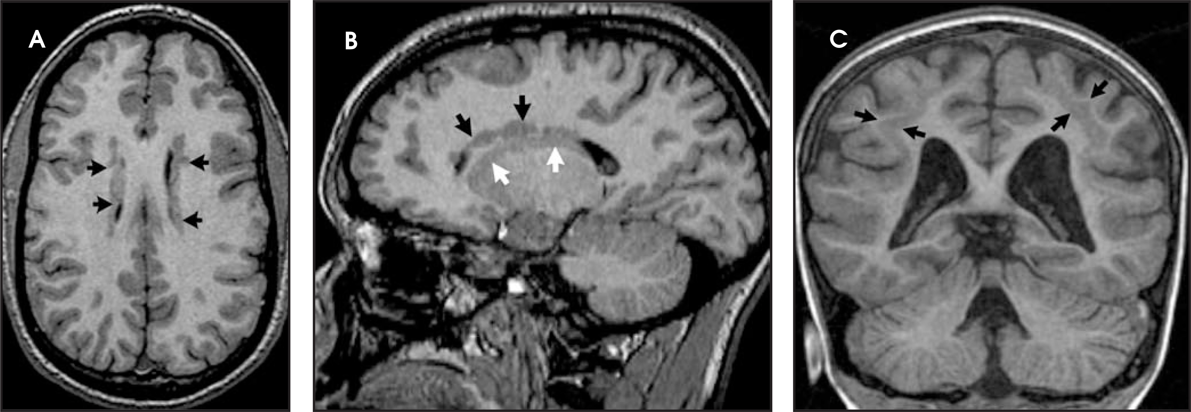
Heterotopia (singular: heterotopion) are abnormal anatomic locations of cortical grey matter which are due to premature arrest of neuronal migration. Typical locations of heterotopia are subependymal, where they are usually asymmetric, at the trigones of the lateral ventricles and subcortical, where they may be focal (Figure 21) or generalized, forming a band or double cortex underlying the normal-appearing cerebral cortices.9
Polymicrogyria
In the later stages of neuronal migration, the 6 layers of the cerebral cortex are organized. When the deep layers of the cerebral cortex form numerous small gyri instead of organized cortical layers, the imaging result appears to be thickening or thinning of the cerebral cortex, which is usually associated with abnormal sulcal formation. MR may also show a nodular appearance of the cortex and normal-to-increased signal in the cortical tissue. There are numerous syndromes and patterns of polymicrogyria, and many have been shown to correspond with chromosomal abnormalities (Figure 22).9,22
Lissencephaly
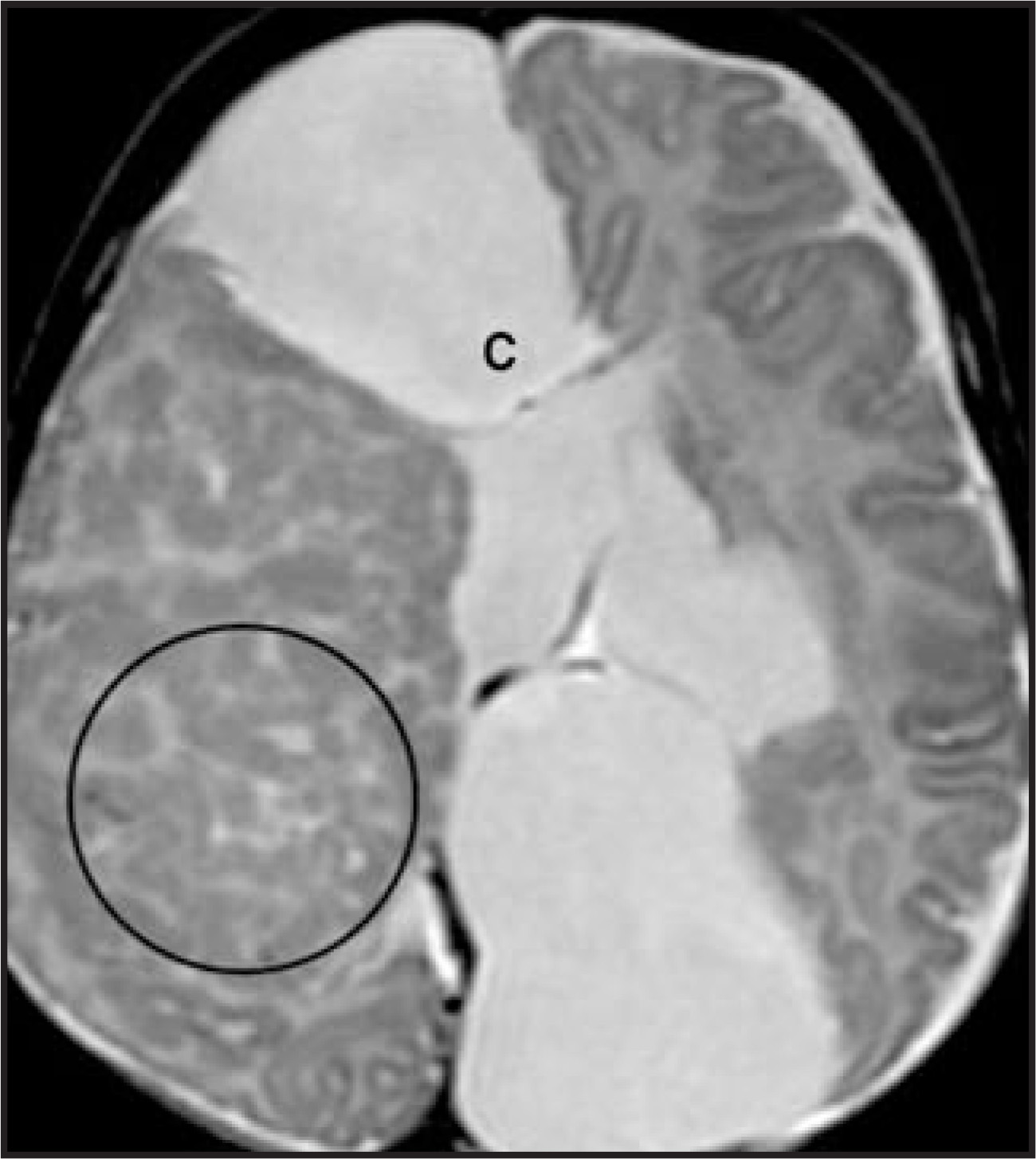
Lissencephaly (smooth brain) describes the malformation with lack of gyral and sulcal development such that the surface of the cerebral hemispheres is smooth, due to arrested neuronal migration. Agyria (complete lissencephaly) or pachygyria (incomplete lissencephaly) as well as a thickened cerebral cortex are seen on imaging studies, differentiating lissencephaly from malformations of neuronal proliferation (Figure 23).9,22
Hemimegalencephaly
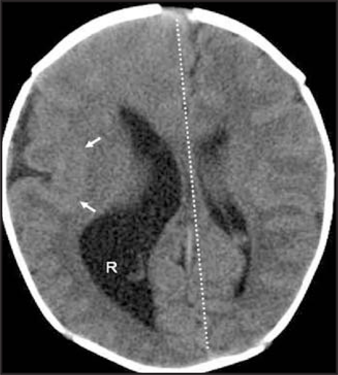
Hemimegalencephaly is unilateral megalencephaly that is isolated, part of a hemihypertrophy syndrome or the result of hamartomatous overgrowth of one cerebral hemisphere. The malformation occurs because of defective neuronal proliferation, migration and cortical organization. The unilateral enlargement of the cerebral hemisphere includes proportionate ventriculomegaly and unilateral enlargement of CN I and CN II which of course are really glial tracts rather than true cranial nerves (Figure 24).9,22
Schizencephaly
Schizencephaly may be the result of abnormal cellular proliferation, migration and/or cortical organization. The malformation could be the result of a focal injury at the germinal matrix as neurons begin to migrate — a transmantle injury later in gestation may be familial or caused by chromosomal mutation. The germinal matrix, located at the caudal thalamic groove is at the margin of the lateral ventricles. A cleft is formed in the cerebral mantle when neurons fail to migrate from a focal area of the germinal matrix.
Characteristic imaging findings may be unilateral or bilateral; when bilateral, the clefts are typically symmetric. The cleft, lined by dysplastic grey matter, extends from the margin of the lateral ventricle to the cerebral cortex and is in communication with the ventricle and the subarachnoid space overlying the cerebral hemisphere. The margins of the cleft may be splayed (open lip) or lie in close apposition (closed lip, Figure 25).9,22
Anomalies of operculization
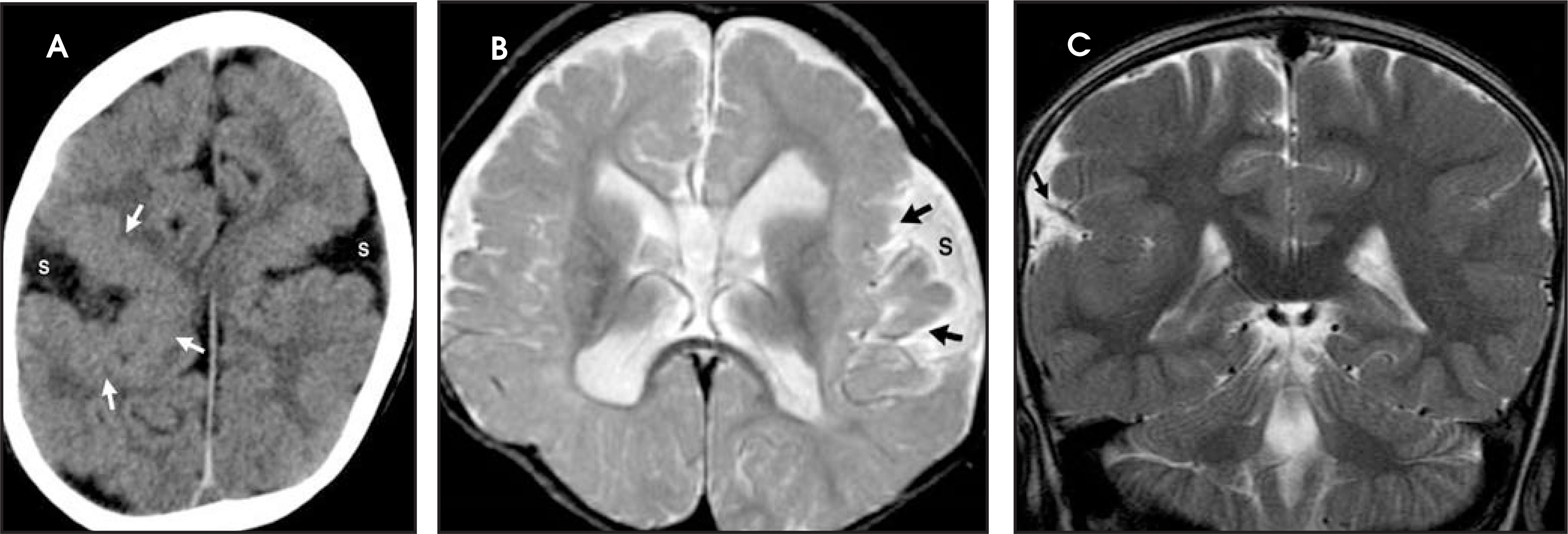
Formation of the Sylvian fissure and insula begins during the 14th week of gestation, between the orbitofrontal and temporal lobes. The insula is defined by infolding of the structures by the 19th week of gestation.12 The process of formation of the Sylvian fissures is called operculization. Disorders of neuronal proliferation and neuronal migration which are limited to the operculum result in abnormal gyration and/or cortical dysplasia which is manifested as abnormalities in the processes managed by these areas, namely speech, language and pseudobulbar palsy.22,23
The imaging appearance can vary from wide Sylvian fissures, thickening of the cortex, localized polymicrogyria of the insula, and thickened or shallow gyri and they may be accompanied by anomalous vessels. When symmetric malformation is found, the brain has a “figure 8” shape on axial images (Figure 26).9,23
Conclusion
Diagnoses in pediatric neuroradiology encompass a broad range of brain malformations, anomalies, and inherited and metabolic disease processes. An understanding of basic brain embryology provides the basis for a more thorough understanding of these pathologic processes.
References
- Anderson D. Dorland’s Pocket Medical Dictionary. 1989.
- Rice D, Barone S. Critical periods of vulnerability for the developing nervous system: Evidence from humans and animal models. Environmental Health Perspectives Supplements. 2000;108(S3).
- Moore K, Wonsiewicz M. The Developing Human; Clinically Oriented Embryology. 1988:379-394.
- Schmalz S. Early embryological development. 1999.
- Neas J, Neas J, Shiao T. The embryology collection. 2009.
- Kier E, Truwit C. The normal and abnormal genu of the corpus callosum: An evolutionary, embryologic, anatomic, and MR analysis. AJNR Am J Neuroradiol. 1996;17(9):1631-1641.
- Kier E, Truwit C. The lamina rostralis: Modification of concepts concerning the anatomy, embryology, and MR appearance of the rostrum of the corpus callosum. AJNR Am J Neuroradiol. 1997;18(4):715-722.
- Toi A, Lister W, Fong K. How early are fetal cerebral sulci visible at prenatal ultrasound and what is the normal pattern of early sulcal development?. Ultrasound in Obstetrics and Gynecology. 2004;24:706-715.
- Barkovich A, Barkovich A. Pediatric Neuroimaging. 2005:291-439.
- Rakic P, Yakovlev P. Development of the corpus callosum and the cavum septi in man. J Comp Neurol. 1968;132:45-72.
- Wong W. “Text Syllabus.”. 2009.
- Kollias S, Ball W, Ball W. Pediatric Neuroradiology. 1997:91-174.
- Osborn A, Blaser S, Salzman K. Diagnostic Imaging: Brain. 2004:4-119.
- Yasari R, Frim D. Evaluation and management of the Chiari malformation type 1 for the primary care physician. Pediatr Clin N Am. 2004;51:477-490.
- Stevenson K. Chiari type II malformation: past, present, and future. Neurosurg Focus. 2004;16:1-7.
- Barkovich A, Barkovich A. Pediatric Neuroimaging. 2005:190-290.
- Barkovich A, Barkovich A. Pediatric Neuroimaging. 2005:659-703.
- Bianachi D, Crombleholme T, D’Alton M, Medina M. Fetology: diagnosis and management of the fetal patient. 2000:117-125.
- Patterson M. Holoprosencephaly: The face predicts the brain; the image predicts its function. Neurology. 2002;59:1833-1834.
- Barkovich A, Simon E, Walsh C. Callosal agenesis with cyst: A better understanding and new classification. Neurology. 2001;56:220-227.
- Patel S, Barkovich A. Analysis and classification of cerebellar malformation. AJNR Am J Neuroradiol. 2002;23:1074-1087.
- Mochida G, Walsh C. Genetic basis of developmental malformations of the cerebral cortex. Arch Neurol. 2004;61:637-640.
- Chi J, Dooling E, Gilles F. Gyral development of the human brain. Ann Neurol. 1977;1:86-93.
Citation
. Pediatric neuroradiology, part 1: Embryologic basis for brain malformation. Applied Radiology. 2009;38(7):29-40. doi:10.37549/AR1703.