Pediatric skeletal trauma—Plain film to MRI
Applied Radiology — Vol. 36 , Issue 8 , pp. 24 -33
DOI: 10.37549/AR1533
Published: August 1, 2007
Categories
Many unique features of the growing skeleton pose specific challenges in imaging skeletal trauma. Differences in the composition and development of the pediatric skeleton (as compared with adults) result in characteristic injuries and fractures. The imaging of these injuries typically begins with plain films, and most cases require no further radiologic evaluation. However, several other imaging modalities may be used in certain cases, depending on the clinical history, physical examination, and initial radiologic studies. Computed tomography (CT) and magnetic resonance imaging (MRI) are the most frequently used adjunctive imaging studies performed in pediatric patients with suspected skeletal trauma.
Unique features of the pediatric skeleton
The unique features of the pediatric skeleton result in injuries and fractures that are characteristic and tend to change with advancing age and skeletal maturity. The pediatric skeleton tends to be more porous and elastic in early childhood.1,2 Children have a thick periosteum that decreases the likelihood that fractures will involve the entire circumference of the bone.3 This allows for the development of incomplete fractures, or fractures that extend only partially through the circumference of the bone. The ligaments and tendons are stronger than the physis and, therefore, are more able to resist stress and torsional forces. As a result, children often suffer fractures of the growth plate in the setting of trauma.4 These features of pediatric skeleton injury, coupled with an increase in muscle strength and rapid growth that accompany puberty, can result in avulsion-type fractures.2,5
Imaging techniques
Plain films
In most cases, imaging of pediatric skeletal trauma begins with radiographs, and very few patients require imaging beyond plain films. This is particularly true in cases of common fractures in which the radiographic findings fit the clinical history and the physical examination findings and treatment are straightforward. Examples of these types of fractures are discussed below.
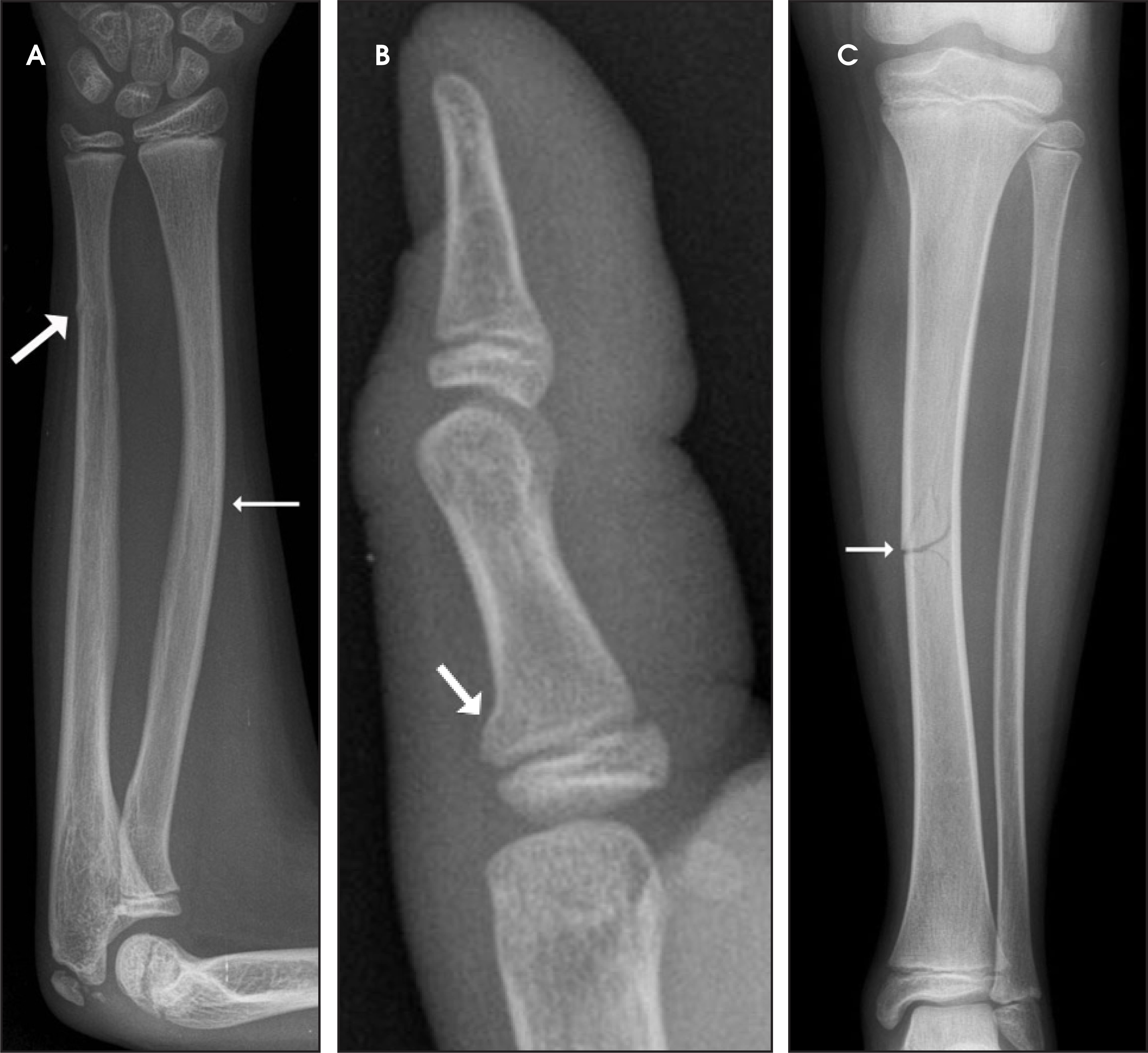
The relative porosity and elastic properties of the pediatric skeleton place it at risk for incomplete fractures. Examples of incomplete fractures include bowing, torus, and greenstick fractures, all of which are uncommon in adults (Figure 1).1,2 Bowing and torus fractures result from longitudinal compressive forces. Bowing fractures are also known as plastic deformities and most frequently involve the ulna. Torus fractures (also known as buckle fractures) usually present as focal angular deformity at the metaphyseal-diaphyseal junction of long bones. Greenstick fractures occur when the bone is bent, which results in a fracture of the convex side while the concave side remains intact.3
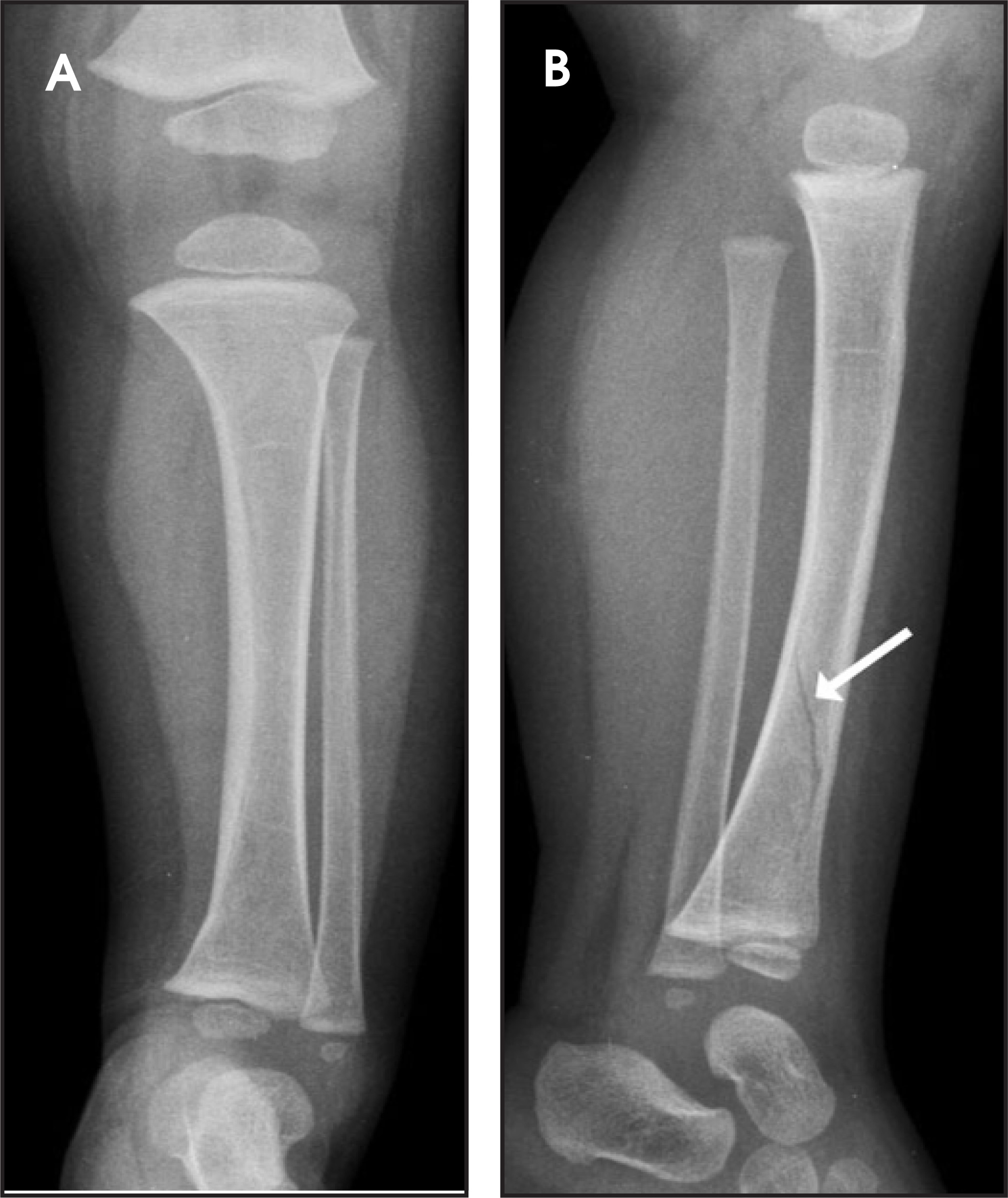
Complete fractures are also commonly seen in the pediatric patient. The so-called toddler’s fracture is a spiral fracture of the tibia that is caused by torsion or rotational force and is usually seen in children when they are learning to walk (Figure 2).3 It is important to note that when imaging skeletal trauma, radiographs should be obtained with at least 2 orthogonal views. In the case of a toddler’s fracture, the addition of oblique views may be helpful, since these fractures are sometimes seen only on a single view.
Approximately 15% of all fractures in children involve the growth plate.6 The Salter-Harris classification continues to be the most commonly used system for characterizing growth plate injuries (Figure 3). Type I Salter-Harris fractures involve the entire physis but do not extend into the adjacent epiphysis or metaphysis. The physis often appears widened on radiographs. Type II fractures involve most of the physis and extend into and through the adjacent metaphysis, creating a triangle-shaped fragment of the metaphysis. Type II fractures make up approximately 75% of all injuries to the physis. Type III fractures involve part of the physis, with vertical extension through the epiphysis. Type IV fractures are vertical fractures that involve the physis as well as the adjacent epiphysis and metaphysis. Type V fractures are due to compression or a crush-type injury of the physis and are usually not visible on initial radiographs.1,2 In general, the prognosis of Salter-Harris fractures worsens and the risk of growth arrest becomes greater with increasing numerical type.5
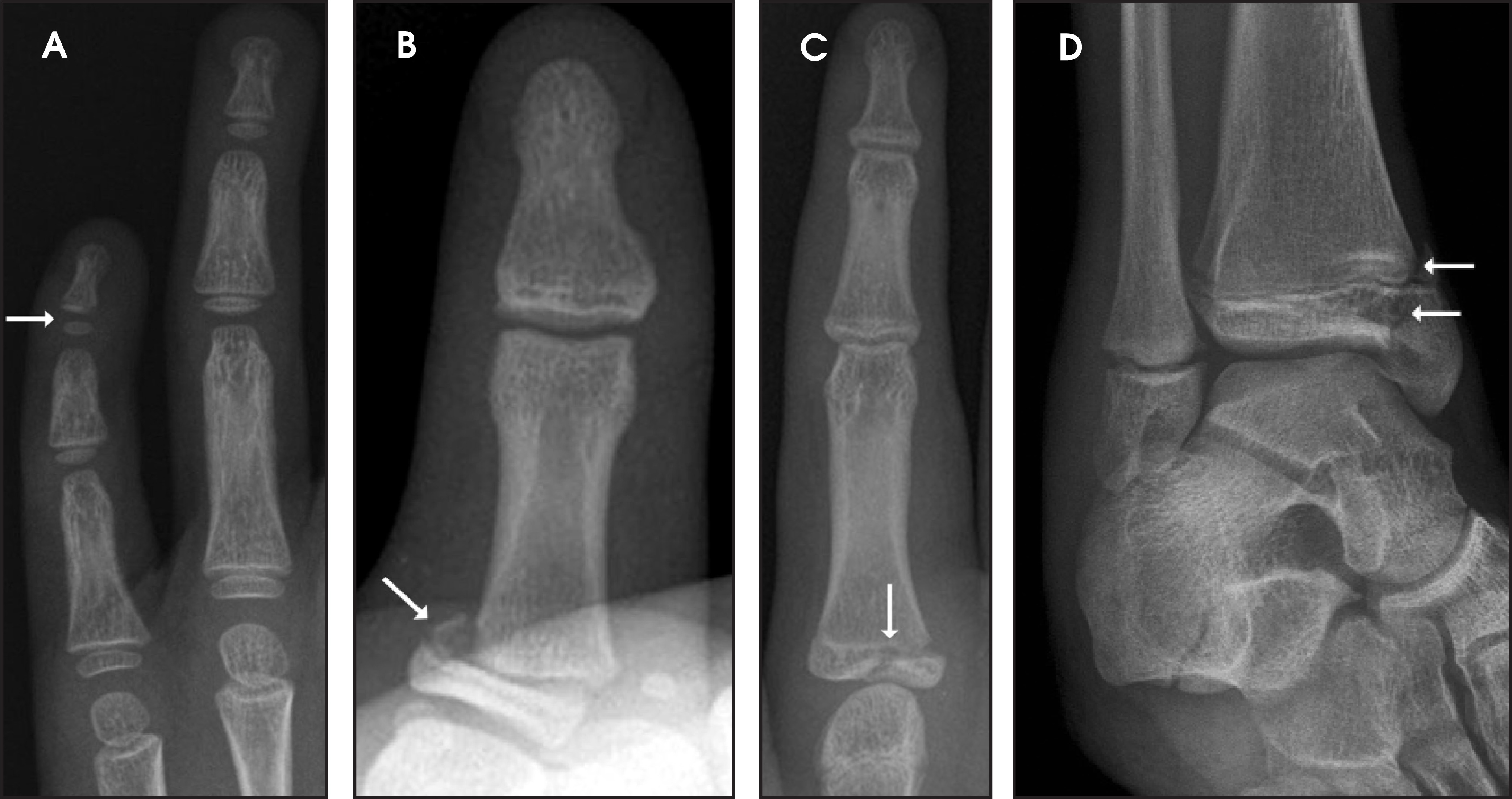
Generally speaking, the pediatric skeleton has strong potential for repair and healing following trauma. However, there is potential for disruption of the vascular supply to the epiphysis that can result in growth disturbances. A bridge of fibrous tissue can develop across the physis and inhibit future bone growth following injury to the growth plate.5 Nearly 15% of all growth plate injuries lead to growth arrest. Growth arrests occur many times more frequently in the lower extremities despite the fact that Salter-Harris fractures are more common in the upper extremities. It is therefore critical to diagnose and treat complications of such injuries early in order to prevent limb shortening or angular deformity.6 Bony bar formation along the central portion of bones tends to lead to limb shortening, whereas peripheral bony bar development is more likely to cause angular deformity of the joint.
Avulsion fractures in children commonly occur during puberty and are usually due to sports injuries involving sudden contraction of the musculotendinous unit pulling at the chondro-osseous site of tendon attachment. Since a child’s growth plate is weaker than the attached ligaments and tendons, avulsion-type fractures often result. Similar mechanisms of injury in the adult patient usually result in tears of ligaments and/or tendons rather than fractures. A common site for avulsion fractures is where major muscle groups have their origins on the pelvis.2,5,7
CT
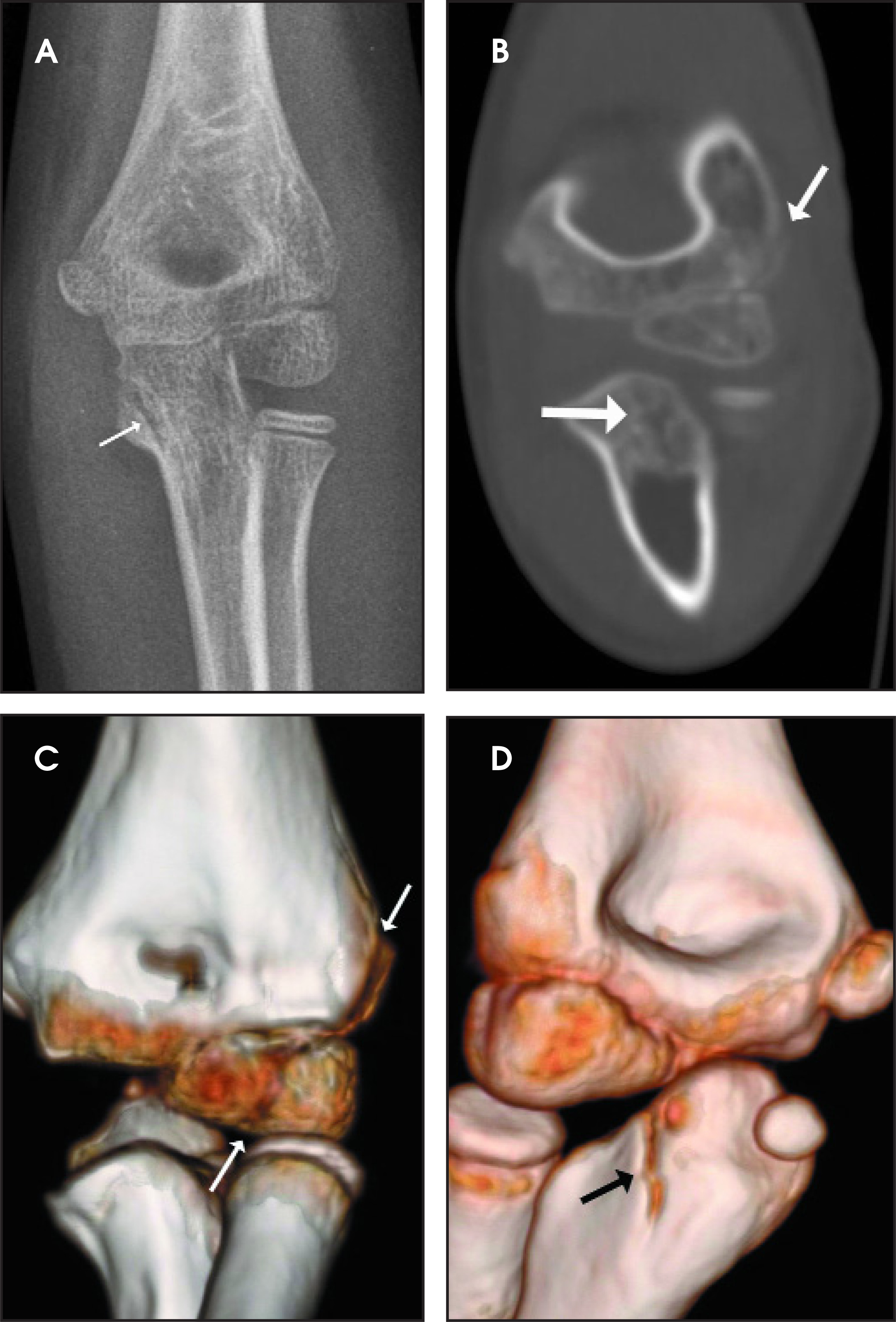
CT continues to be an important tool for the evaluation of pediatric skeletal trauma. Advances in CT technology have reduced imaging time to only seconds, which decreases the need for sedation. The ability to rapidly reconstruct 2-dimensional (2D) and 3-dimensional (3D) images from thin submillimeter volume data sets may obviate the need for additional imaging, thereby decreasing patient radiation exposure.8 CT allows for reconstruction of the data set in views and planes that would be difficult or sometimes impossible to achieve without the use of CT reformatting capabilities. Volume-rendered images can serve as an adjunct to coronal and sagittal 2D reformations that may show fractures that are missed on radiographs (Figure 4). For this reason, CT is often used in the planning of surgical procedures as well as to better define the extent of abnormality. CT is also useful for imaging patients after surgical procedures, since 3D reformatted images can decrease distractions (such as streak artifact) on anatomical structures that may otherwise obscure a region of interest.8 There are specific injuries in the pediatric patient for which the use of CT proves beneficial. Examples of such injuries are discussed below.
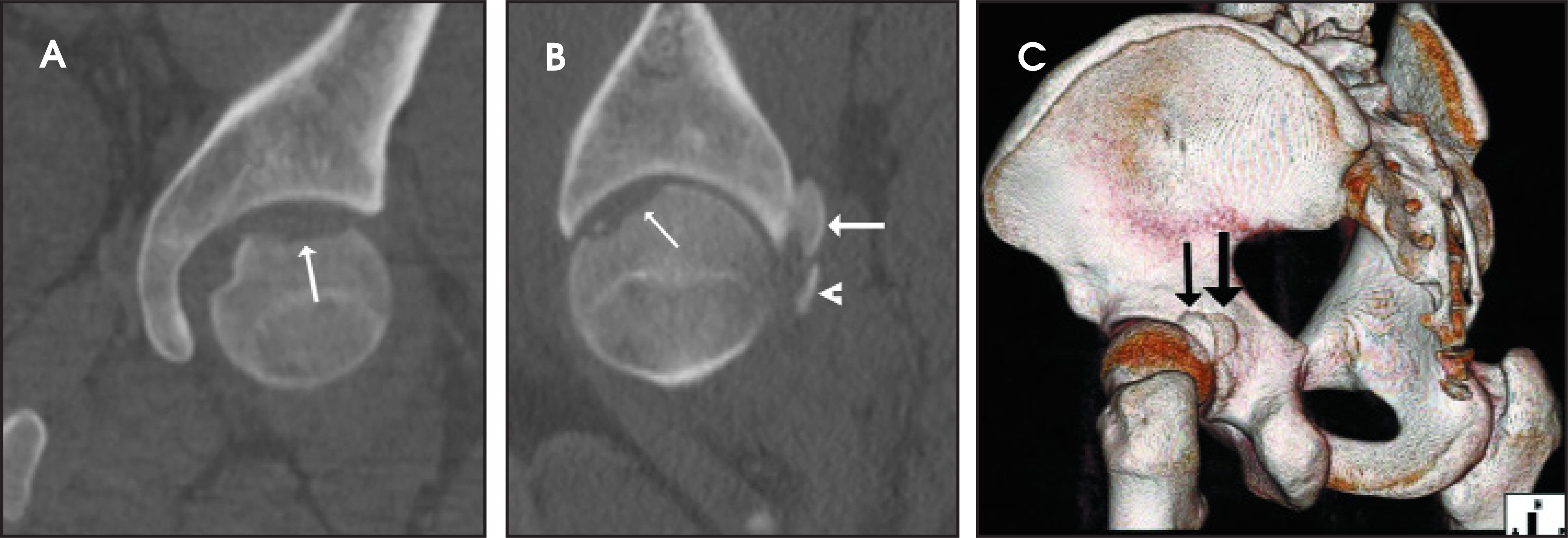
Although plain films may be sufficient to evaluate the pelvis following trauma, CT has been shown to be more sensitive than radiographs for the detection of pelvic fractures. CT has become the gold-standard imaging technique for the evaluation of pelvic trauma. Advantages of CT, multiplanar reformations, and volume-rendered views include their ability to examine the pelvis in multiple planes as well as from multiple perspectives (Figure 5). CT also allows for more detailed visualization of surrounding soft tissues. The additional information provided by CT can have an impact on treatment and surgical planning.9 In fact, many authors have recently questioned the need for plain films in patients who will undergo CT imaging of the abdomen and pelvis.9,10
The ability to make isotropic reconstructions in multiple planes from multidetector CT (MDCT) volume data sets has greatly enhanced our ability to identify triplane fractures (Figure 6). This unique Salter-Harris type IV fracture affects the distal tibia. This fracture typically extends through the epiphysis in the sagittal plane, through the physis in the horizontal plane, and then extends through the posterior metaphysis in the coronal plane. In the axial plane, there is a fracture of the physis that extends anteriorly and laterally from the coronal and sagittal fracture lines, respectively. Thus, this type of fracture may produce 2 to 4 fragments. It resembles a Salter-Harris type III fracture on the anteroposterior view and a type II fracture on the lateral view.1,2,11
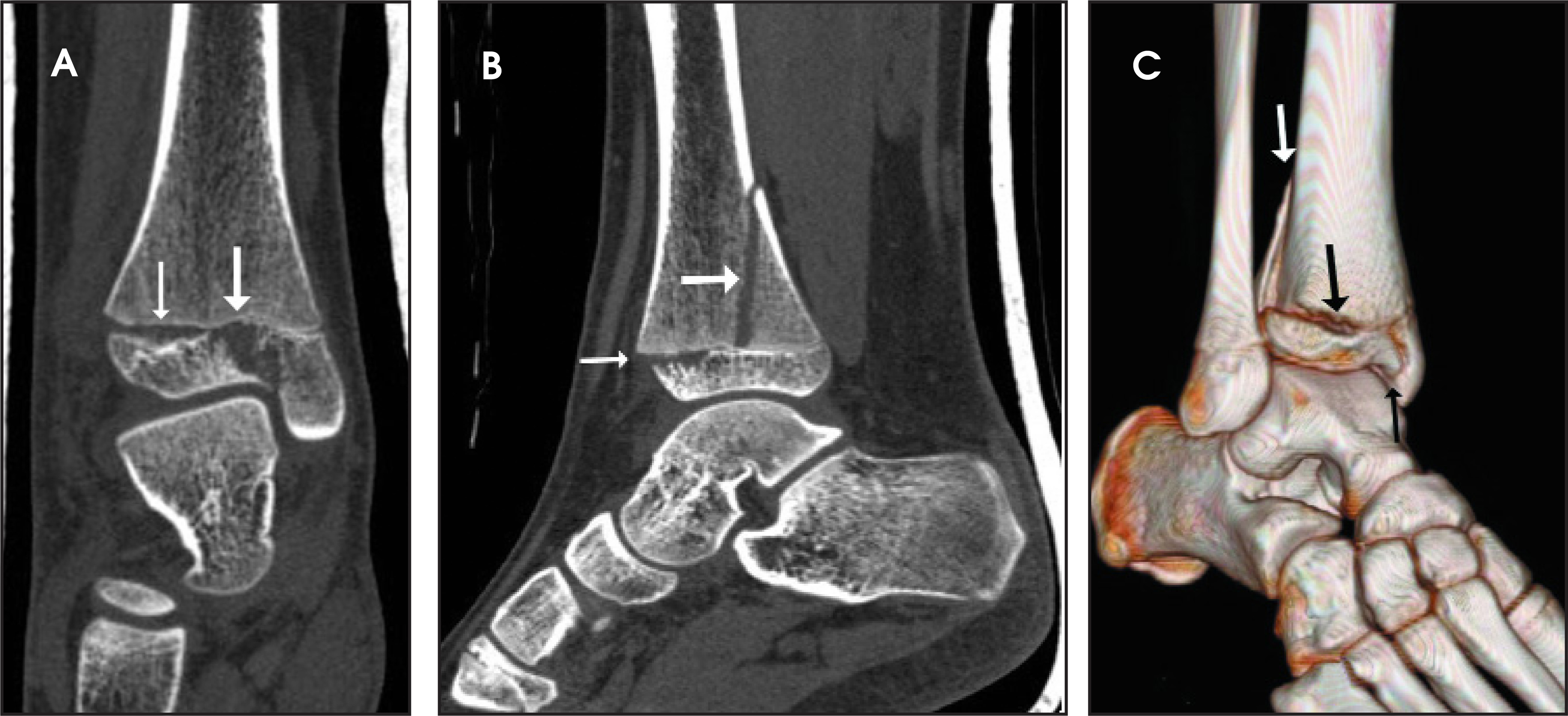
Separation of the physis in triplane fractures is often not apparent on radiographs, partly because of the overlap of the tibia and fibula on lateral views. This finding is more easily appreciated on CT reformats in the sagittal and coronal planes. In addition to aiding in the detection of triplane fractures, reformatted images allow for improved characterization and definition of specific fracture patterns. This, in turn, has important implications for proper diagnosis and for treatment decisions. As new technologies improve the resolution of reformatted images, it is likely that 3D reconstruction will be performed more routinely in such cases.12,13
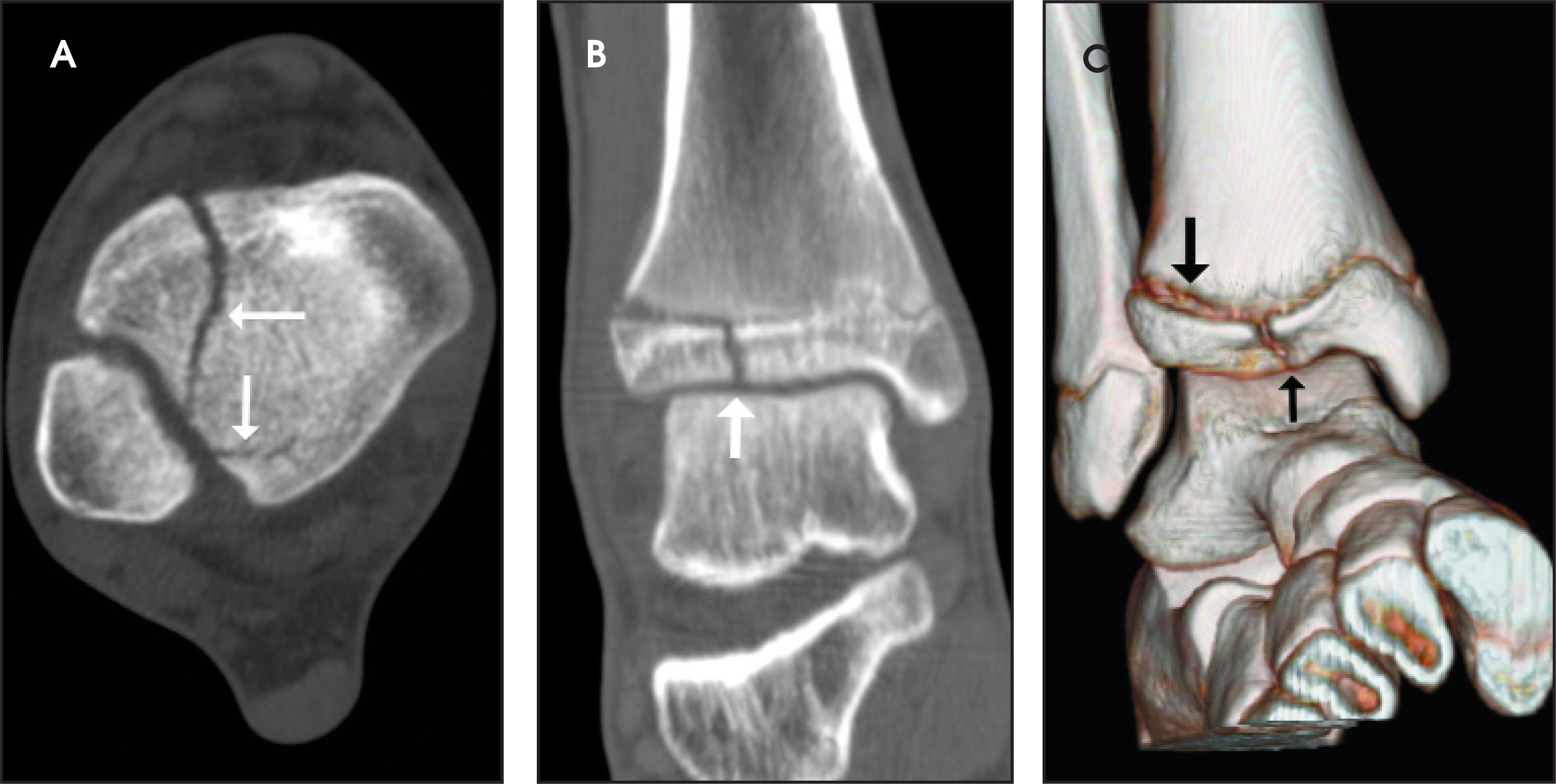
As with triplane fractures, CT is also helpful for imaging Tillaux fractures (Figure 7). These are Salter-Harris type III fractures that involve the physis of the distal tibia.11 CT has been shown to be more sensitive than radiography for detection of Tillaux fractures with >2 mm of displacement. Since 2 mm of displacement is generally considered the cutoff for fractures that require reduction, CT is the imaging modality of choice for the imaging of Tillaux fractures in children.14 Multidetector CT may be preferred to helical CT in these cases because of shorter imaging times and relatively fewer motion artifacts.15
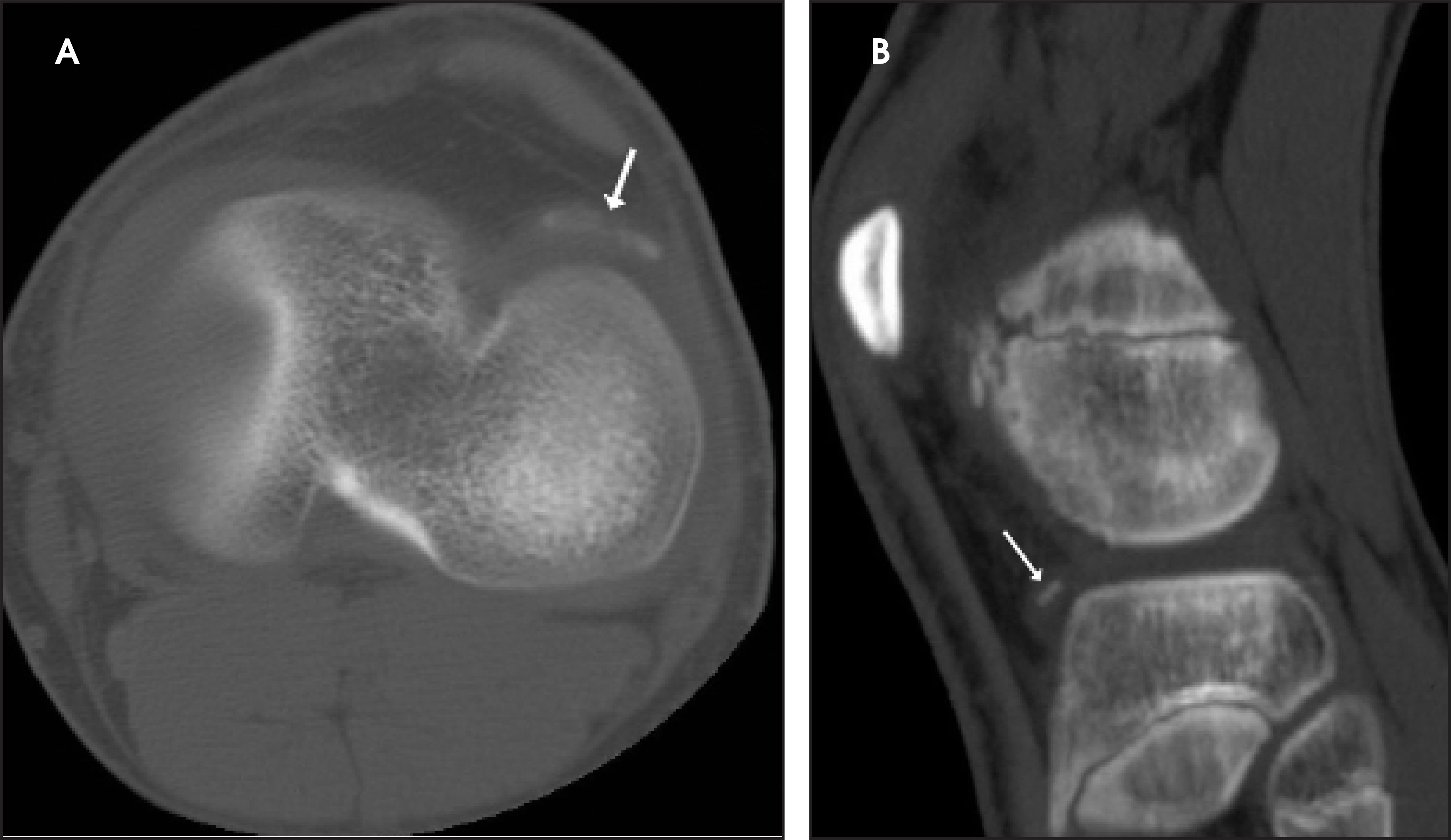
CT can also be particularly useful for the detection of loose bodies or bone fragments following injury (Figure 8).5 Loose bodies with an osseous or calcified component are more easily visualized on CT than those made entirely of cartilage. While cartilaginous loose body assessment is possible on CT, the use of air and/or iodinated contrast introduced into the joint is needed to improve loose body visualization.
MRI
MRI has become an increasingly important imaging technique in the setting of pediatric trauma. Drawbacks of MRI include longer imaging times, dependence on patient cooperation, and frequent need for sedation.8 The relative advantages of MRI include improved soft tissue contrast resolution and multiplanar capabilities.1 Examples of instances in which MRI would be of particular benefit are illustrated below.
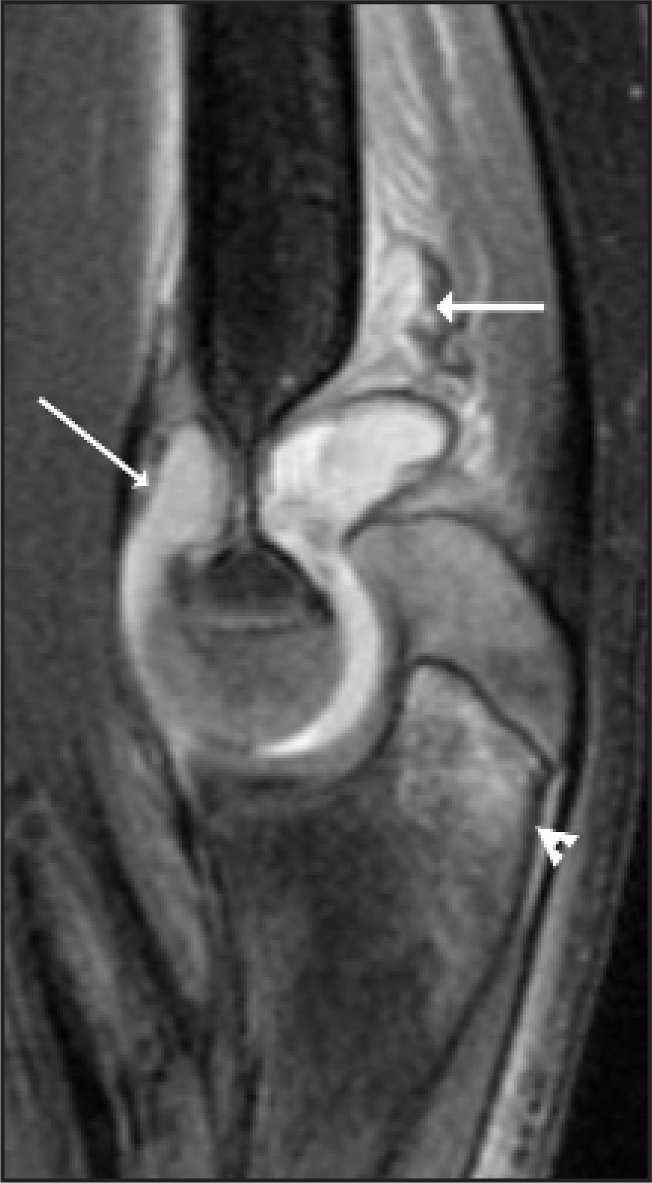
One main advantage of MRI is the identification of fractures that are not clearly seen on plain radiographs. This is explained by the fact that developing bones in children have cartilaginous portions. Cartilage is radiolucent on plain film. MRI is better able to visualize radiographically occult fractures as well as surrounding soft tissue abnormalities and edema in the pediatric patient following trauma (Figure 9).6,16
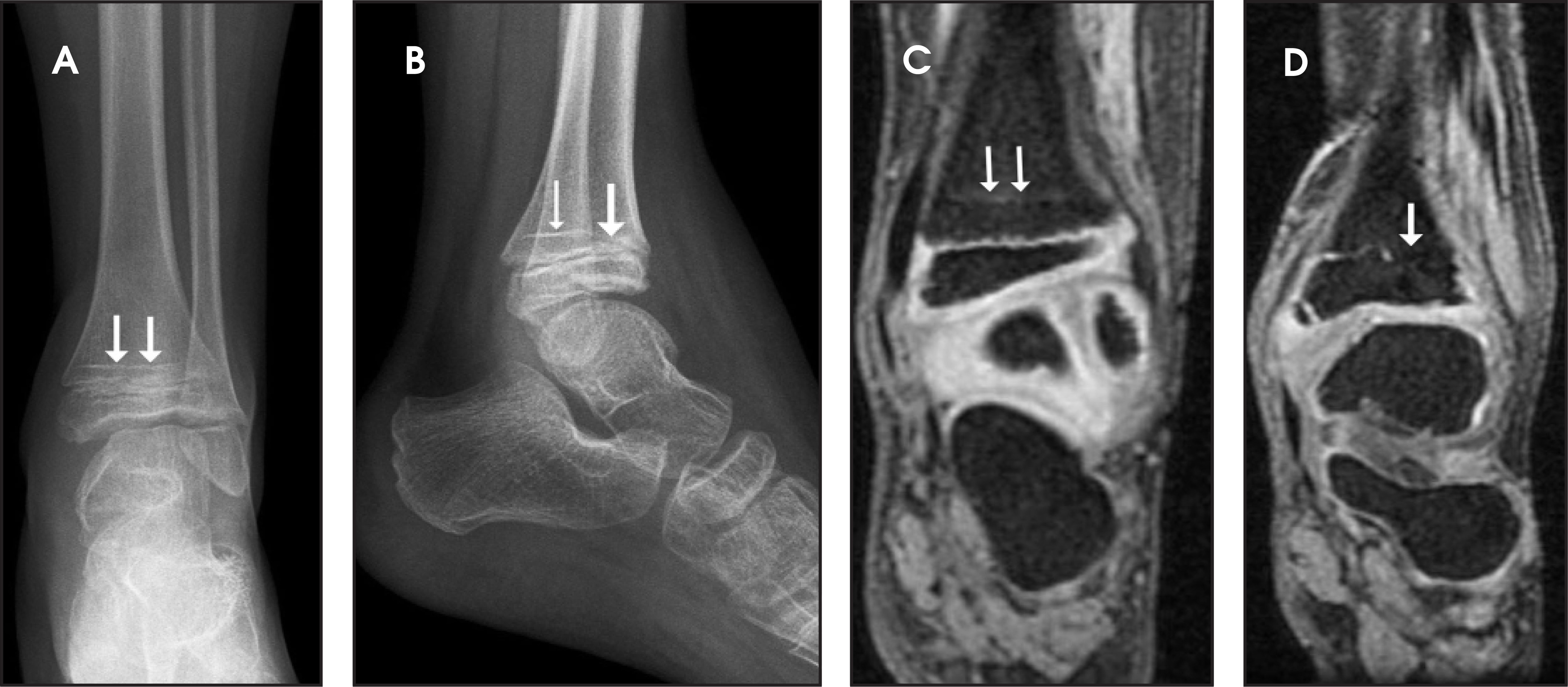
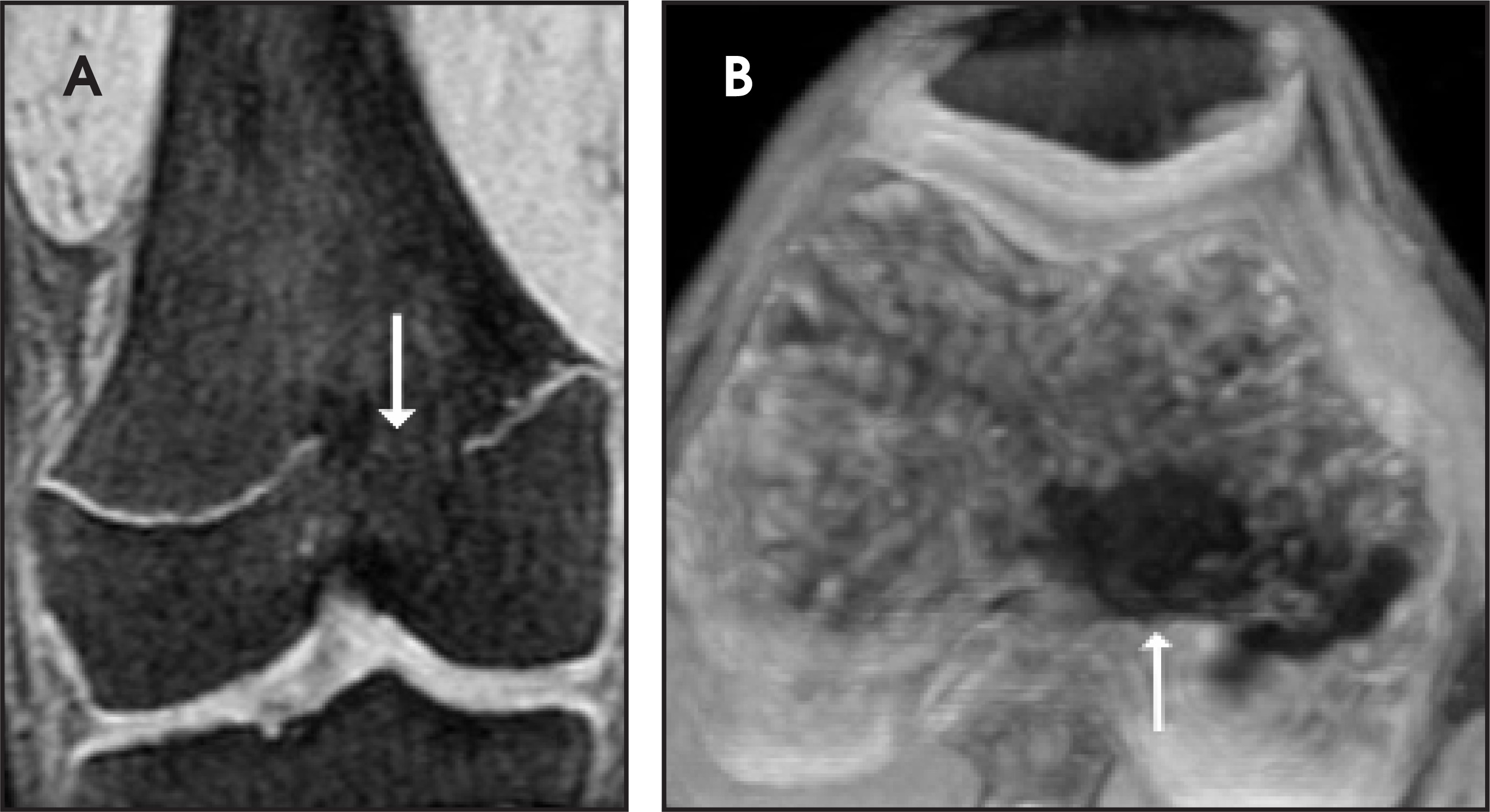
Although plain radiographs are usually the only images necessary for the evaluation of Salter-Harris fractures, injuries to the growth plate are not always visible on plain films.11 In such cases, MRI is often able to delineate physeal abnormality and its associated marrow edema (Figure 10). MRI can alter Salter-Harris classification of growth plate injuries as determined by radiographs.16 Of particular importance, MRI can identify these injuries prior to the development of growth arrest, which can lead to limb length discrepancy and deformity. MRI may guide treatment and surgical planning when a physeal bar is identified (Figures 11 and 12). The generation of maximum-intensity projection, thick-slab reformatted images from 3D fat-saturated, spoiled gradient-recalled acquisition (SPGR), T1-weighted sequences can aid in surgical planning.5,6
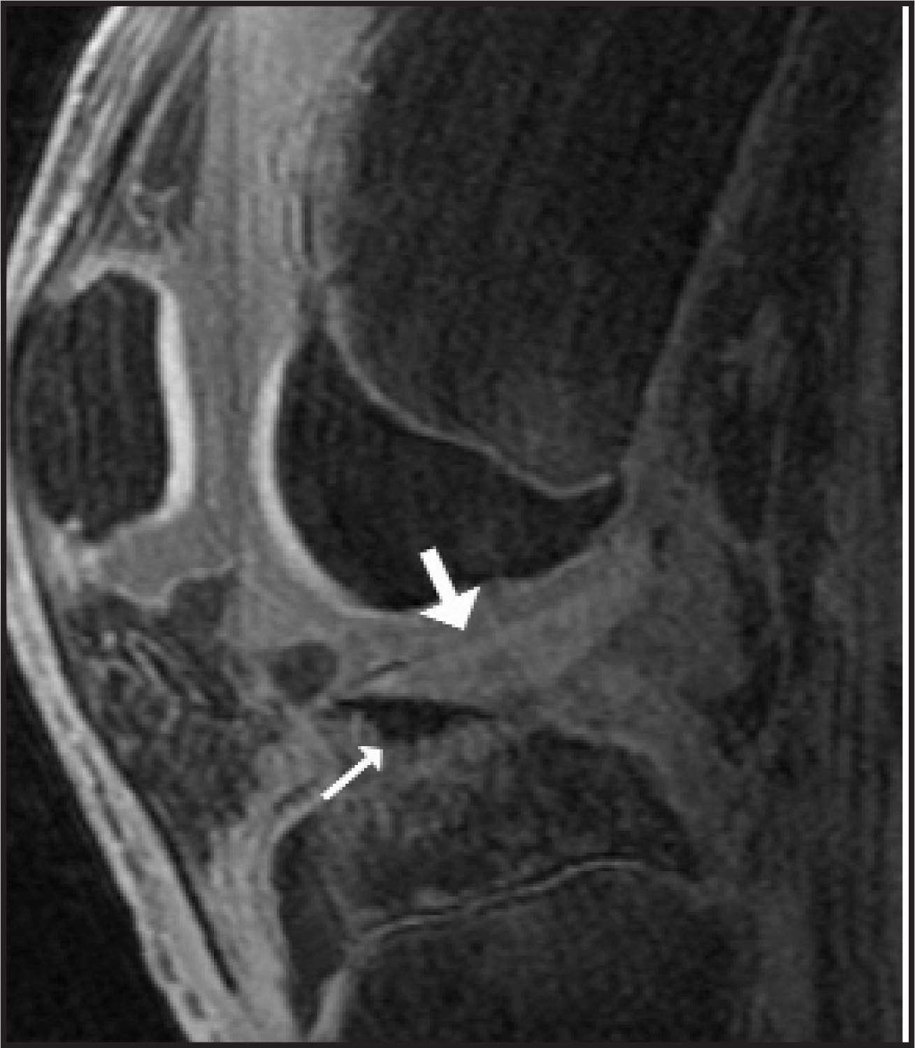
As with fractures of the growth plate, avulsion injuries are usually imaged with plain films alone. MRI can be helpful in the evaluation of avulsion injuries when plain radiographs are atypical or equivocal. Patellar sleeve fractures, for example, often appear normal on radiographs. MRI, however, is more helpful in determining the extent of cartilaginous and soft tissue involvement as well as the presence of edema in surrounding structures.5 Avulsion injuries that frequently require evaluation by MRI include those involving the tibial spines (Figure 13), the lower pole of the patella, and the medial epicondyle of the distal humerus.6
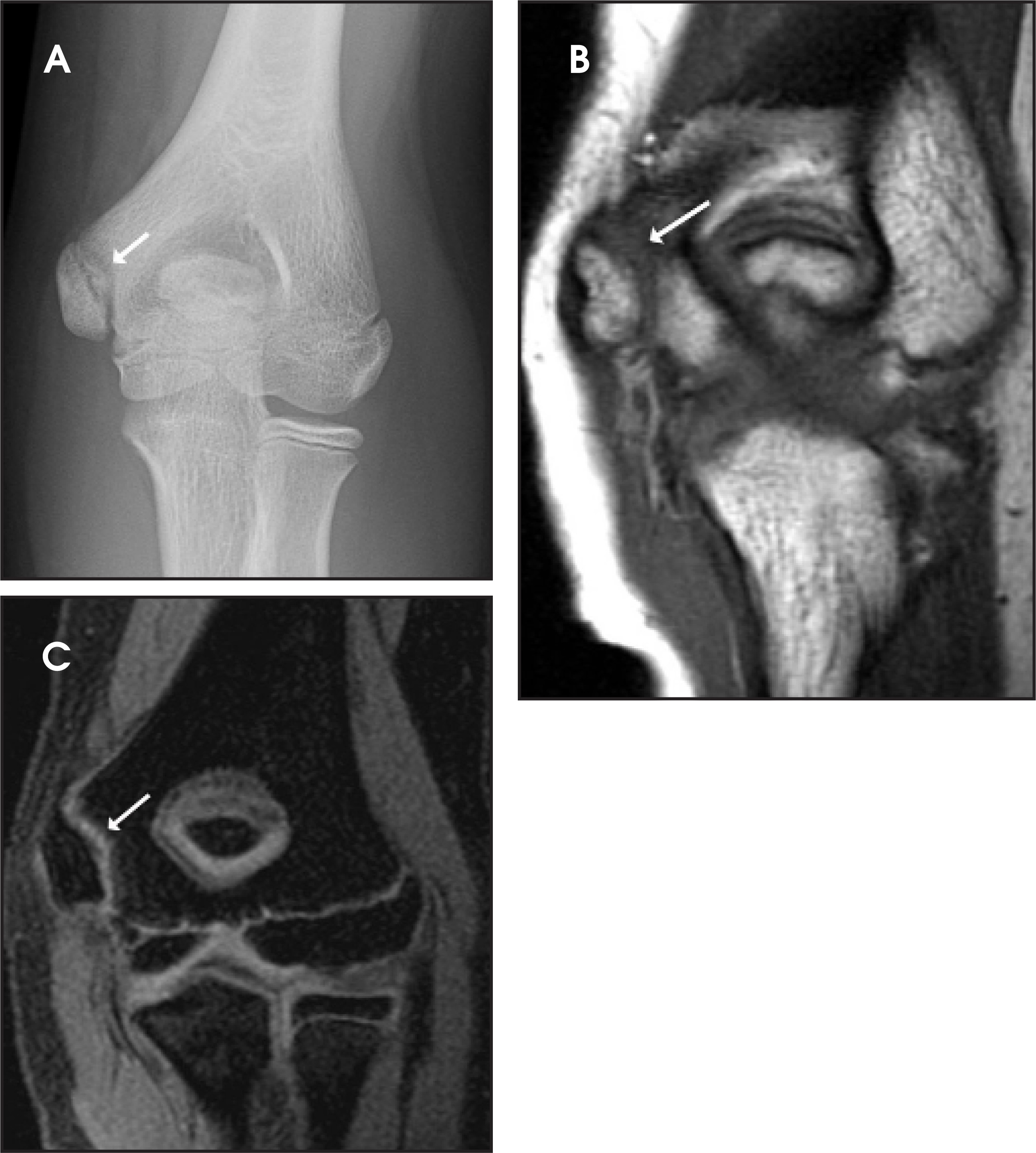
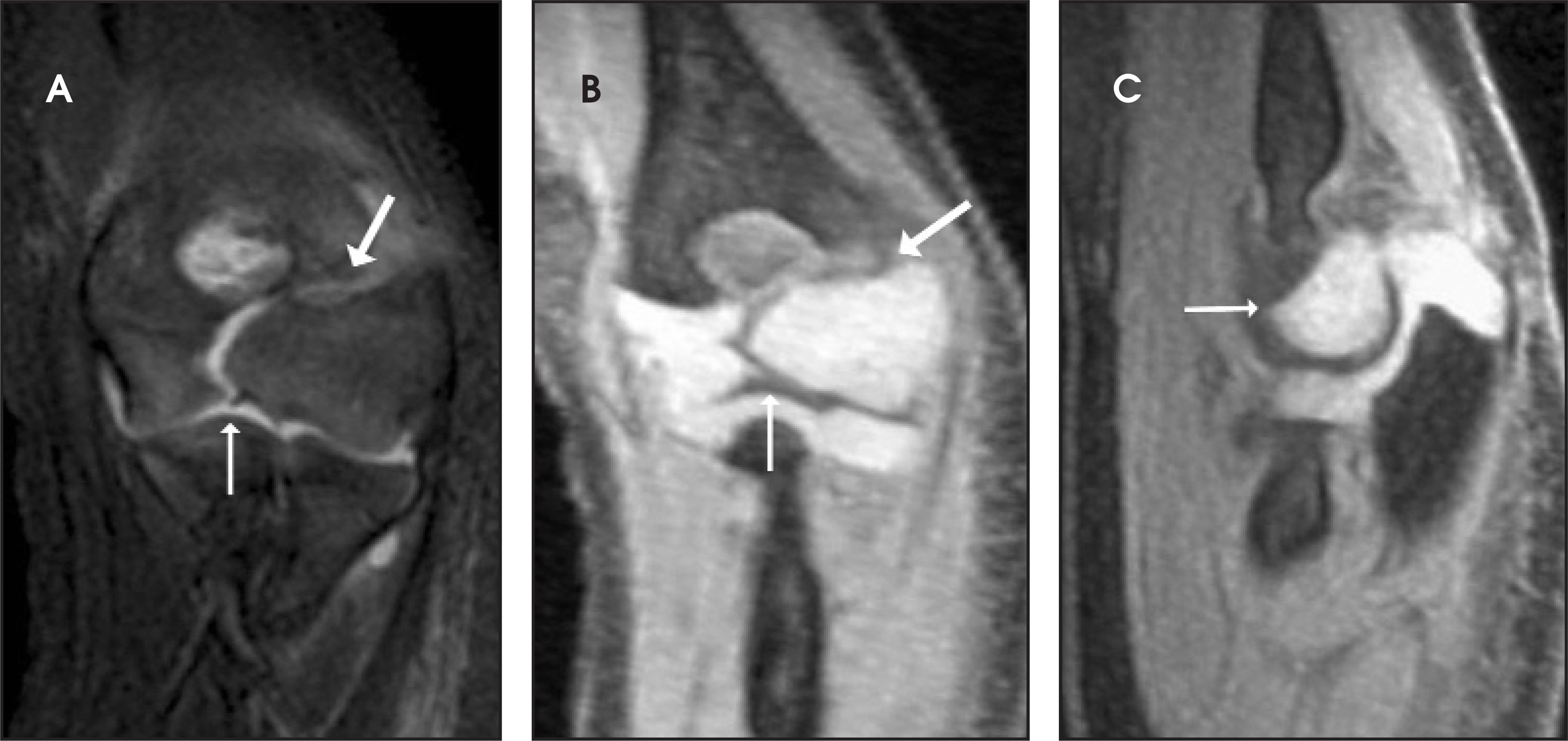
Because of the largely cartilaginous nature of the elbow in young children, it is highly susceptible to fractures that are difficult to recognize on radiographs (Figure 14).17 Therefore, trauma to this region is often best imaged with MRI, which is superior to radiographs in identifying fractures and effusions. MRI is also able to visualize bone bruising and cartilaginous and soft tissue injury that cannot be seen radiographically. The capacity of MRI to detect growth plate injuries that are radiographically occult is another advantage in the setting of pediatric elbow trauma. The use of MRI to evaluate the pediatric elbow following trauma, however, may not result in the alteration of treatment or improved clinical outcomes.18
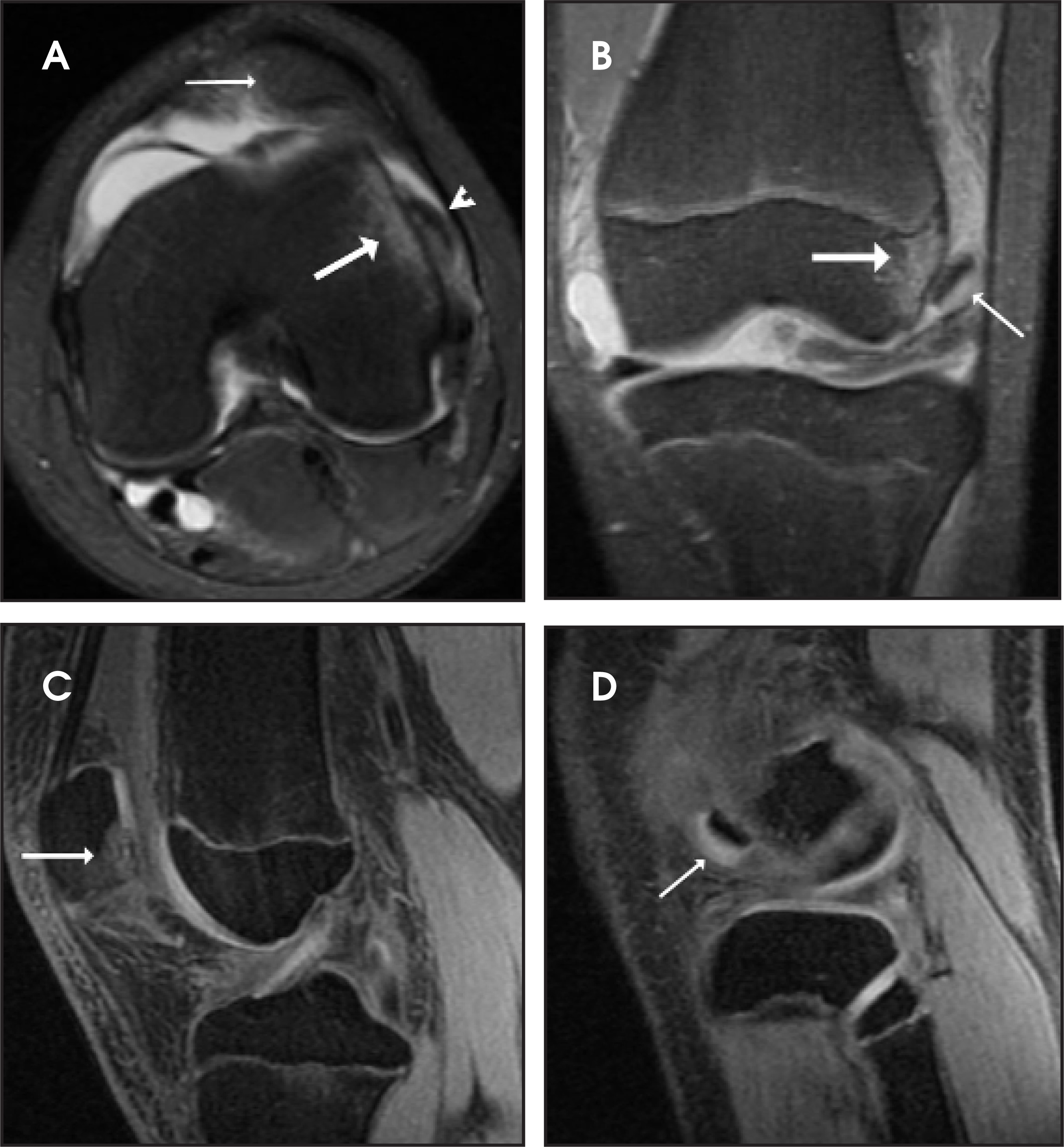
The knee is the most common pediatric joint for which MRI is used. MRI can visualize growth-plate fractures, avulsion fractures, injuries to ligaments, bone-marrow edema, effusion, soft tissue injuries, evidence of patellar dislocation, and meniscal tears (Figure 15).19 In contrast to MRI of the elbow, MRI findings in the pediatric knee will alter management in most cases. Therefore, MRI is indicated in the evaluation of the pediatric knee in cases where plain films are equivocal.20
MRI is also useful in the detection of loose bodies. In contrast to CT, however, MRI is able to detect fragments of bone as well as cartilage fragments. Therefore, we find that loose bodies without a calcified component are better imaged with MRI than with CT. Cartilage-specific sequences (such as 3D volume SPGR) are often helpful in such cases (Figure 15).
Conclusion
Unique features of the pediatric skeleton result in injuries and fractures that are different from those seen in adults. In most cases, imaging of pediatric trauma begins with radiographs, and few patients require further imaging. CT and MRI are the most useful advanced imaging techniques in the evaluation of skeletal trauma in pediatric patients. The advantages of MDCT include reduced imaging time, and the ability to perform 2D and 3D reconstructions in traditional and nontraditional views. MRI allows for excellent evaluation of cartilage, bone marrow, ligaments, tendons, and surrounding soft tissues.
References
- Kao S, Smith W. Skeletal injuries in the pediatric patient. Radiol Clin North Am. 1997;35:727-746.
- Resnik C. Diagnostic imaging of pediatric skeletal trauma. Radiol Clin North Am. 1989;27:1013-1022.
- Rodriguez-Merchan E. Pediatric skeletal trauma: A review and historical perspective. Clin Orthop Relat Res. 2005;432:8-13.
- John S. Trends in pediatric emergency imaging. Radiol Clin North Am. 1999;37:995-1034.
- Jarmillo D, Shapiro F. Musculoskeletal trauma in children. Magn Reson Imaging Clin N Am. 1998;6:521-536.
- Ecklund K. Magnetic resonance imaging of pediatric musculoskeletal trauma. Top Magn Reson Imaging. 2002;13:203-217.
- Rossi F, Dragoni S. Acute avulsion fractures of the pelvis in adolescent competitive athletes: Prevalence, location and sports distribution of 203 cases collected. Skeletal Radiol. 2001;30:127-131.
- Fayad L, Johnson P, Fishman E. Multidetector CT of musculoskeletal disease in the pediatric patient: Principles, techniques, and clinical applications. Radiographics. 2005;25:603-618.
- Guillamondegui O, Mahboubi S, Stafford P, Nance M. The utility of the pelvic radiograph in the assessment of pediatric pelvic fractures. J Trauma. 2003;55:236-239.
- Rees M, Aickin R, Kolbe A, Teele R. The screening pelvic radiograph in pediatric trauma. Pediatr Radiol. 2001;31:497-500.
- Vanhoenacker F, Bernaerts A, Gielen J. Trauma of the pediatric ankle and foot. JBR-BTR. 2002;85:212-218.
- Brown S, Kasser J, Zurakowski D, Jaramillo D. Analysis of 51 tibial triplane fractures using CT with multiplanar reconstruction. AJR AM J Roentgenol. 2004;183:1489-1495.
- Jones S, Phillips N, Ali F. Triplane fractures of the distal tibia requiring open reduction and internal fixation. Pre-operative planning using computed tomography. Injury. 2003;34:293-298.
- Horn B, Crisci K, Krug M. Radiologic evaluation of juvenile tillaux fractures of the distal tibia. J Pediatr Orthop. 2001;21:162-164.
- Haapamaki V, Kiuru M, Koskinen S. Ankle and foot injuries: analysis of MDCT findings. AJR Am J Roentgenol. 2004;183:615-622.
- Carey J, Spence L, Blickman H, Eustace S. MRI of pediatric growth plate injury: Correlation with plain film radiographs and clinical outcome. Skeletal Radiol. 1998;27:250-255.
- John S, Wherry K, Swischuk L, Phillips W. Improving detection of pediatric elbow fractures by understanding their mechanics. RadioGraphics. 1996;16:1443-1460.
- Griffith J, Roebuck D, Cheng J. Acute elbow trauma in children: Spectrum of injury revealed by MR imaging no apparent on radiographs. AJR Am J Roentgenol. 2001;176:53-60.
- Strouse P, Koujok K. Magnetic resonance imaging of the pediatric knee. Top Magn Reson Imaging. 2002;13:277-294.
- Close B, Strouse P. MR of physeal fractures of the adolescent knee. Pediatr Radiol. 2000;30:756-762.
Citation
. Pediatric skeletal trauma—Plain film to MRI. Applied Radiology. 2007;36(8):24-33. doi:10.37549/AR1533.