PET/CT imaging in children: Current techniques and indications
Applied Radiology — Vol. 41 , Issue 6 , pp. 8 -13
DOI: 10.37549/AR1916
Published: June 1, 2012
Categories
Positron emission tomography (PET) imaging of the pediatric patient is being performed with increasing frequency. The common pediatric adage stating that children are not simply little adults holds particularly true with regard to PET imaging. One of the primary differences between pediatric and adult imaging is the underlying pathology being evaluated. While some neoplastic entities are common in children and adults (primarily lymphoma), many are unique to children. Children also have variants of fludeoxyglucose (FDG) uptake that must be known to correctly interpret studies. Patient preparation and radiation dosage are further variables that differ between children and adults.
This paper addresses the unique circumstances involved in PET imaging of children. Patient preparation and image acquisition will be discussed briefly. Normal findings will be reviewed and followed by an overview of the more common uses of PET in children. We presenta particular focus on oncologic, brain, and bone imaging, along with an up-to-date discussion on evolving trends. Finally, we will present abrief overview of some of the more rare indications for PET examinations.
Patient preparation
Preparation for pediatric PET imaging begins well before the actual image acquisition. As in the case of adults, glucose levels are checked prior to FDG administration; levels should be under 150 ng/dL.1 Elevated glucose results in increased insulin release and subsequent muscular FDG uptake. Moreover, in patients with elevated intrinsic glucose levels, native glucose will compete with the radiolabeled glucose, and potentially result in a false negative exam.2 Fasting prior to PET examination is required, and parents are instructed not to feed their children for 4 hours prior to the test. Infants should also fast for 4 hours, but they can be breast- or bottle-fed shortly after radiotracer injection.
Brown fat, while not unique to children, is visualized much more frequently in children than in adults. When FDG uptake is particularly prominent, brown fat can obscure true pathology and make image interpretation difficult. Medications, such as propanolol or fentanyl, have been used to decrease brown fat uptake; however, drug-free methods are also effective. Studies have shown that patients kept in a room witha constant temperature of 24 C for 30 min prior to radiotracer injection and 1 hour following injection, had minimal brown fat uptake.3 This procedure is recommended for children as it minimizes brown fat uptake and eliminates the adverse effects of the aforementioned medications.
Patient cooperation can sometimes be challenging even in adults; thus, the thought of coercing a young child to lie still for an extended time for PET imaging raises obvious concerns. While sedation is utilized more frequently in children, the associated anesthesia risks cannot be ignored. Techniques to minimize patient anxiety can often result in superb image acquisition without anesthesia. Child-friendly rooms and technologists with pediatric experience greatly help to minimize anxiety. Evaluating each child with regard to sedation needs is recommended rather than employing a strict age requirement for sedation.1,2
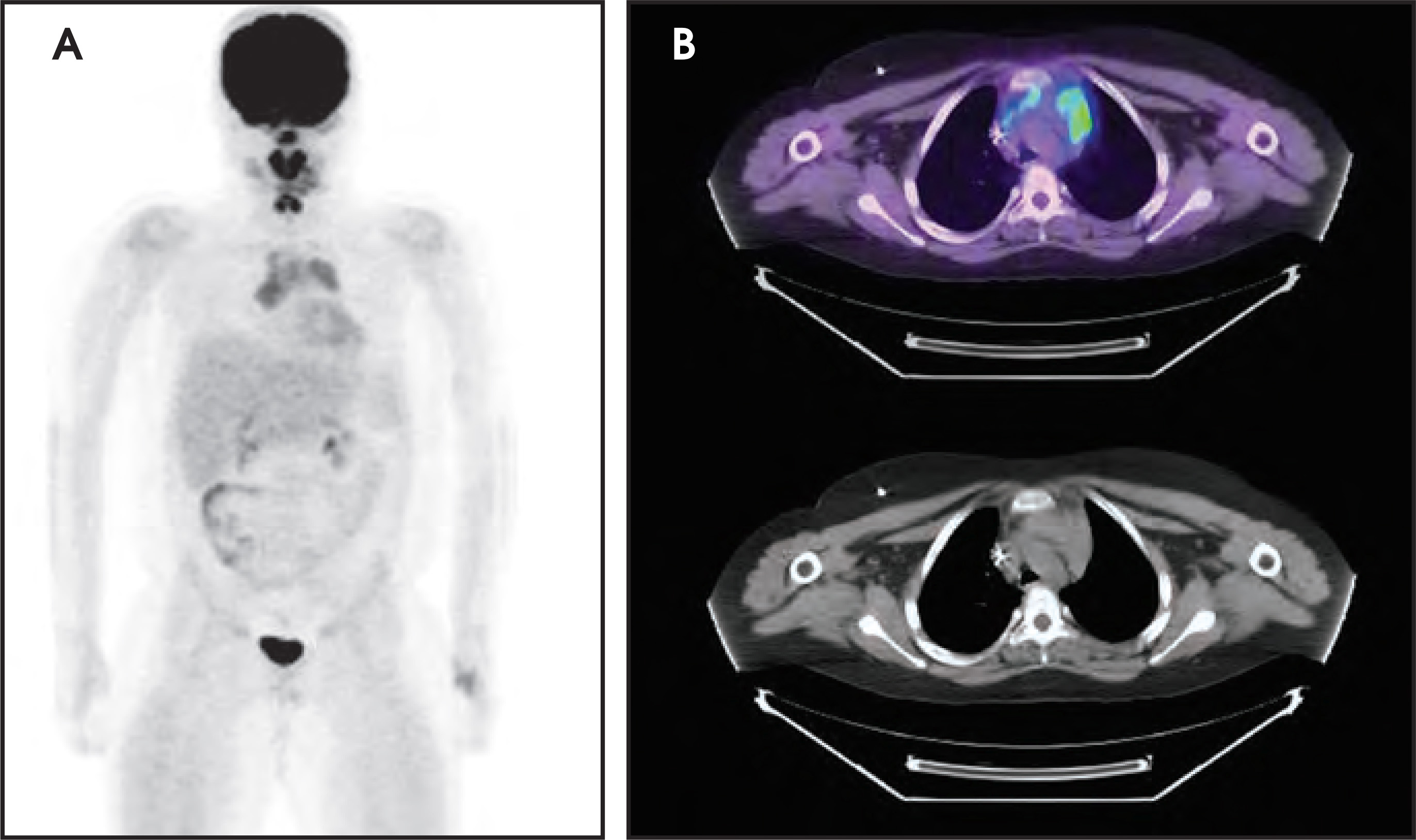
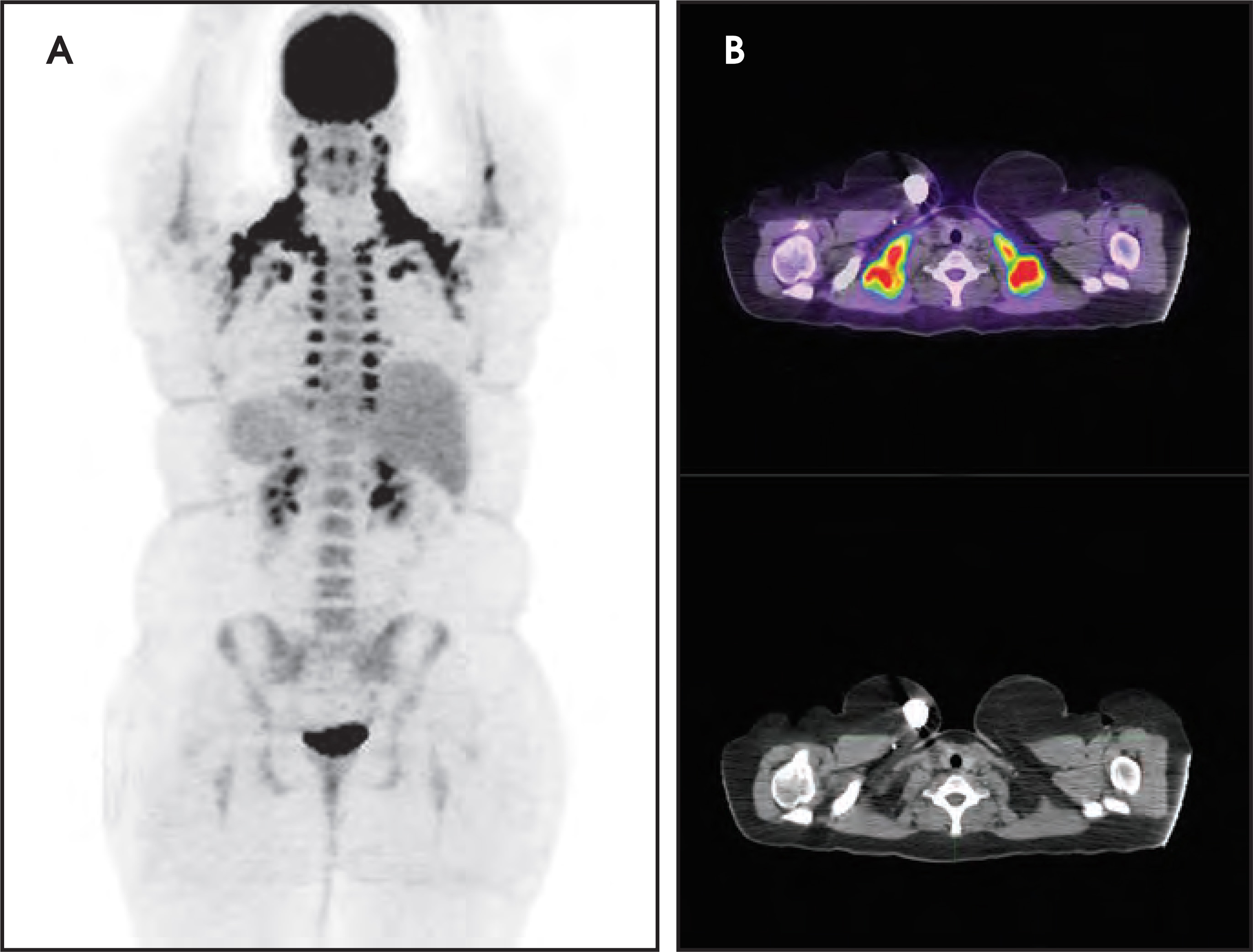
Interpreting pediatric PET exams requires an understanding of normal variants present in children. One of the most confusing areas of potentially normal uptake is the thymus. Normal uptake in the thymus can be detected into adolescence. Differentiating this uptake from neoplastic involvement can be difficult, however, evaluating the shape of the thymus (normally an inverted V) and degree of uptake can be helpful (Figure 1). Tonsillar uptake can frequently be seen in children, and can result in a diagnostic dilemma in lymphoma patients. Benign uptake within the tonsils is typically symmetric; lymphomatous involvement is usually irregular/asymmetric. Brown adipose uptake is common in children, and knowing its distribution is critical for image interpretation (Figure 2).4 Visualization of uptake in the cervical cords is common, especially if the child talks or cries after FDG injection.
Oncologic imaging
As is the case with adults, the most common use of PET in children is oncologic imaging. PET has many uses in tumor evaluation, including initial staging, evaluating therapy response and disease recurrence, and aiding in therapy planning.5 Occasionally, PET is used to evaluate tumors with atypical behavior. For instance, PET has been found useful in evaluating metaiodobenzylguanidine (MIBG) negative neuroblastoma6,7 or radioiodine-negative thyroid carcinomas.8 Another rare use of PET is to search for occult malignancy.
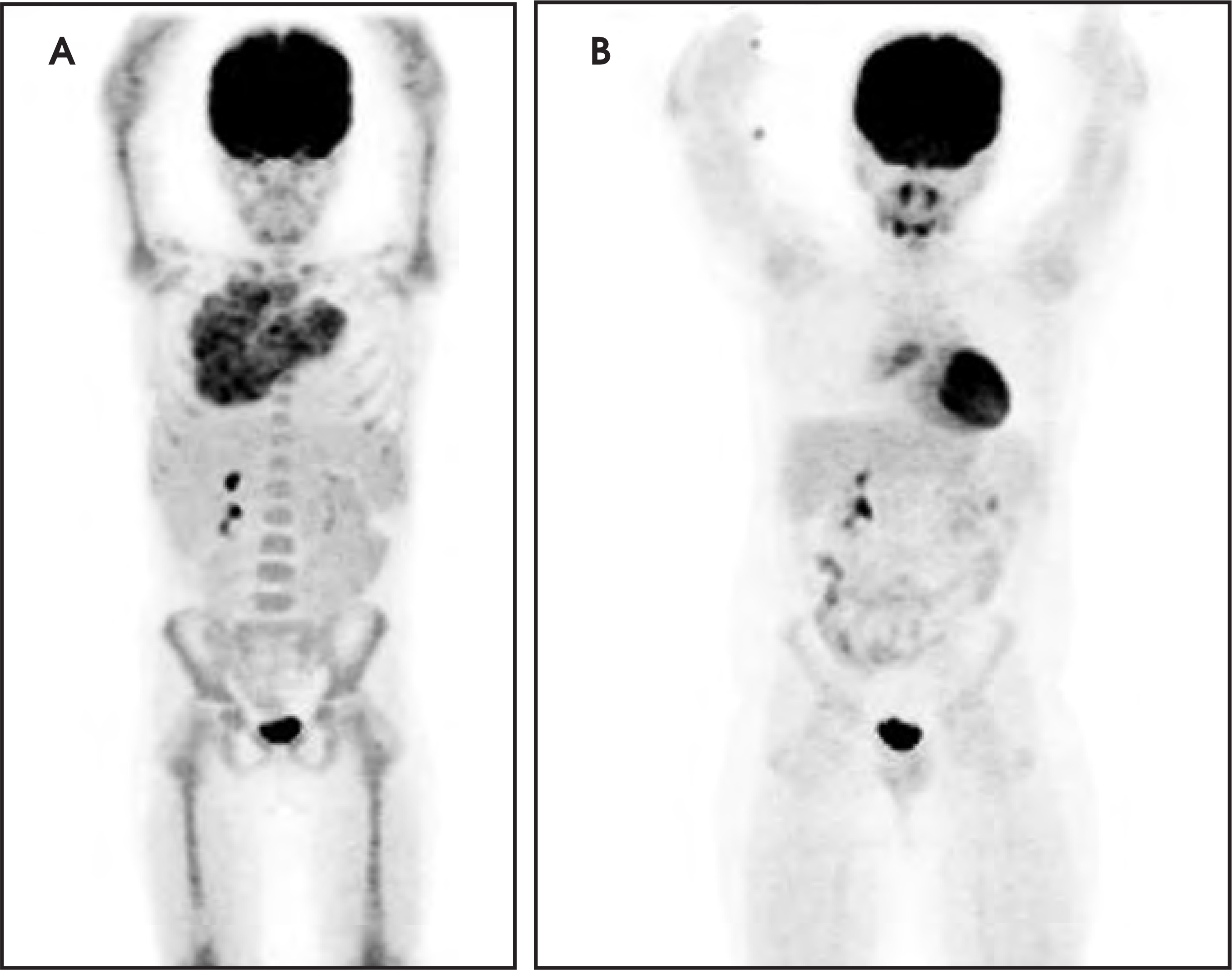
The most frequently imaged neoplasm in the child is lymphoma.9 Pediatric lymphoma is a highly curable disease,10 but determining disease extent is critical to proper therapy. PET is used for initial staging and frequently repeated following a short interval to ensure that an appropriate response to chemotherapy has occurred (Figure 3). This allows for rapid therapy alterations in children not demonstrating a robust response, and for improvement in survival.
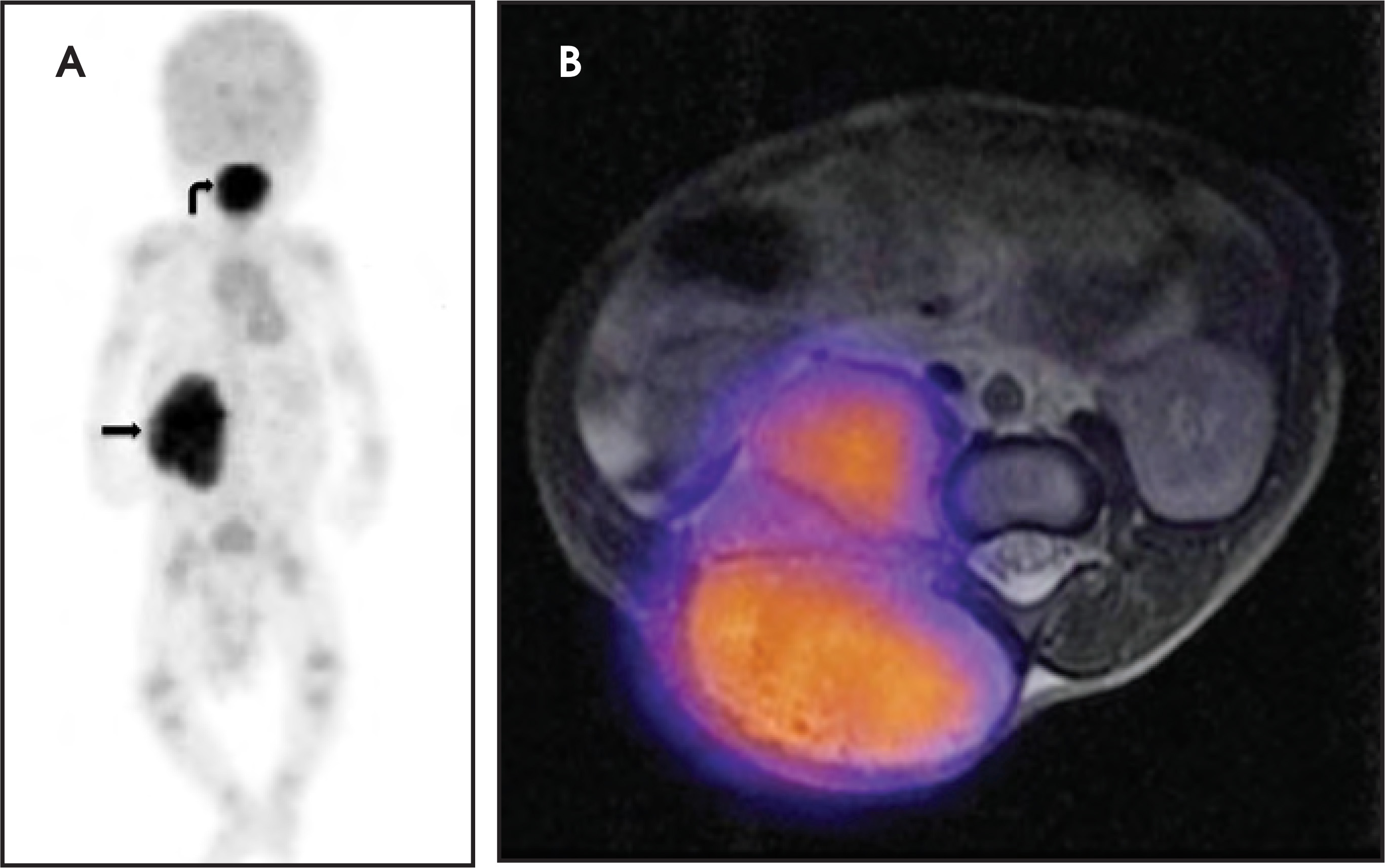
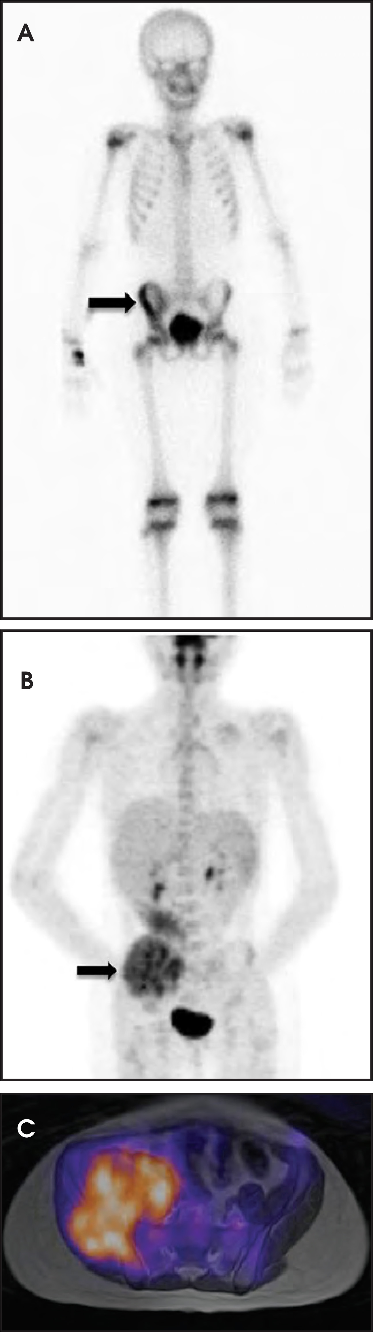
Sarcomas are also frequently imaged in children. Rhabdomyosarcoma is the most frequent soft-tissue malignancy in children. While it arises from all cells in the body, excluding only the brain parenchyma, in children it most commonly arises from the genitourinary tract and muscular system. PET is extremely useful in evaluating the extent of disease in these patients (Figure 4). PET can sometimes better demonstrate disease extent in osteosarcoma and Ewing’s sarcoma as well. (Figure 5).11
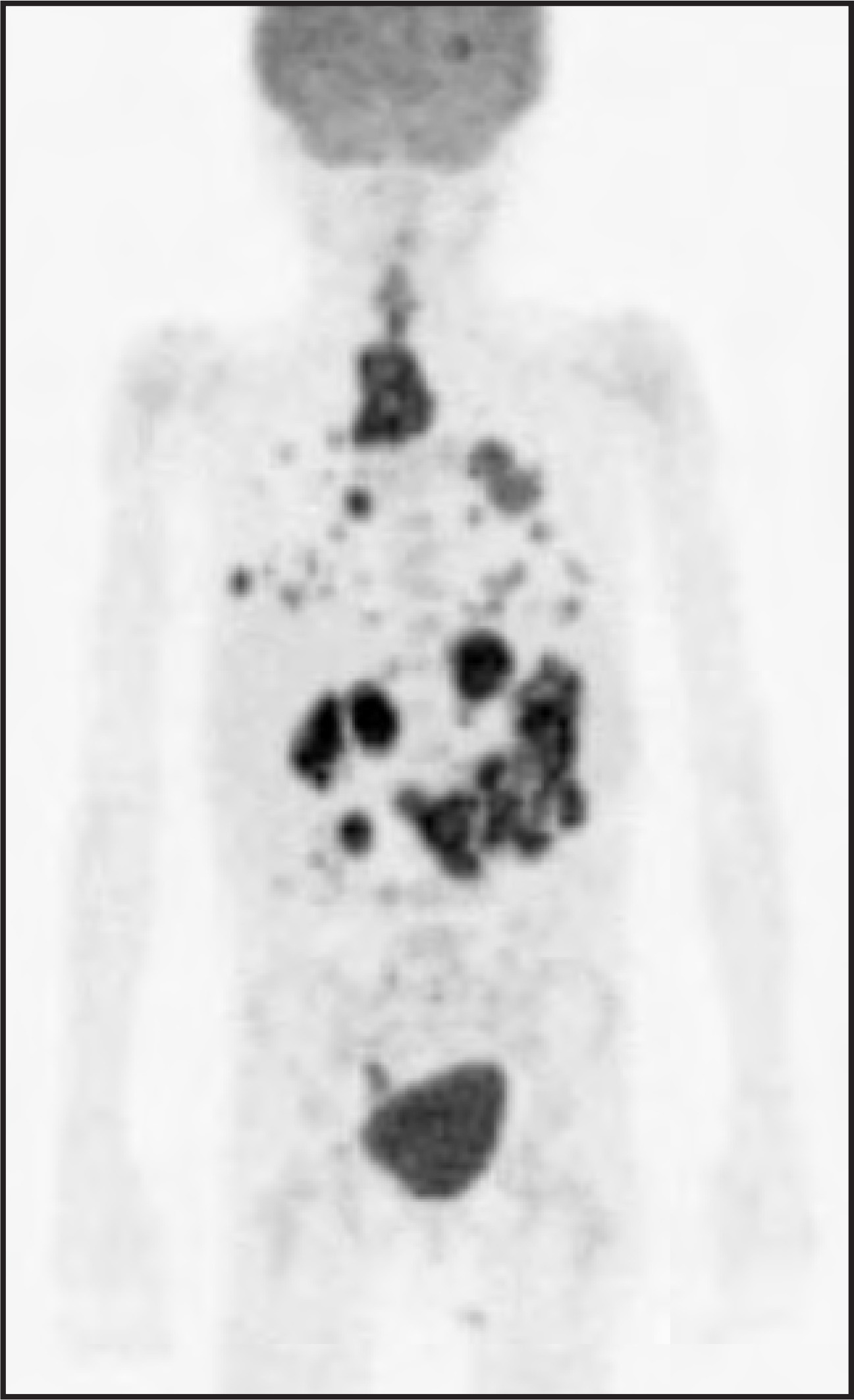
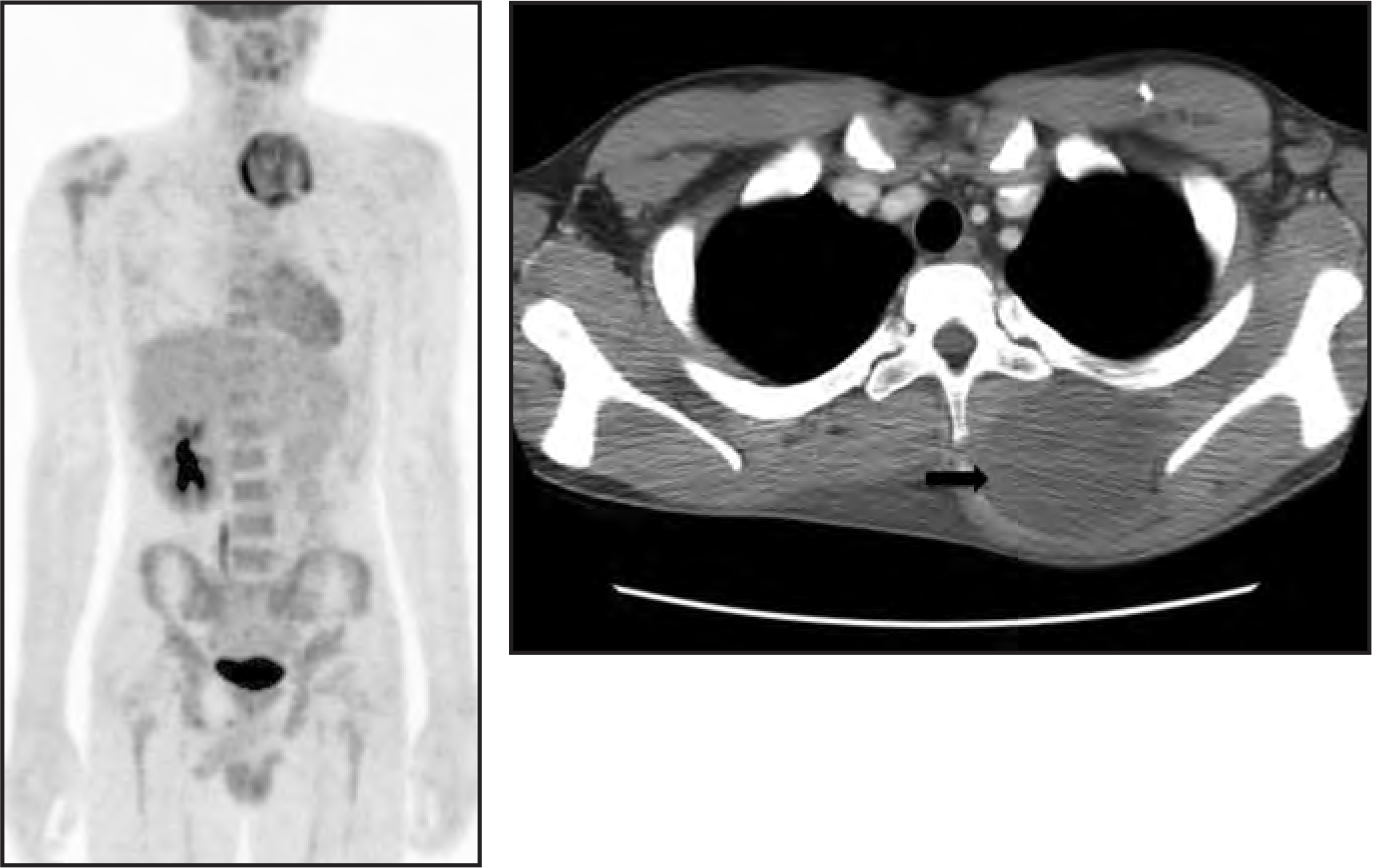
PET has been used to evaluate other rare pediatric neoplasms, including central nervous system neoplasms (discussed below), post-transplant lymphoproliferative disorder (PTLD) (Figure 6) leukemia, Wilm’s tumor, hepatoblastoma, retinoblastoma, and neurofibromatosis/nerve sheath tumors (Figure 7). Continued research in specific pediatric tumors is needed to evaluate PET’s efficacy in these malignancies.For instance, PET evaluation in adult patients with neurofibromatosis type I is beneficial in differentiating between malignant and nonmalignant neoplasms in adults.12 Essentially, any FDG-avid neoplasm can be imaged with PET; however, clinical investigation is necessary to determine its appropriate use in children.
While FDG is, by far, the most commonly employed radiopharmaceutical used in tumor evaluation, other agents are available. F-18 sodium fluoride is useful in imaging skeletal sarcomas (see bone section below). Radiolabeled amino acids, such as C-11 methionine and F-18 fluorothymidine,13,14 are being investigated for brain tumor evaluation secondary to their significant improvement in targeting to background ratio. With continued research, additional agents will likely be available in the future.
Bone imaging
18F-labeled sodium fluoride (NaF) was initially introduced in the early 1960’s. It was infrequently used, however,secondary due to the technical limitations of imaging 511 kEv photons with a gamma camera, as well as to the difficulty with radiotracer preservation.15 The development of PET imaging in the early 1990’s has resulted in a resurrection in NaF use.16 Several studies have reported improved detection of both benign and malignant bone lesions with NaF PET compared to technetium methylene diphosphonate(TcMDP), although most of these were performed on adults. Importantly, the dosimetry of NaF PET is similar to that of the MDP bone scan.15
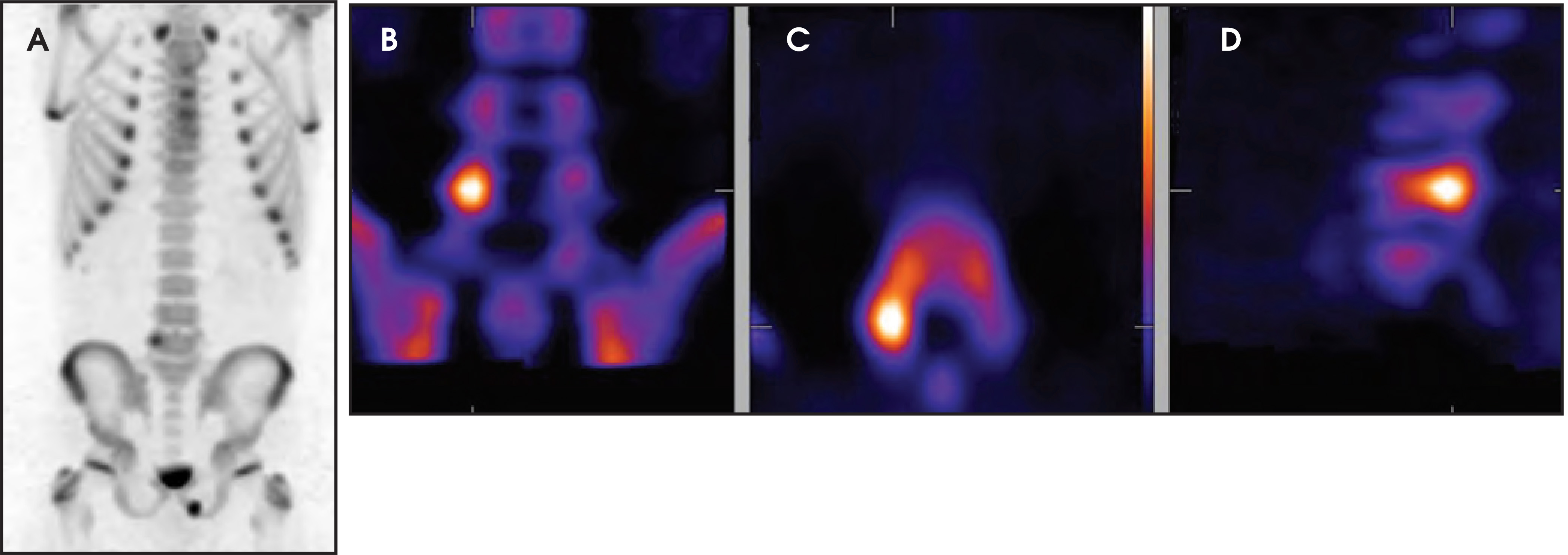
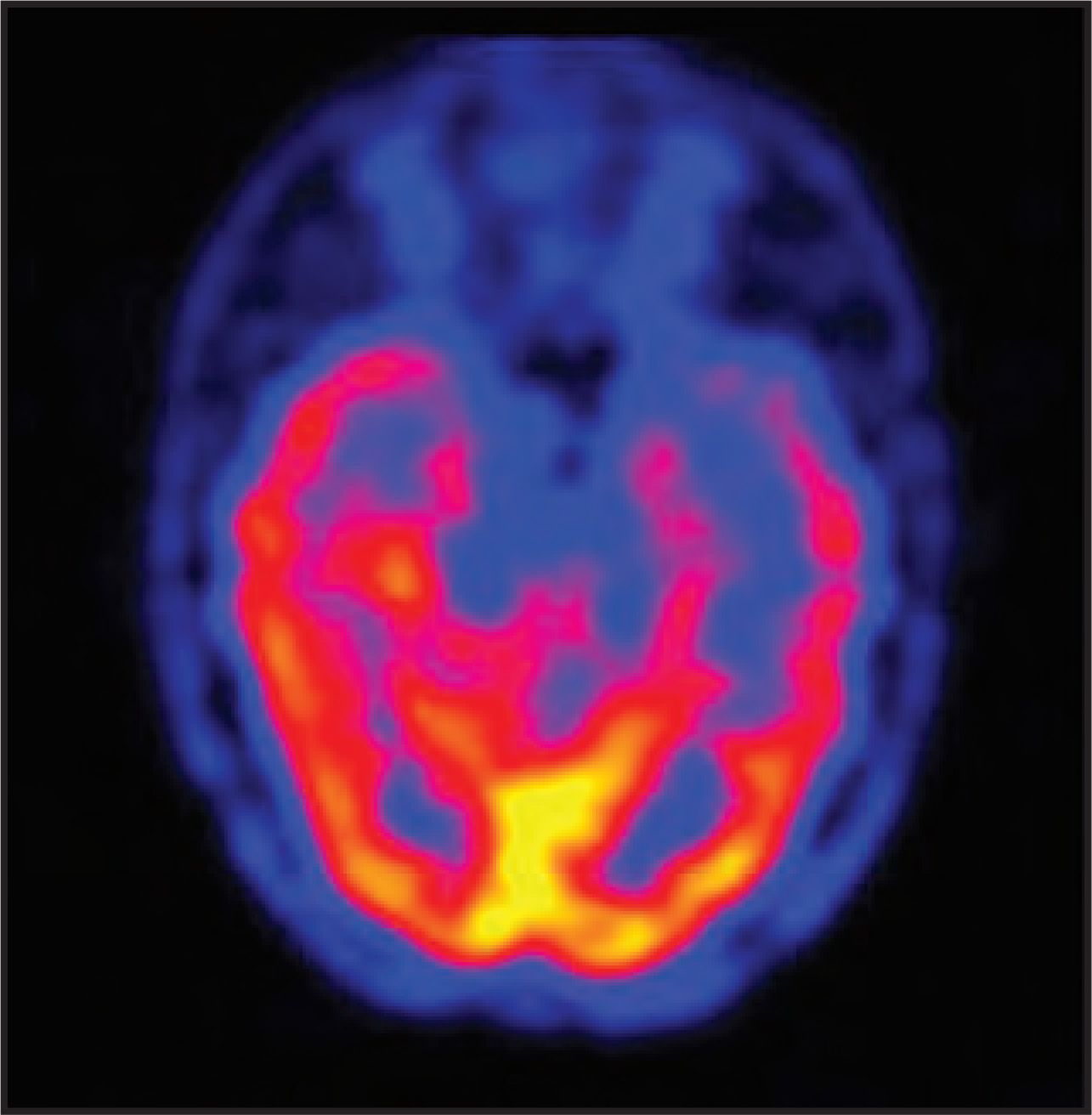
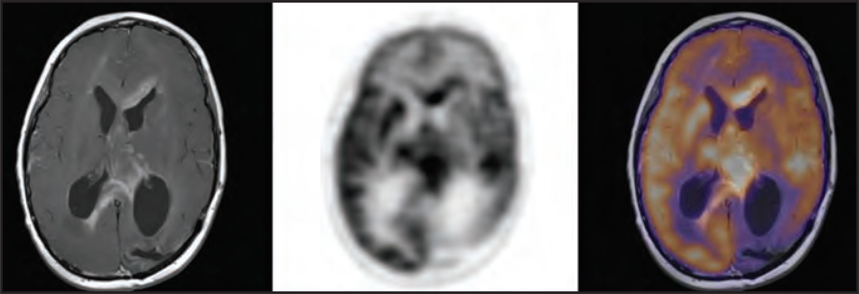
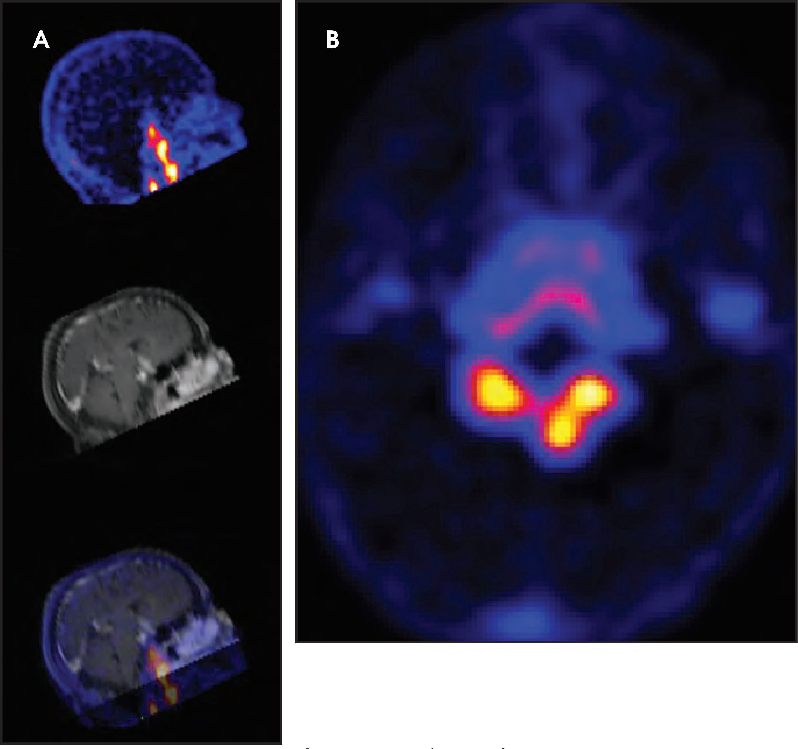
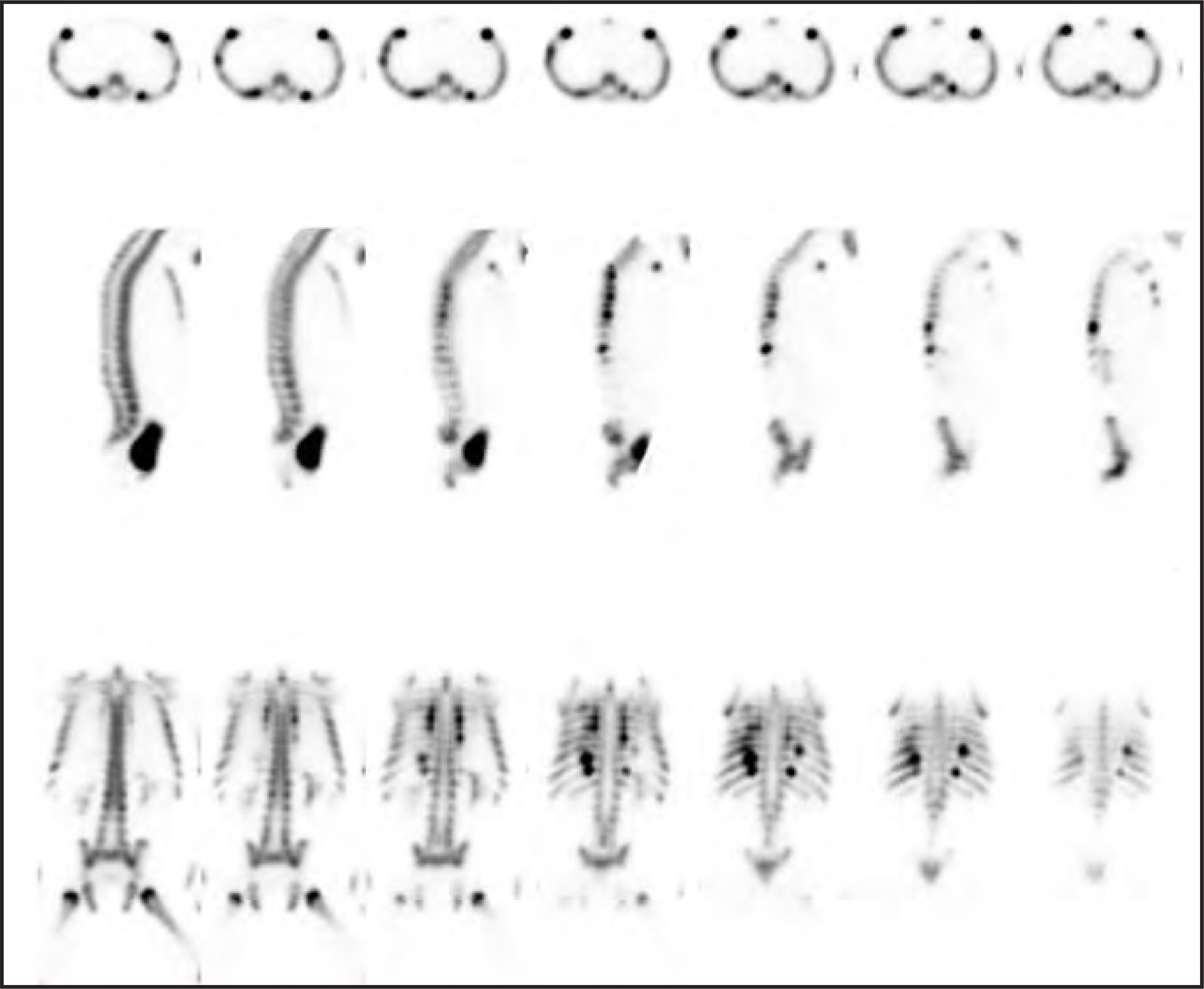
18F-labeled NaF is useful in the evaluation of back pain.16,17 Spondylolysis is a common cause of back pain in children, and PET imaging can produce detailed images of a pars defect (Figure 8). In general, NaF PET has been shown to have increased sensitivity in lesion detection with radiation doses similar to those of MDP bone scans. NaF has been shown to have particular benefit in the evaluation of child abuse.Recent studies have shown NaF PET to have improved sensitivity in detection of all fractures except classic metaphyseal lesions.18 It is particularly useful in early detection of posterior rib fractures (Figure 9). Often NaF PET can be used to supplement a skeletal survey in questionable cases. As research continues, growing acceptance of this agent and its increased utilization for various pathologies is likely.
Brain imaging
PET is highly useful in pediatric neuroimaging, primarily in seizure evaluation. There are multiple etiologies to be considered in the child or infant presenting with a seizure: infantile spasms, structural abnormalities (ie, cortical dysplasia), tuberous sclerosis/phakamotsis, metabolic conditions, trauma, and neoplasms, to name a few. When routine medical and imaging evaluation does not provide answers, further investigation with PET imaging, and typically single photon emission computed tomography (SPECT), is often employed.
PET of the brain is performed on patients with seizure disorders during the interictal phase, and it has been found to be more sensitive in lesion detection versus interictal SPECT. Regions of decreased FDG uptake on interictal exams correlate to seizure foci (Figure 10).19 PET and magnetic resonance imaging (MRI) fusion is particularly useful in localizing a lesion, and when fusion software is available, multiple modalities can be summed to best localize a lesion.
The other main indication for PET in brain imaging is in tumor evaluation. FDG uptake in neoplasms can be difficult to evaluate secondary to extensive normal background uptake in the brain. Some neoplasms demonstrate marked FDG avidity, and can be easily visualized on PET (Figure 11).20 PET can be beneficial in differentiating between residual tumor and radiation necrosis. Amino acid-labeled agents have shown promise for improved brain parenchyma imaging as there is minimal background uptake in normal brain obscuring neoplastic uptake (Figure 12).13,14
Conclusion
PET imaging in children is continuously evolving. It is being used to evaluate new pathologies, such as LCH, moyamoya, and fibromyxoid tumors. New radiopharmaceuticals are being developed and will likely provide clinical benefits in the future. Moreover, the fundamental technology is on the verge of advancing with PET/MR availability approaching the clinical arena. The future of PET imaging in children will undoubtedly continue to advance, and with this comes an exciting opportunity for the interpreting physician.
References
- McQuattie S. Pediatric PET/CT imaging: Tips and techniques. J Nucl Med Technol. 2008;36:171-178.
- Roberts E, Shulkin B. Technical issues in performing PET studies in pediatric patients. J Nucl Med Technol. 2004;32.
- Zukotynski K, Fahey F, Laffin S. Seasonal variation in the effect of constant ambient temperature of 24°C in reducing FDG uptake by brown adipose tissue in children. Eur. J. Nucl. Med. Mol. Imaging. 2010;37:1860-1854.
- Shammas A, Lim R, Charron M. Pediatric FDG PET/CT: Physiologic uptake, normal variants, and benign conditions. Radiographics. 2009;29:1467-1486.
- Chen Z, Li X, Li F. Evolving role of 18F-FDG-PET/CT for the body tumor and metastases in pediatrics. Euro J Radiol. 2010;75:329-335.
- Sharp S, Shulkin B, Gelfand M. 123I-MIBG scintigraphy and 18F-FDG PET in neuroblastoma. J Nucl Med. 2009;50:1237-1243.
- Taggart D, Han M, Quach A. Comparison of iodine-123 metaiodobenzylguanidine (MIBG) scan and [18F] fluorodeoxyglucose positron emission tomography to evaluate response after iodine-131 MIBG therapy for relapsed neuroblastoma. J Clin Oncol. 2009;27:5343-5349.
- Schlüter B, Bohuslavizki K, Beyer W. Impact of FDG PET on patients with differentiated thyroid cancer who present with elevated thyroglobulin and negative 131I scan. J Nucl Med. 2001;42:71-76.
- Stauss J, Franzius C, Pfluger T. Guidelines for 18F-FDG PET and PET-CT imaging in paediatric oncology. Eur. J. Nucl. Med. Mol. Imaging. 2008;35:1581-1588.
- Hudson M, Krasin M, Kaste S. PET imaging in pediatric Hodgkin’s lymphoma. Pediatr Radiol. 2004;34:190-198.
- McCarville M, Christie R, Daw N. PET/CT in the evaluation of childhood sarcomas. AJR Am J Roentgenol. 2005;184:1293-1304.
- Bredella M, Torriani M, Hornicek F. Value of PET in the assessment of patients with neurofibromatosis type 1. AJR Am J Roentgenol. 2007;189:928-935.
- Jacobs A, Thomas A, Kracht L. 18F-fluoro-L-thymidine and 11C-methylmethionine as markers of increased transport and proliferation in brain tumors. J. Nucl. Med. 2005;46:1958-1948.
- Rueger M, Ameli M, Li H. [18F]FLT PET for non-invasive monitoring of early response to gene therapy in experimental gliomas. Mol Imaging Biol. 2010;13:547-557.
- Grant F, Fahey F, Packard A. Skeletal PET with 18F-fluoride: Applying new technology to an old tracer. J Nucl Med. 2007;49:68-78.
- Lim R, Fahey F, Drubach L. Early experience with fluorine-18 sodium fluoride bone PET in young patients with back pain. J Pediatr Orthop. 2007;27:277-282.
- Ovadia D, Metser U, Lievshitz G. Back pain in adolescents: assessment with integrated 18F-fluoride positron-emission tomography-computed tomography. J Pediatr Orthop. 2007;27:90-93.
- Drubach L, Sapp M, Laffin S, Kleinman P. Fluorine-18 NaF PET imaging of child abuse. Pediatr Radiol. 2008;38:776-779.
- Sood S, Chugani H. Functional neuroimaging in the preoperative evaluation of children with drug-resistant epilepsy. Childs Nerv Syst. 2006;22:810-820.
- Kim S, Salamon N, Jackson H. PET imaging in pediatric neuroradiology: Current and future applications. Pediatr Radiol. 2010;40:82-96.
Citation
. PET/CT imaging in children: Current techniques and indications. Applied Radiology. 2012;41(6):8-13. doi:10.37549/AR1916.