Photon-counting digital mammography detects rare breast lesion
Applied Radiology — Vol. 42 , Issue 4 , pp. 28 -30
DOI: 10.37549/AR1986
Published: April 1, 2013
Categories
Case summary
A healthy 55-year-old woman presented for her annual screening mammogram. She had no personal or family history of breast cancer or heightened risk factors for breast cancer.
IMAGING FINDINGS
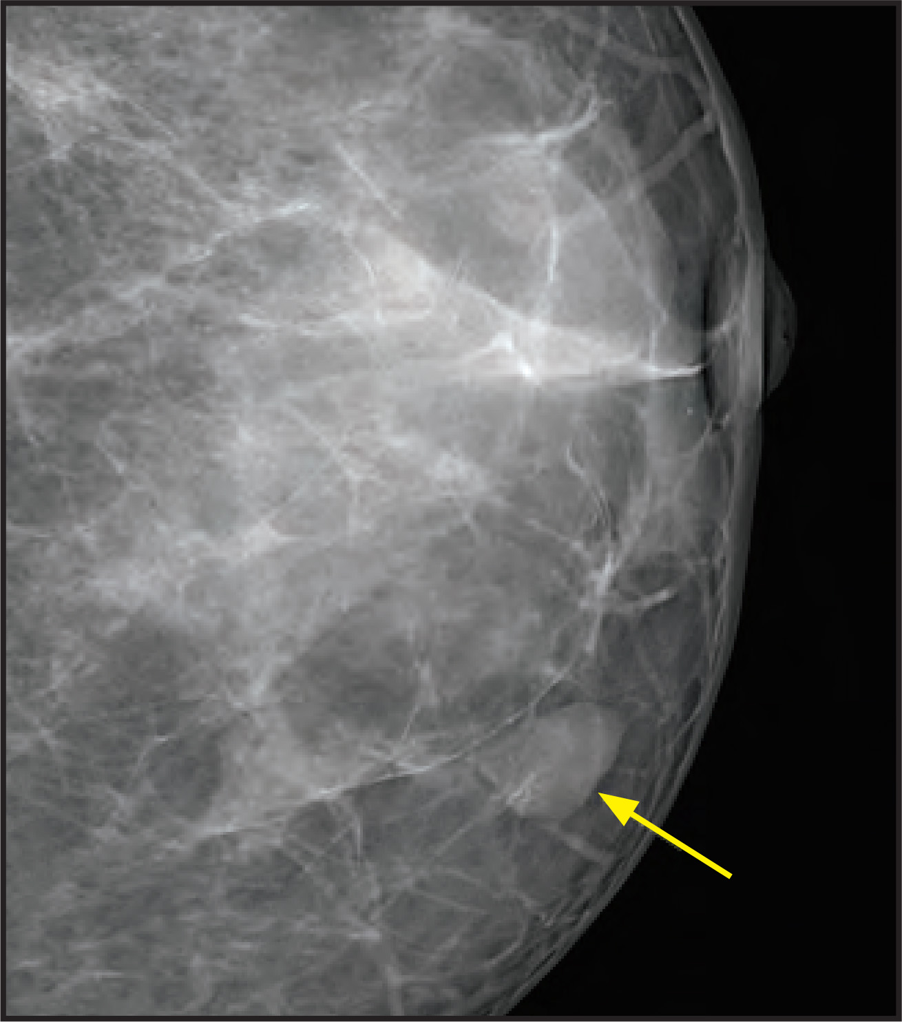
Screening mammography revealed a 1-cm focal asymmetry with a very subtle lobular shape and smooth, microlobulated margin at the inferomedial quadrant of the left breast, 2 cm from the nipple (Figure 1). No fatty hilus or malignant calcifications were identified.
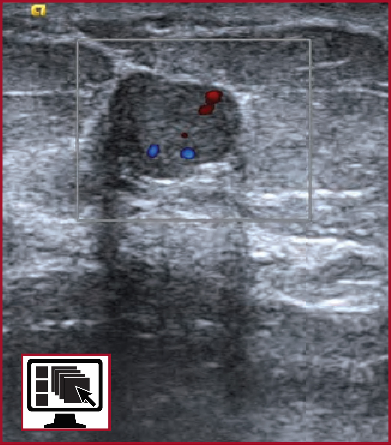
The patient was recalled from screening and additional diagnostic images and directed sonography confirmed an oval, well-circumscribed solid mass with smooth borders, and color Doppler flow (Figure 2). An uncomplicated ultrasound-guided core needle biopsy was completed.
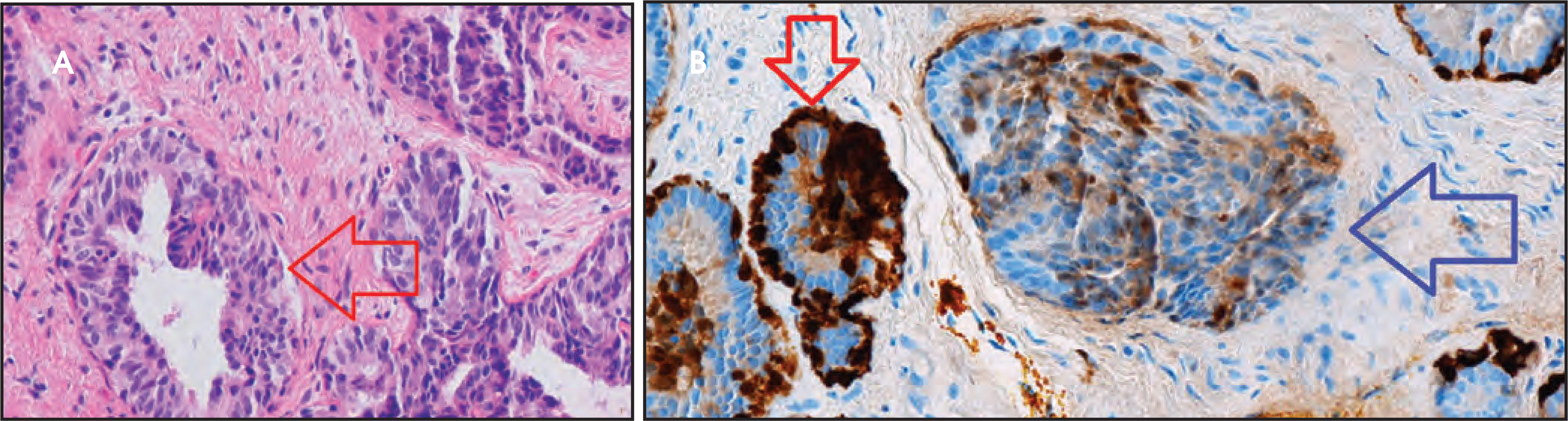
Hemotoxylin-eosin, S100, and MA903 histologic stains identified myoepithelial nuclear and cytoplasmic elements consistent with adenomyoepithelioma (Figure 3).
Diagnosis
Adenomyoepithelioma
Discussion
The Breast Imaging Reporting and Data System (BI-RADS) developed by the American College of Radiology enables the use of consistent language in the radiology report. The uniform description facilitates standardized communication between and among breast imagers. Mass shape (round, oval, lobular, irregular), margin (circumscribed, microlobulated, indistinct, ill defined, spiculated) and density (high, equal, low, fat-containing) are defined in BI-RADS.1 This case illustrates the importance of lesion characterization in a small, otherwise benign mass. The border lobulation in a nonaggressive-appearing mass triggered further evaluation. Though the lesion was small, its shape and margin were accurately identified on mammography. The sonographic features of uniformly iso- to hypoechogenic, circumscribed margins with no through transmission and positive vascularity on color Doppler were consistent with a solid mass.
We present the first published case of a lesion containing adenomyoepithelioma imaged with photon-counting digital mammography. Photon-counting digital mammography is the most recent fundamental improvement in mammographic x-ray detector technology. The 50-micron detector resolution and up to 50% dose reduction are unique to MicroDose (Philips Healthcare, Andover, MA). The very subtle characteristics of the mass shape and margin were accurately identified with the photon-counting mammography system; such subtle features in this case could easily have been misinterpreted as normal glandular tissue if the images were of the lower resolution (up to 4 times lower) produced by standard mammographic systems (personal communication).
Adenomyoepithelioma is a rare myoepithelial breast lesion, with only 150 cases documented in the literature. Patients range from 26 to 81 years.2 Most adenomyoepitheliomas are benign and occur in women.3 They do have malignant potential to metastasize hematogenously to the brain and lungs; however, only 40 of the 150 cases have been identified as having malignant potential.
The mass may present as a central, nontender, palpable abnormality or as an incidental finding on screening mammography. These tumors are characterized by a biphasic proliferation of epithelial and myoepithelial elements.4 The typical histologic appearance of benign adenomyoepithelioma consists of acinar structures, an inner layer of epithelial cells with eosinophilic cytoplasm, and a peripheral layer of myoepithelial cells.5 The myoepithelial lesions are immunoreactive with dark-brown staining for alpha-smooth muscle actin, calponin, and myosin. In the case of our patient, the specimen was positive for S100, a stain for epithelial cells, and MA903, a stain for keratins on myoepithelial cells. These markers are indicative of the adenomyoepithelioma histology (Figure 3).
This case illustrates the importance of accurate border and margin evaluation despite initial nonaggressive imaging features in routine screening mammography. Though the lesion appeared nonaggressive and lacking a fatty hilus on mammography or sonography, thereby excluding an intramammary lymph node, tissue diagnosis is imperative to evaluate other benign, nonaggressive masses. The margins were exquisitely sharp on photon-counting digital mammography and there were no microcalcifications. The differential diagnosis as described in the literature includes fibroadenoma, malignant myoepithelioma, spindle-cell carcinoma, aggressive fibromatosis, and various other myofibroblastic lesions (Table 1).6
The lesion margins must be accurately characterized as circumscribed, indistinct, or spiculated. In small lesions, subtle features may not be readily apparent on standard mammography. The nonaggressive lobulated margin is a key feature of adenomyoepitheliom; nonetheless it is as nonspecific as any other nonaggressive lesion.5 Variability in these characteristics is based on the cellular composition of the tumor. In our patient’s sonogram, the mass was circumscribed with no posterior sonographic enhancement. There was vascularity on color Doppler (Figure 2). This lesion is in contrast to typically mammography-malignant lesions, which have poorly defined margins and architectural distortion.5 As in our case, surgical excision is recommended even if the diagnosis at needle biopsy is benign adenomyoepithelioma. Pleomorphism may include fibroadenoma and malignant adenomyoepithelial elements. There is a risk of local recurrence several years after the first surgical excision.7
ACKNOWLEDGEMENT
The authors wish to thank Ted Williams, RT(R), Director of Radiology, United Medical Center, Washington, DC, and the Women’s Imaging Center team.
References
- Breast imaging reporting and data system, breast imaging atlas. 2003.
- Park Y, Okuyama N, Hatano Y. Adenomyoepithelioma of the breast: A case report and a review of literature. Breast Cancer. 1996;3:65-69.
- Ruiz-Delgado M, López-Ruiz J, Eizaguirre B. Benign adenomyoepithelioma of the breast: Imaging findings mimicking malignancy and histopathological features. Acta Radiol. 2007;48:27-29.
- Ahmed A, Heller D. Malignant adenomyoepithelioma of the breast with malignant proliferation of epithelial and myoepithelial elements: A case report and review of the literature. Arch Pathol Lab Med. 2000;124:632-636.
- Rosen P. Adenomyoepithelioma of the breast. Hum Pathol. 1987;18:1232-1237.
- Howlett D, Mason C, Biswas S. Adenomyoepithelioma of the breast: Spectrum of disease with associated imaging and pathology. AJR Am J Roentgenol. 2003;180:799-803.
- Loose J, Patchefsky A, Hollander I. Adenomyoepithelioma of the breast: A spectrum of biologic behavior. Am J Surg Pathol. 1992;16:868-876.
Citation
. Photon-counting digital mammography detects rare breast lesion. Applied Radiology. 2013;42(4):28-30. doi:10.37549/AR1986.