Pigmented villonodular synovitis of the elbow
Applied Radiology — Vol. 37 , Issue 9 , pp. 42 -43
DOI: 10.37549/AR1647
Published: September 1, 2008
Categories
CASE SUMMARY
A 73-year-old woman presented to the authors’ institution with increasing elbow pain and swelling. She denied any previous arthritic condition. The physical examination revealed nonspecific diffuse joint tenderness. Conventional radiography of the elbow showed nonspecificswelling around the joint and suggested a small joint effusion (not shown). Further evaluation was obtained with magnetic resonance imaging(MRI).
IMAGING FINDINGS
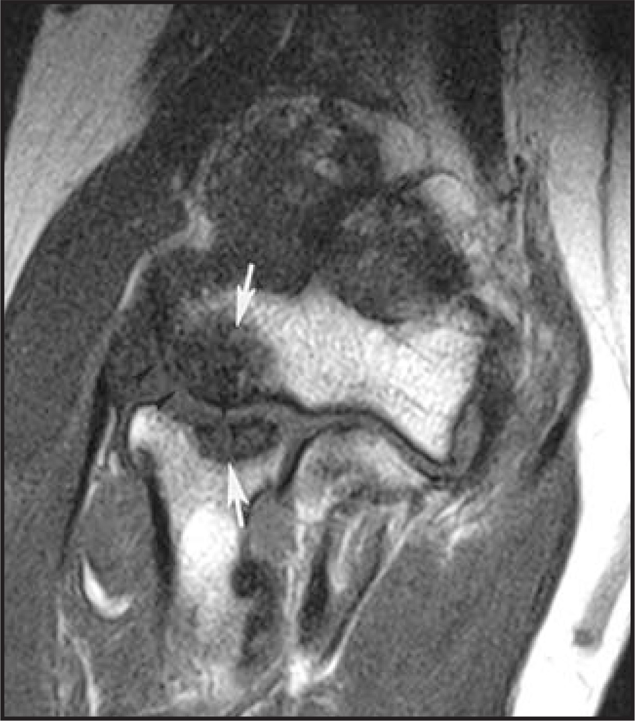
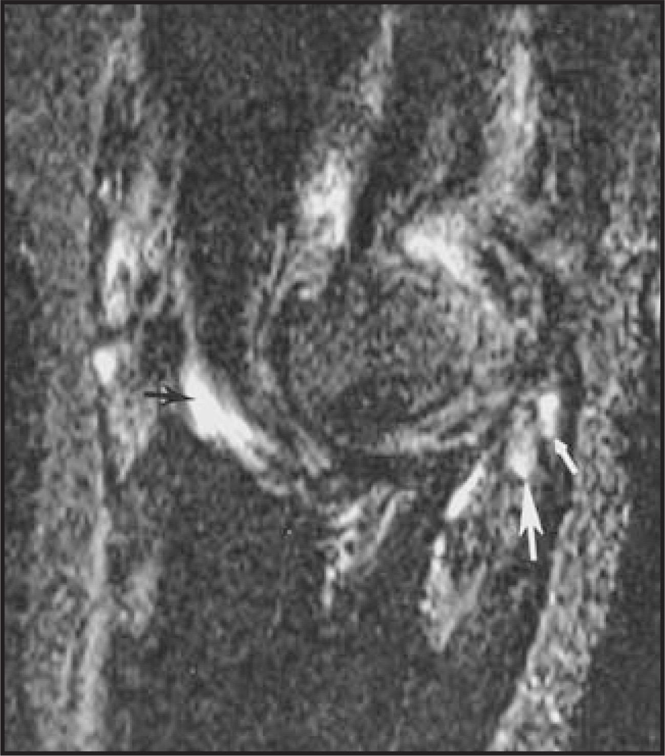
MRI revealed erosions of the articular surfaces of the radial head and capitellum and decreased subchondral marrow signal that is isointenseto the surrounding muscle (Figure 1). The ulnotrochlear joint shows subchondral cystic change involving the olecranon process (Figure 2). Thereis fluid surrounding the radial head just distal to the proximal radioulnar joint. There are also areas of decreased signal within the fluid, signifyingcomplex fluid and/or a synovial reaction.
DIAGNOSIS
Pigmented villonodular synovitis (PVNS) of the elbow
DISCUSSION
Pigmented villonodular synovitis is an uncommon disease that affects primarily tendon sheaths and less commonly joints and bursae.Characterized by hyperplastic synovium, large effusions, and bone erosions, the synovial proliferation may be either villous or nodular (or both).The form affecting the tendon sheath is known as the localized form of PVNS (LPVNS), and the diffuse form (DPVNS) affects the synovial membrane around joints.1
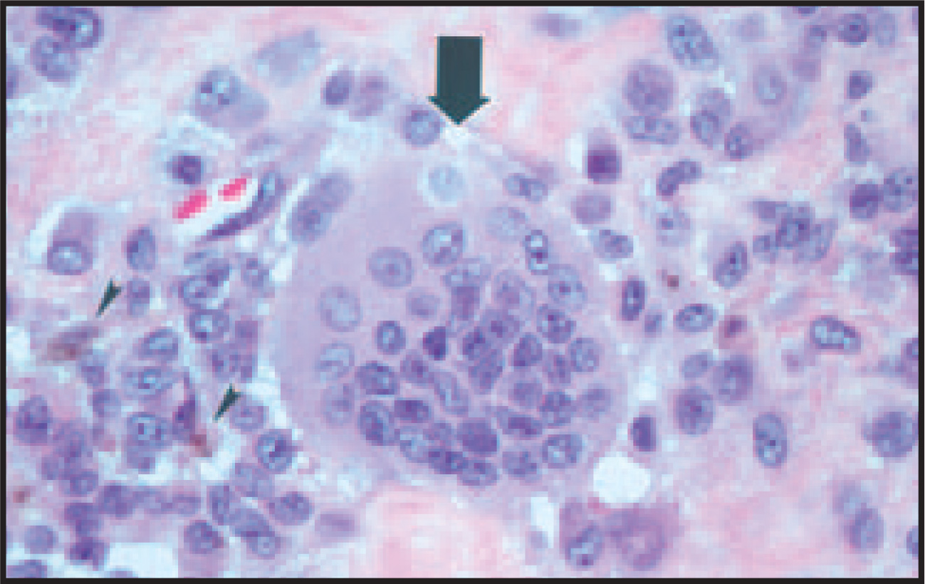
Grossly, the diffuse form has a mossy or nodular texture, spongy cut surfaces, and is most often rust colored. Microscopically, the synoviumis composed of fingerlike or rounded masses of fibrous stoma covered by hyperplastic lining cells. Large numbers of foamy macrophages in the stroma account for the yellow coloration, and the rusty color is caused by hemosiderin deposits in the stroma and in the cytoplasm of macrophages and synovial lining cells.2
Pigmented villonodular synovitis generally occurs between the ages of 20 and 45 years, but it has been found in patients as young as 11 years and as old as 70 years. The average duration of signs and symptoms before presentation is 2 to 3 years, with the total duration of symptoms totalling from 6 months to as long as 25 years.2 The cause of PVNS is unknown, but possibilities include a neoplastic or inflammatory process, a localized abnormal lipid metabolism with secondary inflammatory and traumatic changes, and a reactive response to chronic trauma and repeated hemorrhage. Most authors believe PVNS is either a localized aggressive neoplasm, or reactive synovitis.3
Limitation of motion may be described in 50% of patients.1 Joint enlargement, due to joint effusion or synovial masses, and tenderness to palpation are also commonly reported.1,4 The physical examination may, however, be noncontributory. Initial patient presentation mostcommonly includes complaints of monoarticular pain and swelling, most commonly at the knee (80% of cases), followed by the hip, ankle, small joints of the hands and feet, shoulder, and elbow.1 The treatment of choice is synovectomy, and recurrence rates range from 21% to48%.
Elbow involvement is rare. A literature review yielded only 18 case reports of PVNS that involved the elbow. It is both the site of involvement and the patient’s advanced age that makes this case unique, as this case is the oldest known case of elbow PVNS.
Fifteen of the 18 previously reported cases of PVNS were classified as the diffuse type and 1 case was classified as the focal type. The remaining 2 cases were unclassified. MRI of this case showed erosions on both sides of the joint, which is consistent with a synovial process (Figure 1). The pattern of involvement in this case may be categorized as diffuse, similar to the majority of cases reported previously.
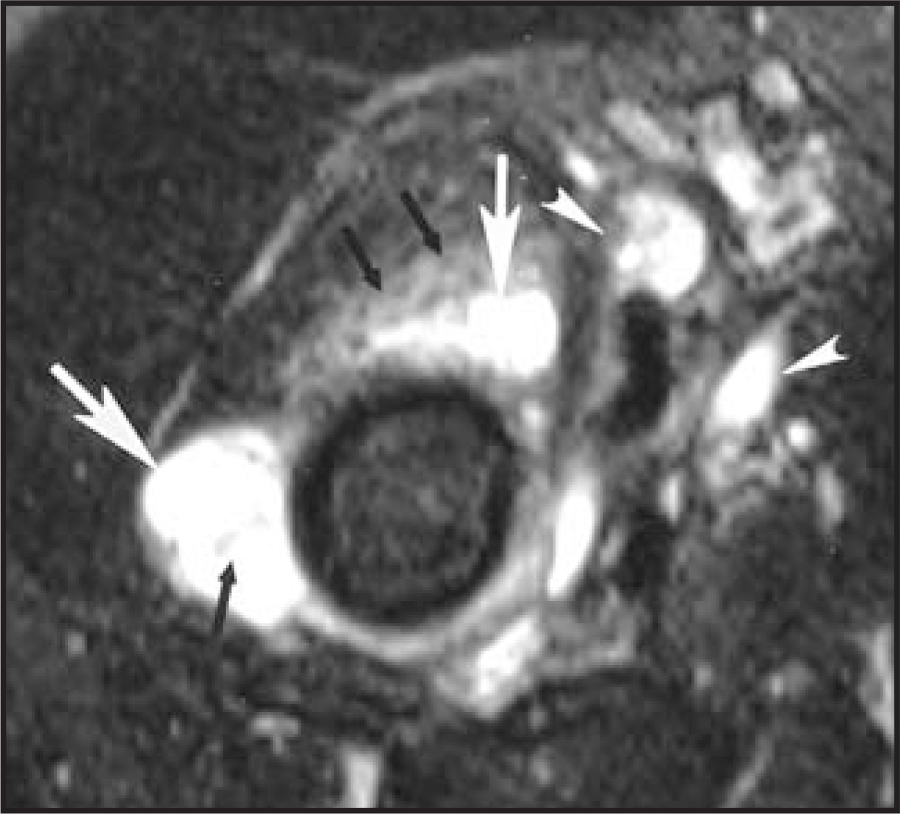
In 13 of 18 of the published cases of elbow PVNS, imaging depicted bony erosion, which was also found in this case (Figure 1).5 Small bony erosions must also be differentiated from subchondral cystic change caused by degenerative arthritis (PVNS also predisposes to degenerative arthritis). In this case, there was evidence of degenerative arthritis along with subchondral cystic change (Figure 2), in addition to the previously described erosions. Although hemosiderin is commonly seen in the diffuse form of PVNS, it may not be consistently present as seen in this case. PVNS may also surround the tendons (Figure 3).
The diagnosis of PVNS is often difficult, and the final diagnosis of PVNS should be confirmed by biopsy (Figure 4). This will allow confident differentiation from other conditions that may mimic PVNS, such as rheumatoid arthritis, osteoarthritis, and other inflammatory andneoplastic processes of the synovial lining.
CONCLUSION
Pigmented villonodular synovitis is an uncommon disease that affects tendon sheaths most commonly, as well as joints and bursae. While the etiology is unclear, it is commonly believed that PVNS is either a localized aggressive neoplasm or reactive synovitis. The use of MRI can aid in the diagnosis of PVNS. Biopsy should be performed to confirm the diagnosis histologically.
References
- Frassica F. Pigmented villonodular synovitis of the hip and knee. Am Fam Physician. 1999;60:1404-1410.
- Dorwant R, Genant H, Johnston W, Morris J. Pigmented villonodular synovitis of synovial joints: Clinical, pathologic, and radiologic features. AJR Am J Roentgenol. 1984;143:877-885.
- Jaffe H, Lichtenstein L, Sutro C. Pigmented villonodular synovitis, bursitis, and tenosynovitis. Arch Path. 1941;31:731-765.
- Bravo S, Winalski C, Weissman B. Pigmented nodular synovitis. Radiol Clin North Am. 1996;34.
- Pimpalnerkar A, Barton E, Sibly T. Pigmented villonodular synovitis of the elbow. J Shoulder Elbow Surg. 1998;7:71-75.
Citation
. Pigmented villonodular synovitis of the elbow. Applied Radiology. 2008;37(9):42-43. doi:10.37549/AR1647.