Positron emission tomography imaging of lung and esophageal cancer
Applied Radiology — Vol. 31 , Issue 6 , pp. 9 -17
DOI: 10.37549/AR1097
Published: June 1, 2002
Categories
Positron emission tomography (PET) imaging is strongly indicated for the diagnosis of solitary pulmonary nodules and for the diagnosis, staging, and restaging of both non–small-cell lung cancer and esophageal cancer. PET is also used in guiding treatment plans, monitoring therapeutic response, and detecting tumor recurrence. PET imaging can also be used to guide invasive diagnostic procedures by determining the most readily accessible and metabolically active lesions. In surgically high-risk patients, PET can be an alternative to biopsy or surgical evaluation. PET is as sensitive as transthoracic needle aspiration (TTNA) biopsy with less risk in identifying malignant pulmonary lesions.1,2 Other uses of PET include planning radiotherapy fields, measuring tumor aggressiveness, and assessing prognosis.
These applications stem from the metabolically key glucose analogue, fluorine-18-labeled fluoro-2-deoxy-D-glucose (FDG). This molecule is similar enough to glucose to be transported through the cell membrane and to be phosphorylated by hexose-6-phosphate. The cellular enzymatic machinery that processes glucose cannot further metabolize FDG, and the molecule becomes trapped inside tumor cells.3 Image region localization that appears greater than blood pool activity is often associated with malignancy, especially for foci smaller than 1.5 cm in size. The amount of FDG localized in a tumor is characterized by comparing its uptake to the total body administered dose. The standardized uptake value (SUV) or standardized uptake ratio is defined as the FDG concentration in the region of interest to the average FDG concentration in the body (injected dose divided by [lean] body mass). The factors that can affect the SUV include the body surface area (distribution of FDG is higher in muscle than in fat), the time after the FDG injection, the partial volume effects, and the blood glucose level at the time of injection. An SUV >2.5 is sensitive and specific for malignant lesions.2,4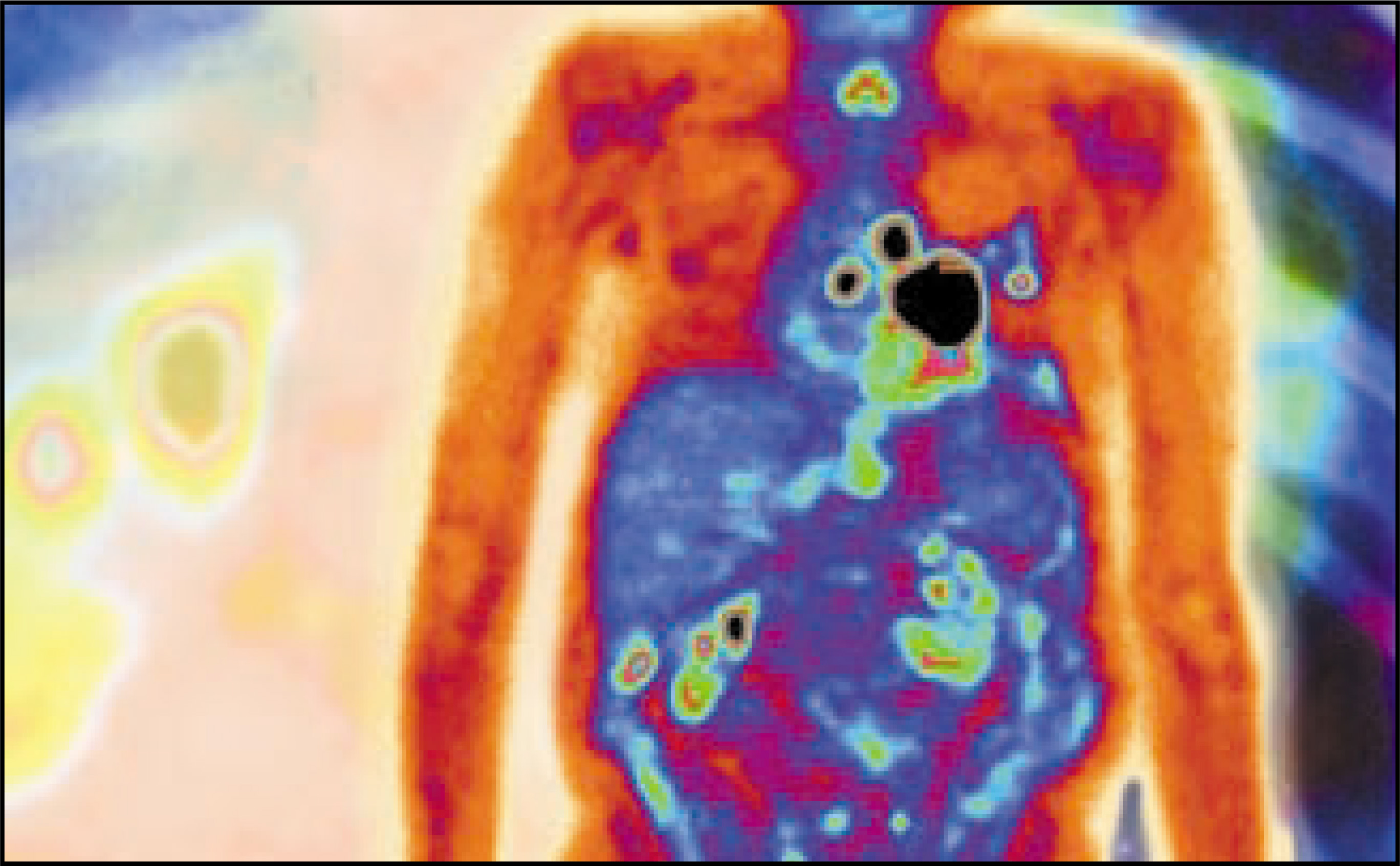
The following discussion focuses on the use of PET in three important areas in thoracic oncology: Evaluation of solitary pulmonary nodule (SPN), the staging and management of non–small-cell lung cancer (NSCLC), and the workup of patients with esophageal cancer.
Solitary pulmonary nodule
Patients with an SPN rarely have symptoms attributable to the nodule, and so the detection of the SPN is usually serendipitous. The plain chest radiograph usually defines the presence and appearance of the SPN, unless it was discovered on CT or other radiographic imaging performed for another purpose. The lesion must be singular, surrounded by normal lung tissue, and not be involved with obstructive atelectasis or hilar enlargement. There are many benign and malignant processes that may present as a solitary pulmonary nodule (SPN) on a chest radiograph (Table 1). The most common benign causes of SPNs are granulomas from histoplasmosis, coccidioidomycosis, and mycobacteria. Hamartomas are the most common benign neoplasms and constitute approximately 10% of benign nodules.
Bronchogenic carcinoma is, by far, the most common malignant lesion in surgical series of SPNs. Adenocarcinoma and large-cell carcinoma account for more peripheral nodules than squamous and small-cell carcinomas, although all histologic types of lung cancer may present as an SPN. Metastatic lesions from non-lung primary tumors constitute about 10% to 30% of resected malignant nodules. The most frequent sources of metastasis are squamous carcinomas of the head and neck and adenocarcinomas of the breast, kidney, and colon.
Most stage I lung cancers (T1-2,N0,M0) are within the definition of SPN (Figure 1). The 5-year survival for resection of stage I bronchogenic carcinoma is 75% and is more than 80% for lesions <3 cm. Ideally, all malignant SPNs would be resected shortly after detection, and all benign lesions would be identified without surgical intervention. The overall goal in the evaluation of the SPN is to resect potentially curable cancers expeditiously and to avoid surgical resection of benign nodules. As many nodules are indeterminate in appearance, the presence of an SPN presents a diagnostic dilemma.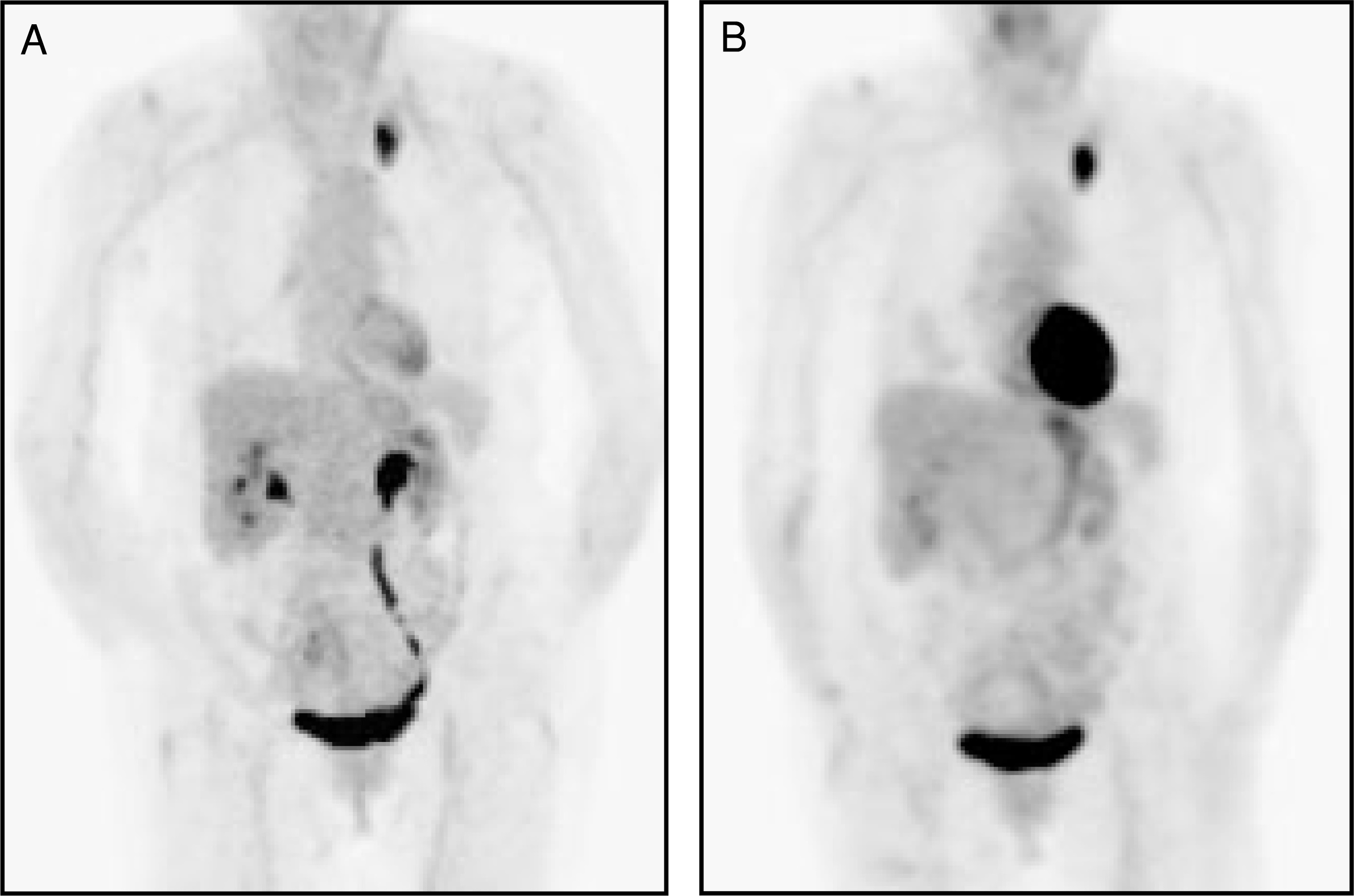
Increasing incidence of SPN malignancy has been demonstrated with advancing age. The probability that a nodule is malignant also increases with increasing size of the nodule. Approximately 93% to 99% of nodules >3 cm in CT diameter are malignant. The rate of enlargement is also important. Malignant pulmonary nodules have doubling times between 21 and 400 days. Shorter times are usually related to infections and longer times are nearly always benign growths. How a nodule looks (ie, its size, shape, pattern of calcification, and whether there are any surrounding or “satellite” lesions) provides important clues about whether or not it is cancerous. Radiographically, malignant SPNs tend to have lobulated or shaggy borders, and there is usually some distortion of the adjacent blood vessels. A calcification pattern that appears irregular or spotty is a good indication of malignancy. Other characteristic features that may appear on the radiograph include a tail on the lesion and a corona radiata (a soft halo around the lesion). The presence of calcification within a nodule on plain film, tomography, or a CT scan is a reliable indicator that the nodule is benign. Granulomas classically may show a laminated or a concentrically ringed calcification pattern. Other benign calcification patterns include central, diffuse, and “popcorn ball,” which may be seen with hamartomas. Unfortunately, about 10% of malignant lesions show evidence of calcification on plain chest film.
With the possible exception of the heavy central calcification characteristic of an old granuloma, lesion morphology is not a reliable indicator of whether a nodule is benign or malignant. Absolute CT density is also unreliable and irreproducible. CT contrast enhancement may provide important help. In a study of 163 patients, Swensen5 reported a sensitivity of 100%, a specificity of 76.9%, and an overall accuracy of 93% in identifying malignant neoplasms by the amount of CT contrast enhancement. Malignant neoplasms enhanced significantly more (>20 Hounsfield units) than granulomas and benign neoplasms, although hamartomas and active granulomas also showed high enhancement. The degree of enhancement was related to the amount of central vascular staining in histologic evaluation of surgical specimens.
Given a radiographically indeterminate nodule <3 cm in diameter, patients can be managed by observation, biopsy, or thoracotomy. Observation involves careful follow-up with serial chest radiographs every 3 months for the first year, every 6 months for the second year, and yearly thereafter, if necessary, to exclude the possibility of a slow-growing malignancy. Whether life expectancy changes if malignant SPNs are observed for growth is unknown. Some studies suggest that the prognosis (outcome) is the same whether immediate action involves no action at all, surgery, or biopsy. On the other hand, survival appears to be longer among patients following resection of small malignant nodules compared with larger ones. Further, it is difficult for most patients to live with the uncertainty of whether a malignancy is being left untreated. Unfortunately, immediate resection of indeterminate nodules requires expensive and invasive thoracotomies for a large number of benign lesions that might have been identified by observation or biopsy techniques.
Biopsy by means of TTNA has a diagnostic yield of 43% to 97% in peripheral pulmonary lesions. For malignant lesions <2 cm in size, the yield of a positive tissue diagnosis is about 60%. Pneumothorax, the most frequent complication of TTNA, is in the range of 15% to 30%, with approximately half requiring tube thoracostomy. Although bronchoscopy with transbronchial biopsy is a low-risk procedure, the likelihood of obtaining a diagnostic specimen is approximately 10% for nodules <2 cm in diameter and 40% to 50% for nodules 2 to 4 cm in diameter.
An SPN can be removed via video-assisted thoracoscopic surgery (VATS) if it is smaller than 2 to 3 cm in diameter and is located <2 cm from the pleural surface. However, nearly half of the lesions removed using VATS are benign. Considering the expense and potential morbidity of thoracoscopy, PET appears to offer a less expensive, less invasive, and a more specific diagnostic alternative.
FDG PET imaging can be used to determine, based on its metabolic utilization of glucose, whether an SPN is benign or malignant. Dewan et al6 showed that PET scanning was able to identify malignant lesions with a sensitivity of 95% to 100% and a specificity of 80% to 89%, respectively. Using an SUV ≥ 2.5 as an indicator of malignancy, FDG PET has a sensitivity and specificity ranging from 83% to 100% and 63% to 90%, respectively (Figure 2). Tumors with high FDG uptake (SUV >10) and diameter >3 cm have the worst prognosis, with a survival time of less than 6 months. Studies suggest a strong association between PET and cell differentiation, which in turn correlates with prognosis.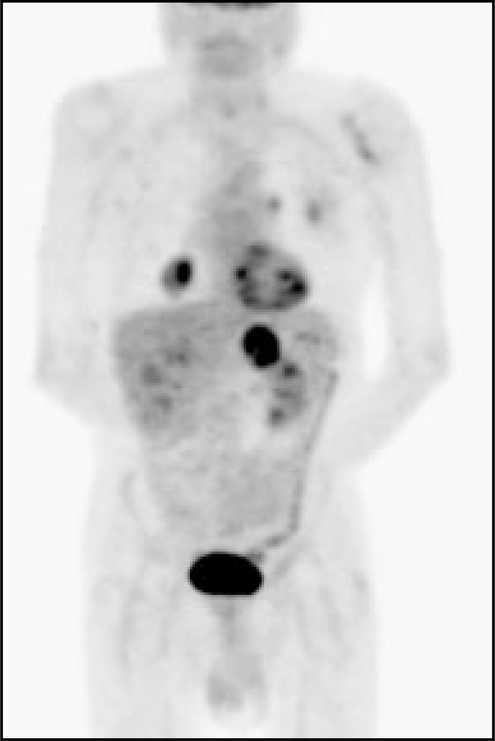
PET may be used to guide invasive diagnostic procedures by determining the most readily accessible and metabolically active lesions. In surgically high-risk patients, PET can be an alternative to biopsy or surgical evaluation. PET is as sensitive as TTNA biopsy in identifying malignant pulmonary lesions with less risk.
False-positive results can be seen with granulomas (tuberculosis, histoplasmosis, aspergillosis, cryptococcosis, and inflammatory pseudotumor), or inflammatory processes (sarcoid, Wegener’s), and rheumatoid nodules. In these conditions, increased FDG uptake may be related to enhanced glycolytic activity in activated macrophages.
False-negative examinations can occur in small lesions (under 0.7 to 0.8 cm in size) or in neoplasms having low metabolic activity (ie, bronchoalveolar cell carcinoma and carcinoid tumors). Competitive inhibition from high-serum glucose levels (>250 to 300 mg/dL) interferes with tumor cell FDG uptake. This is more pronounced in acute hyperglycemia while a chronic increase in glucose level results in less inhibition. PET imaging should be postponed until the serum glucose is <200 mg/dL.
PET in NSCLC
Non–small-cell lung cancer includes different histopathological cell types (adenocarcinoma, squamous-cell carcinoma, large cell, and mixed cell histologies) and comprises 75% to 80% of all new cases of lung cancer.7 Squamous-cell carcinoma associated with major bronchi is the most common type, followed by adenocarcinoma, the most common type of lung cancer in people who have never smoked. This usually arises in the peripheral regions of the lung under the bronchial mucosa. Large-cell carcinoma also presents in the lung periphery.
In NSCLC, tumor stage is the most important prognostic factor that guides treatment planning. Patients with metastasis to the mediastinal lymph nodes have an average 5-year survival rate of approximately 10% compared with a survival rate of 50% in the absence of mediastinal metastases.8 Unfortunately, surgery with curative intent is an option in only 30% of patients. Patients with nonresectable but loco-regionally confined disease may have prolonged survival and even cure with radical radiotherapy. The combination of radiation to 60 to 66 Gy and platinum-based chemotherapy is a common approach in inoperable patients.
Unfortunately, conventional staging commonly underestimates the true extent of non–small-cell lung cancer. PET has proven to be more sensitive and specific compared with conventional imaging of NSCLC in several important areas, principally in staging of the mediastinum and in the detection of distant metastases (Figure 3).1,10-17 A recent analysis of 40 studies showed that PET is a highly accurate noninvasive imaging test for the diagnosis of pulmonary nodules and larger mass lesions. The mean sensitivity and specificity were 96.8% and 77.8%, respectively.18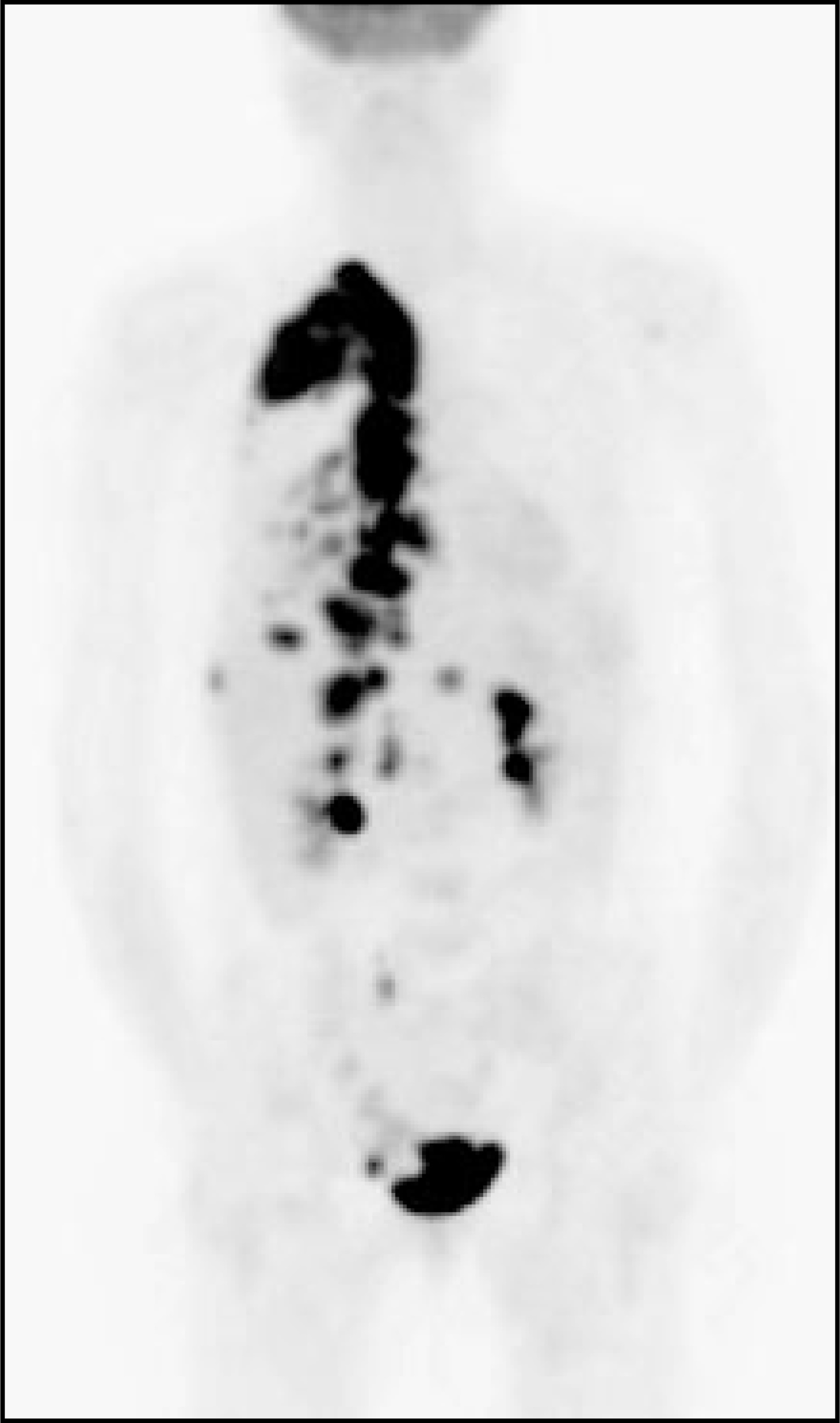
FDG PET imaging can have a significant impact on patient management by heightening suspicion for pulmonary malignancy, identifying unsuspected sites of disease, and by guiding selection of a biopsy site. Similarly, a negative PET can indicate a low likelihood of malignancy and supports the use of conservative management and follow-up. PET scans influence treatment in 65% of patients with NSCLC and offer new information in 85% of patients.14-16,18
FDG PET has been found to be superior to CT, MRI, and mediastinoscopy in the nodal staging of bronchogenic carcinoma. In suspected or proven lung cancer, PET is equally accurate and reliable for detecting disease in small (<1 cm) and large (>3 cm) lymph node lesions, with better accuracy than CT. In a study by Wahl,19 the diagnostic accuracy for PET was 92% versus 75% for CT. The positive predictive value for PET was 90% versus 50% for CT and the negative predictive value was 93% for PET versus 85% for CT. A meta-analysis of 14 PET studies and 29 CT studies showed PET to be superior to CT in mediastinal imaging with a mean sensitivity and specificity of 79% and 91%, respectively, for PET in contrast to 60% and 77%, respectively, for CT.19 Vansteenkiste et al20 also found a high negative predictive value (86%) of FDG PET for disease in the mediastinal lymph nodes. It has been suggested that negative PET results can be used as a basis for proceeding to potentially curative thoracotomy even though a small fraction of patients have lymph node involvement undetected by FDG PET.20
One of the benefits of FDG PET is that the whole body can be imaged without additional radiation exposure. At least 10% of patients are found to have metastatic disease on PET scanning when routine CT scan fails to show evidence of metastasis (Figure 4).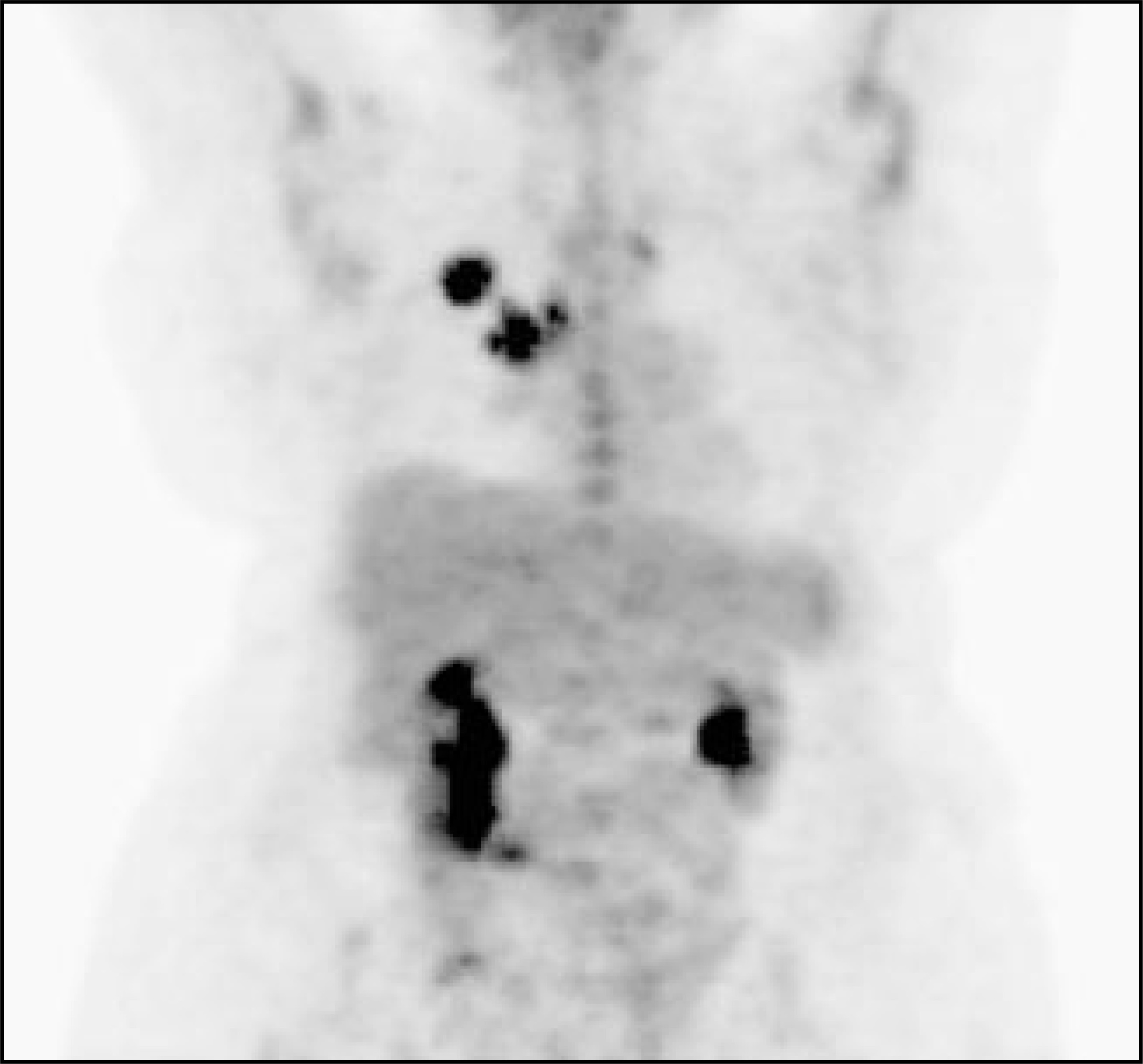
Between 30% and 50% of patients with resected non–small-cell lung cancer will develop recurrent tumor. FDG uptake in NSCLC has been correlated with tumor growth rate, aggressiveness, and proliferation capacity. The higher the SUV, the higher the aggressiveness of the tumor and the worse the prognosis.21
Determination of the extent of the primary tumor and of nodal involvement is crucial for successful surgery and radical radiotherapy. Most patients treated with radical chemotherapy relapse with disease progression in the thorax or with distant metastasis, suggesting that, in many cases, the initial staging assessment underestimated the true extent of disease. Accurate staging helps to avoid futile surgery or radical radiotherapy in patients with incurable extensive disease. Bradley22 reported that gross tumor volume was the sole independent predictor of survival in NSCLC treated with conformal radiotherapy indicating the importance of accuracy in tumor delineation. High-dose radiotherapy is of little value if existent tumor is not included in the target volume.
PET imaging has been very useful in assessing the response to chemotherapy or radiation therapy in patients with advanced NSCLC (Figure 5). Decrease in FDG uptake after treatment may prove to be a better indicator of a favorable response rather than change in tumor size. In a recent study, all patients with negative post-therapy PET findings were alive 2 years after completion of treatment, whereas in the group with residual hypermetabolism, 50% of patients died.23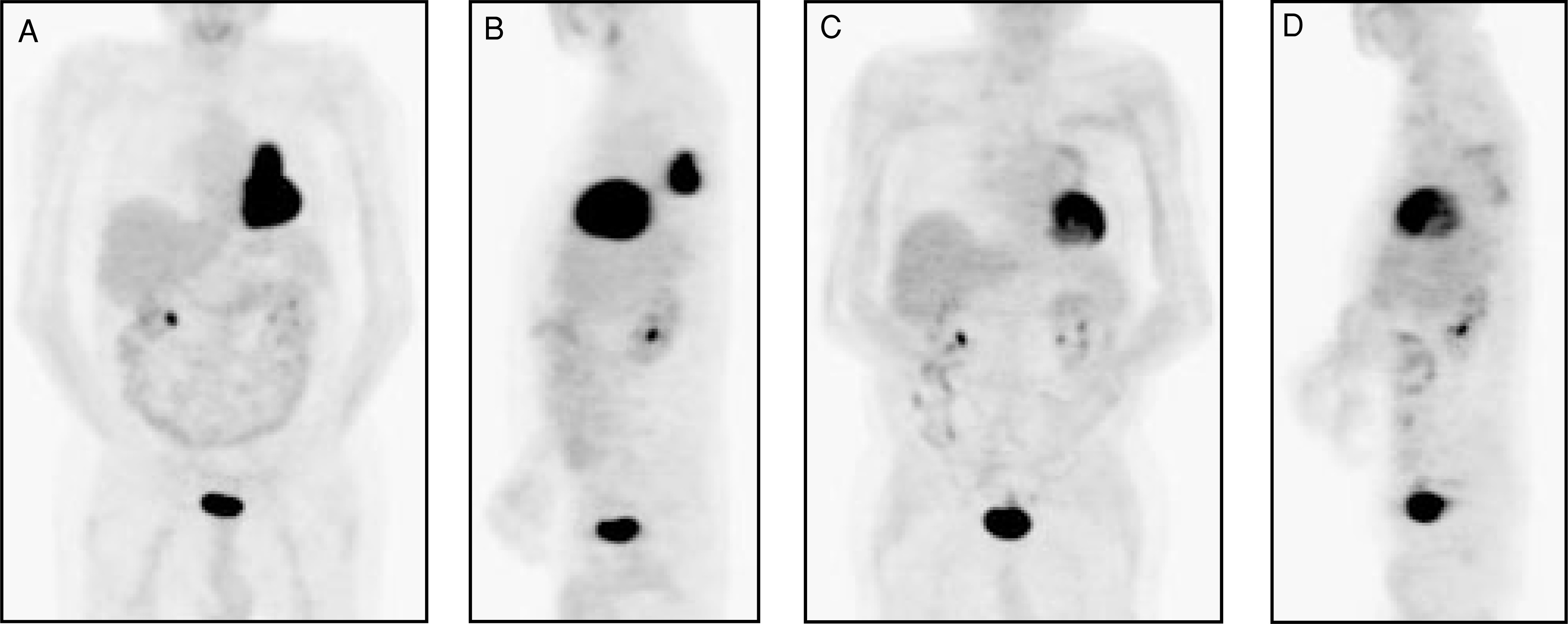
PET imaging is very sensitive and highly accurate in distinguishing recurrent malignancy from scarring or fibrosis and from radiation-induced benign pleural thickening (Figure 6). It has been shown to have a sensitivity of 98% to 100% for the differentiation of posttreatment scar from tumor recurrence. There are potential pitfalls when PET is used for the purpose of differentiating hypermetabolic inflammatory changes induced by radiation therapy (Figure 7) from recurrent tumor. Radiation produces a diffuse mildly elevated FDG accumulation within the tissues, which is due to the inflammatory changes caused by the radiation. This activity decreases over time (3 to 6 months).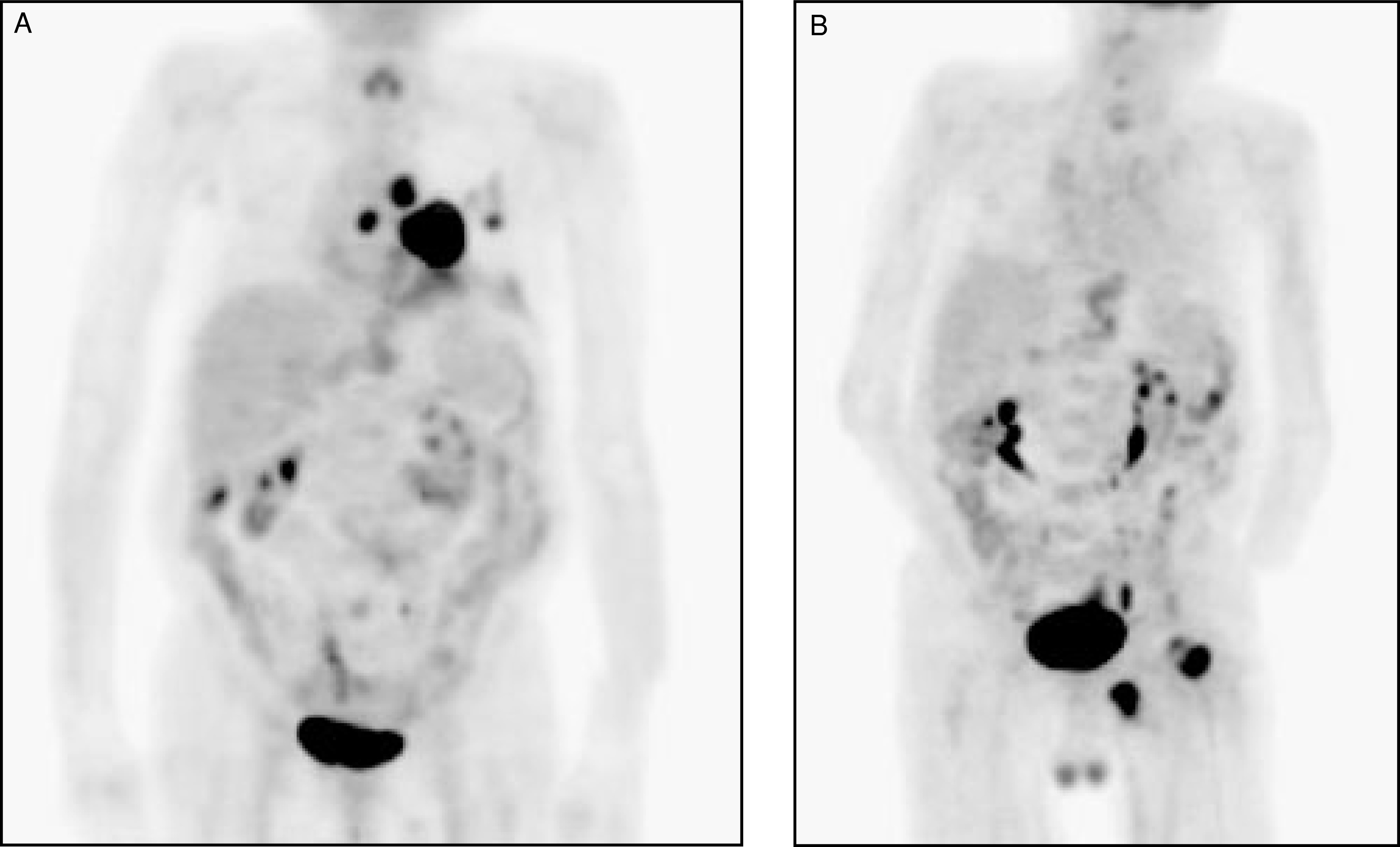
PET in esophageal cancer
Approximately 13,200 Americans are diagnosed with esophageal cancer and 12,500 die from this malignancy annually.24 There are two histologic types of esophageal carcinoma that account for the majority of malignant cases: Squamous-cell carcinoma (>75% to 90%) and adenocarcinoma. Esophageal cancer tends to be aggressive in its behavior. It invades locally, spreads to local lymph nodes, and then metastasizes throughout the body. Approximately 15% of esophageal cancers occur in the upper third of the esophagus, 45% in the middle third of the esophagus, and 40% in the distal third of the esophagus.
Patients with esophageal carcinoma have a poor prognosis. Although it is a disease that can be treated, it can rarely be cured. By the time the patient becomes symptomatic, their disease is usually at an advanced stage. The overall 5-year survival rate in patients who undergo surgery ranges from 5% to 20%, while the 5-year survival rate in patients with lymph node metastases (nonsurgical patients) ranges from 0% to 7%.24 Once the diagnosis of esophageal cancer has been made, staging is the next critical step in determining the most appropriate treatment plan for the patient.
One of the major difficulties in planning treatment for patients with esophageal cancer is the lack of precise preoperative staging. Noninvasive imaging modalities include CT, endoscopic ultrasound (EUS), and FDG PET. The overall staging accuracy of EUS in esophageal cancer is 85% to 90%, as compared with 50% to 80% for CT.25 The reported sensitivities for FDG PET imaging is between 91% and 100%. False-positive uptake can occur due to inflammation, and there can be normal mild FDG activity from muscular contractions. The accuracy of regional nodal staging is 70% to 80% for EUS, while CT has an accuracy of 40% to 73% for the detection of pathologic mediastinal nodes (using a 1-cm size criteria).25 The reported accuracy of FDG PET in the staging of regional lymph node metastases ranges from 24% to 90%.25 The major limitation of FDG PET with regard to the detection of nodal metastases adjacent to the primary tumor is its relatively poor spatial resolution (approximately 6 mm for a dedicated PET scanner), which reduces sensitivity.
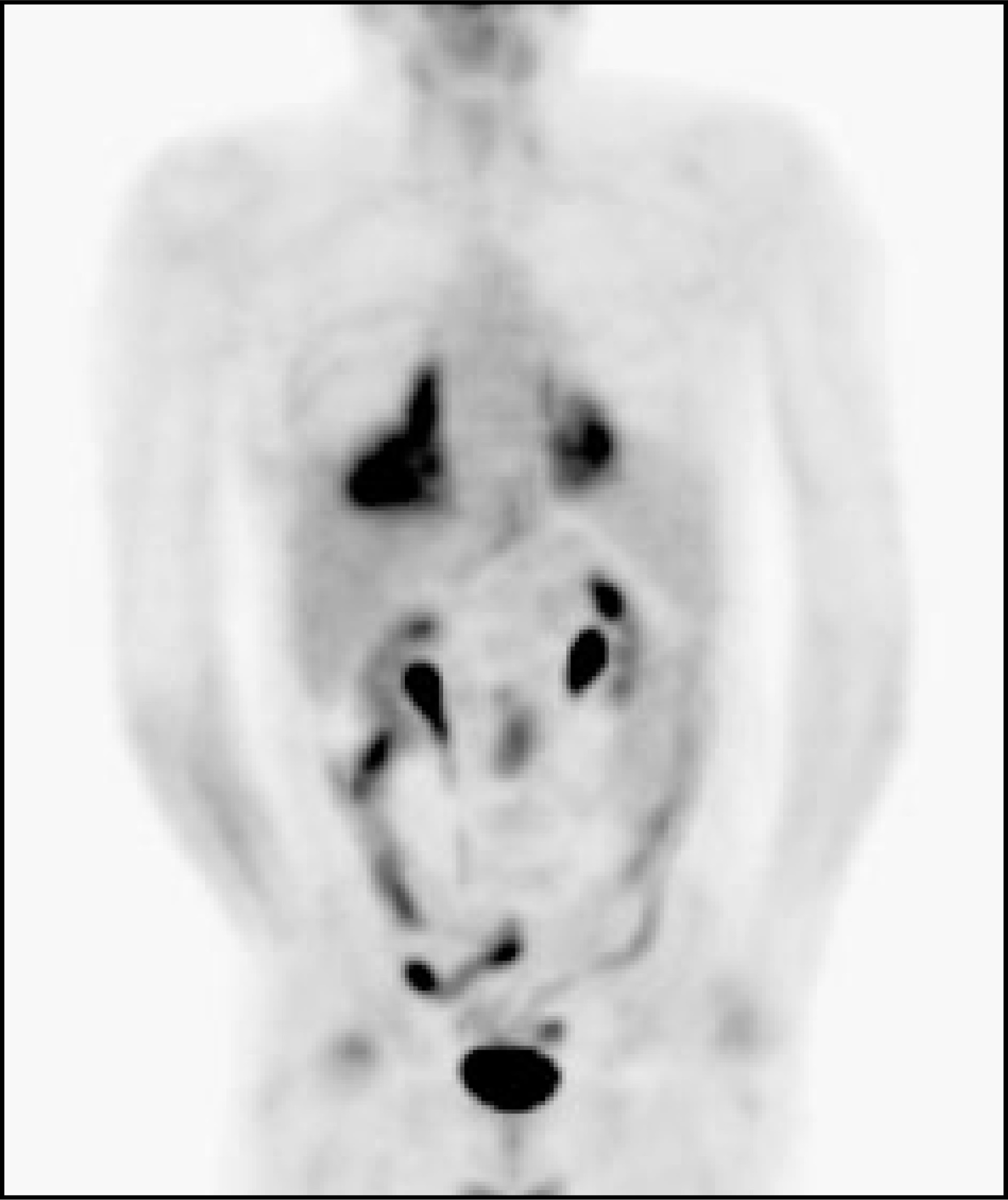
The major advantage of FDG PET over conventional imaging is its ability to detect distant metastases to facilitate treatment planning (Figures 8 and 9). Distant metastatic disease has a significant impact on patient management because these patients are no longer eligible for surgical resection. FDG PET has a reported sensitivity of 69% to 100%, a specificity of 84% to 90%, and an accuracy of 84% to 91% for the evaluation of distant metastases, while the sensitivity of CT for distant metastases has been reported to be lower.25,26 FDG PET scans have also excluded metastatic disease at sites considered abnormal on conventional imaging.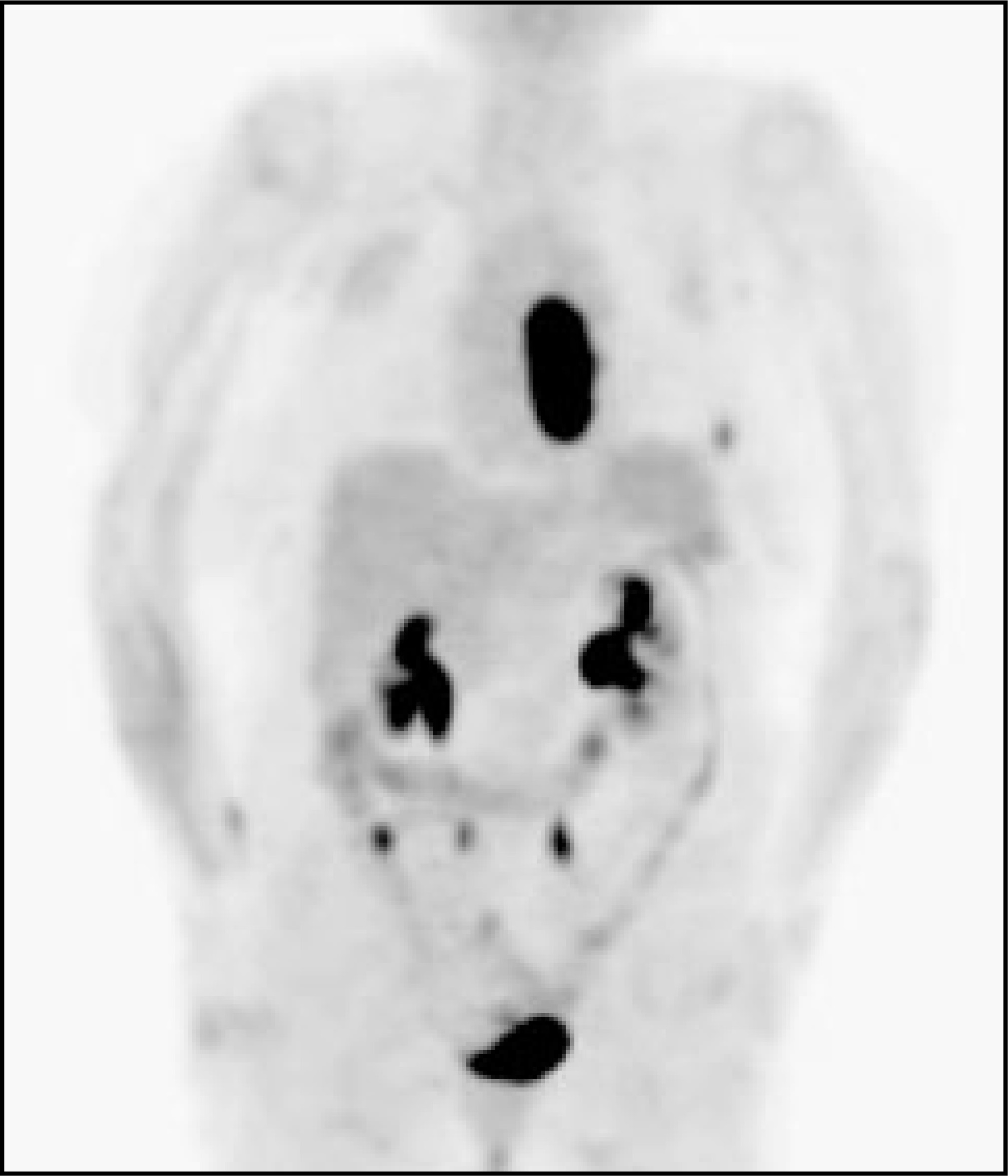
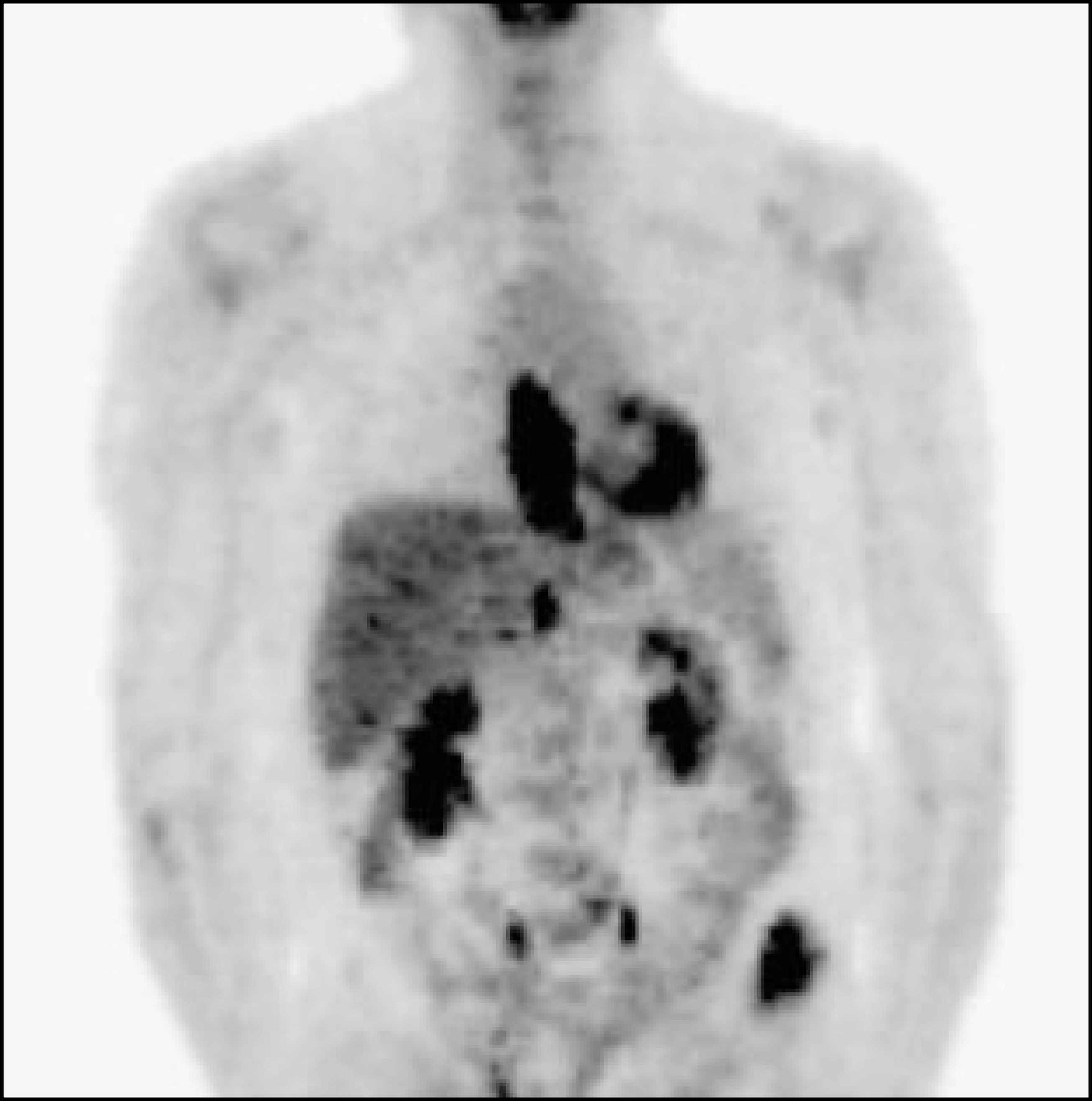
Primary treatment modalities include surgery, or chemotherapy with radiation therapy. Combined modality therapy, which includes chemotherapy plus surgery, or chemotherapy and radiation therapy plus surgery, is another form of treatment. Palliative therapy includes various combinations of surgery, chemotherapy, radiation therapy, photodynamic therapy, endoscopic therapy, and stent placement.
Two-thirds of patients with esophageal carcinoma have recurrence within 1 year after primary operation and the majority of recurrences are distant metastases (Figure 10).27 For the diagnosis of regional and distant recurrences, FDG PET has a sensitivity of 94%, a specificity of 82%, and an accuracy of 87% (compared with 81%, 82%, and 81% for conventional imaging).27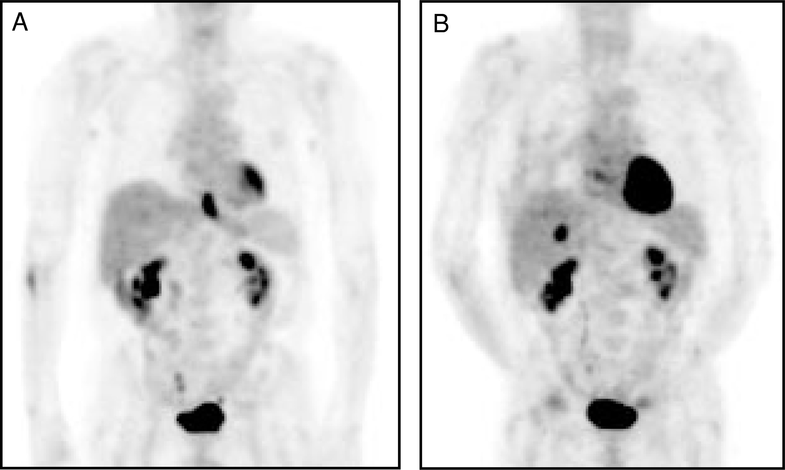
Conclusions
Positron emission tomography has had a significant impact on the evaluation and management of patients with lung and esophageal cancer. It provides an important noninvasive diagnostic modality for localizing nodal involvement, primary tumor extent, and distant metastases. The metabolic foundation of FDG PET imaging provides the sensitivity to better define the fields for radiation therapy and allow assessment of the effectiveness of either chemotherapy or radiation therapy. With the use of combined functional and anatomic imaging devices (PET/CT), this modality will become even more valuable. The future will bring continued expansion of clinical applications and new positron radiopharmaceuticals that should greatly enhance the care delivered to patients with thoracic malignancies. AR
References
- Dwamena B, Sonnad S, Angobaldo J, Wahl R. Metastases from non-small cell lung cancer: Mediastinal staging in the 1990s—Meta-analytic comparison of PET and CT. Radiology. 1999;213:530-536.
- Lowe V, Fletcher J, Gobar L. Prospective investigation of positron emission tomography in lung nodules. J Clin Oncol. 1998;16:1075-1084.
- Vesselle H, Schmidt R, Pugsley J. Lung cancer proliferation correlates with [F-18] fluorodeoxyglucose uptake by positron emission tomography. Clin Cancer Res. 2000;6:3837-3844.
- Knight S, Delbeke D, Stewart J, Sandler M. Evaluation of pulmonary lesions with FDG-PET. Comparison of findings in patients with and without a history of prior malignancy. Chest. 1996;109:982-988.
- Swensen S, Brown L, Colby T, Weaver A. Pulmonary nodules: CT evaluation of enhancement with iodinated contrast material. Radiology. 1995;194:393-398.
- Dewan N, Reeb S, Gupta N. PET-FDG imaging and transthoracic needle lung aspiration biopsy in evaluation of pulmonary lesions. A comparative risk-benefit analysis. Chest. 1995;108:441-446.
- Carney D, Hansen H. Non–small-cell lung cancer--stalemate or progress?. N Engl J Med. 2000;343:1261-1262.
- Kadri M, Dussek J. Survival and prognosis following resection of primary non–small-cell bronchogenic carcinoma. Eur J Cardiothorac Surg. 1991;5:132-136.
- Deslauriers J, Ginsberg R, Dubois P. Current operative morbidity associated with elective surgical resection for lung cancer. Can J Surg. 1989;32:335-339.
- Weder W, Schmid R, Bruchhaus H. Detection of extrathoracic metastases by positron emission tomography in lung cancer. Ann Thorac Surg. 1998;66:886-892.
- Erasmus J, Patz E, McAdams H. Evaluation of adrenal masses in patients with bronchogenic carcinoma using 18F-fluorodeoxyglucose positron emission tomography. AJR Am J Roentgenol. 1997;168:1357-1360.
- Bury T, Barreto A, Daenen F. Fluorine-18 deoxyglucose positron emission tomography for the detection of bone metastases in patients with non-small cell lung cancer. Eur J Nucl Med. 1998;25:1244-1247.
- Schumacher T, Brink I, Mix M. FDG-PET imaging for the staging and follow-up of small cell lung cancer. Eur J Nucl Med. 2001;28:483-488.
- Weng E, Tran L, Rege S. Accuracy and clinical impact of mediastinal lymph node staging with FDG-PET imaging in potentially resectable lung cancer. Am J Clin Oncol. 2000;23:47-52.
- Saunders C, Dussek J, O’Doherty M, Maisey M. Evaluation of fluorine-18-fluorodeoxyglucose whole body positron emission tomography imaging in the staging of lung cancer. Ann Thorac Surg. 1999;67:790-797.
- Scott W, Gobar L, Terry J. Mediastinal lymph node staging of non-small-cell lung cancer: A prospective comparison of computed tomography and positron emission tomography. J Thorac Cardiovasc Surg. 1996;111:642-648.
- Gupta N, Graeber G, Rogers J, Bishop H. Comparative efficacy of positron emission tomography with FDG and computed tomographic scanning in preoperative staging of non-small cell lung cancer. Ann Surg. 1999;229:286-291.
- Gould M, Maclean C, Kuschner W. Accuracy of positron emission tomography for diagnosis of pulmonary nodules and mass lesions: A meta-analysis. JAMA. 2001;285:914-924.
- Wahl R. Targeting glucose transporters for tumor imaging: “Sweet” idea, “sour” result. J Nucl Med. 1996;37:1038-1041.
- Vansteenkiste J, Stroobants S, De Leyn P. Lymph node staging in non-small-cell lung cancer with FDG-PET scan: A prospective study on 690 lymph node stations from 68 patients. J Clin Oncol. 1998;16:2142-2149.
- Higashi K, Ueda Y, Arisaka Y. 18F-FDG uptake as a biologic prognostic factor for recurrence in patients with surgically resected non-small cell lung cancer. J Nucl Med. 2002;43:39-45.
- Bradley J, Ieumwananonthachai N, Purdy J. Gross tumor volume, critical prognostic factor in patients treated with three-dimensional conformal radiation therapy for non-small-cell lung carcinoma. Int J Radiat Oncol Biol Phys. 2002;52:49-57.
- Hebert M, Lowe V, Hoffman J. Positron emission tomography in the pretreatment evaluation and follow-up of non-small cell lung cancer patients treated with radiotherapy: Preliminary findings. Am J Clin Oncol. 1996;19:416-421.
- Greenlee R, Hill-Harmon M, Murray T, Thun M. Cancer statistics, 2001. CA Cancer J Clin. 2001;51:15-36.
- Skehan S, Brown A, Thompson M. Imaging features of primary and recurrent esophageal cancer at FDG PET. Radiographics. 2000;20:713-723.
- Lowe V, Naunheim K. Current role of positron emission tomography in thoracic oncology. Thorax. 1998;53:703-712.
- Flamen P, Lerut A, Van Cutsem E. The utility of positron emission tomography for the diagnosis and staging of recurrent esophageal cancer. J Thorac Cardiovasc Surg. 2000;120:1085-1092.
Citation
. Positron emission tomography imaging of lung and esophageal cancer. Applied Radiology. 2002;31(6):9-17. doi:10.37549/AR1097.