Post-Traumatic “Floating Fat” in the Extensor Tendon Sheaths of Wrist
Applied Radiology — Vol. 54 , Issue 3 , pp. 1 -2
Published: June 1, 2025
1 Department of Radiodiagnosis, Dr. D. Y. Patil Medical College, Hospital and Research Center, Dr. D. Y. Patil Vidyapeeth, Sant Tukaram Nagar, Pimpri, Pune, Maharashtra, India
* Corresponding author: Parag Vijaysingh Patil (drparagpatil@yahoo.com)
Abstract
Not Applicable
Keywords
Floating fat, Extensor tendon sheaths, Wrist, Trauma
Categories
Case Summary
An adult presented with pain and swelling of the right wrist after a fall.
Imaging Findings
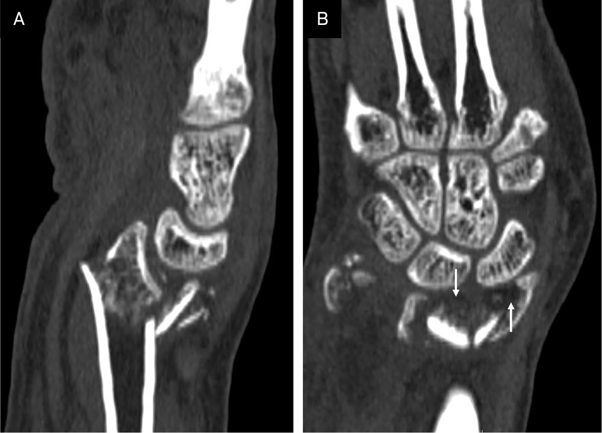
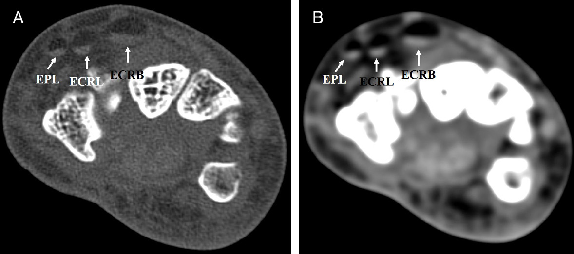
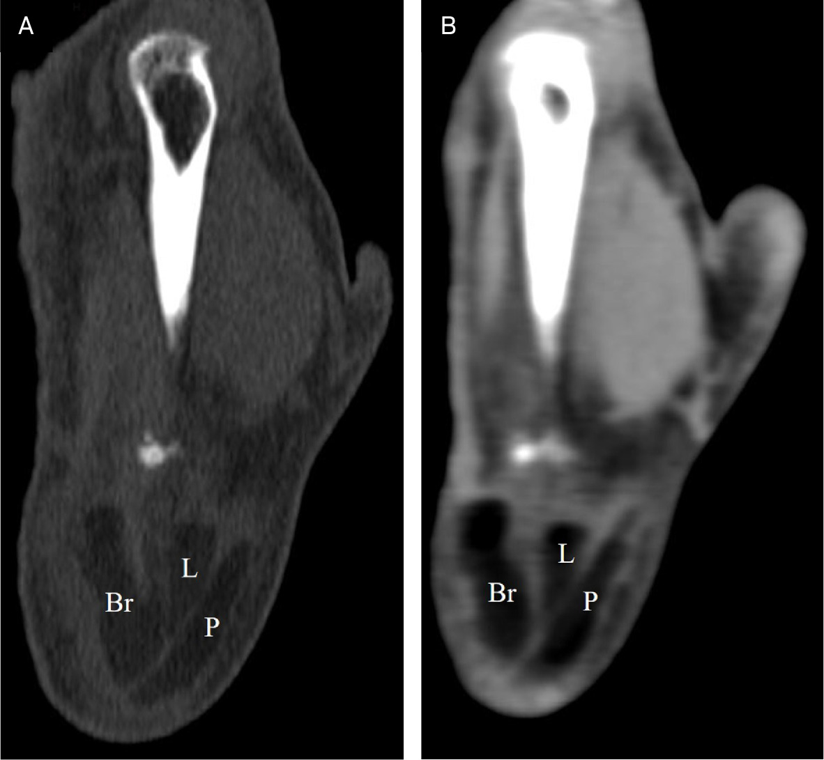
Sagittal reformatted CT of right wrist (Figure 1) demonstrated a comminuted intra-articular fracture of the distal end of the radius (Frykman type VIII). Fat density was noted in the wrist joint space. Axial CT images (Figure 2) demonstrated fat-fluid levels in the extensor pollicis longus (EPL), extensor carpi radialis longus (ECRL), and extensor carpi radialis brevis (ECRB) tendon sheaths. Coronal reformatted CT images (Figure 3) also showed fat densities in EPL, ECRL, and ECRB tendon sheaths.
Diagnosis
Post-traumatic floating fat in the extensor tendon sheaths of the wrist. Differential diagnosis includes a true synovial lipoma and lipoma arborescens.
Discussion
Post-traumatic fat-density effusion in the extensor tendon sheath on CT was first reported by Le Corroller et al in 2010.1 Ali et al observed a similar finding in multiple patients and thought it was an underreported finding.2 They also suggested that floating fat is a secondary sign of an intra-articular fracture or an occult fracture. In 2017, von Schneider-Egestorf et al retrospectively analyzed MDCT scans of individuals with distal radial intra-articular fractures and found that most of the individuals demonstrated fat-fluid levels in the extensor tendon sheaths.3 They also found that fracture severity was not related to frequency and severity of tendon sheath involvement.
Bone marrow fat leakage in joints with torn tendon sheaths has been postulated as a cause of fat-density effusion in the joint and tendon sheaths.3 Tendons of the second extensor compartment (ECRL, ECRB) of the wrist are most often involved, followed by the tendons of the third compartment (EPL). Involvement of both compartments is also common, owing to the existence of a normal foramen between the EPL and ECRB tendon sheaths.4
Involvement of the tendon sheaths can be assessed using the Likert scale of semi-quantitative grading from 0 to 2.3
Conclusion
Although floating fat in the extensor tendon sheaths is not uncommon, the condition is an important finding that can indicate the intra-articular extension of a distal radial fracture or an occult fracture and should be recognized.
References
- Le Corroller T, Parratte S, Zink J, Argenson J, Champsaur P. Floating fat in the wrist joint and in the tendon sheaths. Skeletal Radiol. 2010;39(9):931-933. doi:10.1007/s00256-010-0929-x.
- Ali S, Huebner S, Groshek F, Schaffer A. The floating fat sign of trauma. Can Assoc Radiol J. 2014;65(2):106-112. doi:10.1016/j.carj.2013.02.008.
- von Schneider-Egestorf A, Meyer B, Wacker F, Rosenthal H, von Falck C. Systematic evaluation of concomitant extensor tendon sheath injury in patients with distal intra-articular radial fractures in MDCT using the floating fat sign. Eur Radiol. 2017;27(10):4345-4350. doi:10.1007/s00330-017-4787-y.
- Cvitanic O, Henzie G, Adham M. Communicating foramen between the tendon sheaths of the extensor carpi radialis brevis and extensor pollicis longus muscles: imaging of cadavers and patients. AJR Am J Roentgenol. 2007;189(5):1190-1197. doi:10.2214/AJR.07.2281.
Disclosures
The authors have no conflicts of interest to disclose. None of the authors received outside funding for the production of this original manuscript and no part of this article has been previously published elsewhere.
Citation
. Post-Traumatic “Floating Fat” in the Extensor Tendon Sheaths of Wrist. Applied Radiology. 2025;54(3):1-2. doi:10.37549/AR-D-25-0080.