Primary osteosarcoma of the breast
Applied Radiology — Vol. 46 , Issue 12 , pp. 28 -30
DOI: 10.37549/AR2437
Published: December 1, 2017
1 Medical Student at Wayne State University School of Medicine, Detroit, MI
2 PGY5 in the Department of Radiology, Detroit, MI
3 PGY3 in the Department of Pathology and Laboratory Medicine, Detroit, MI
4 Division of Breast Imaging at Henry Ford Hospital; and a Clinical Associate Professor at Wayne State University School of Medicine, Detroit, MI
Categories
CASE SUMMARY
A 94-year-old woman presented with a lump in her right breast. Her clinical exam showed a large, hard, palpable mass measuring approximately 6 cm in diameter in the upper outer quadrant. Initial mammogram and ultrasound findings were consistent with a benign calcified fibroadenoma. However, upon return to clinic for reevaluation, the patient reported unremitting right breast pain, and imaging findings were highly suggestive of a malignancy. The patient reported no family history of breast cancer. Biopsy revealed an ER/PR- and HER-2-negative malignant neoplasm with osseous differentiation. A right modified radical mastectomy was performed, and no further tests were administered due to the patient’s age and family requests.
IMAGING FINDINGS
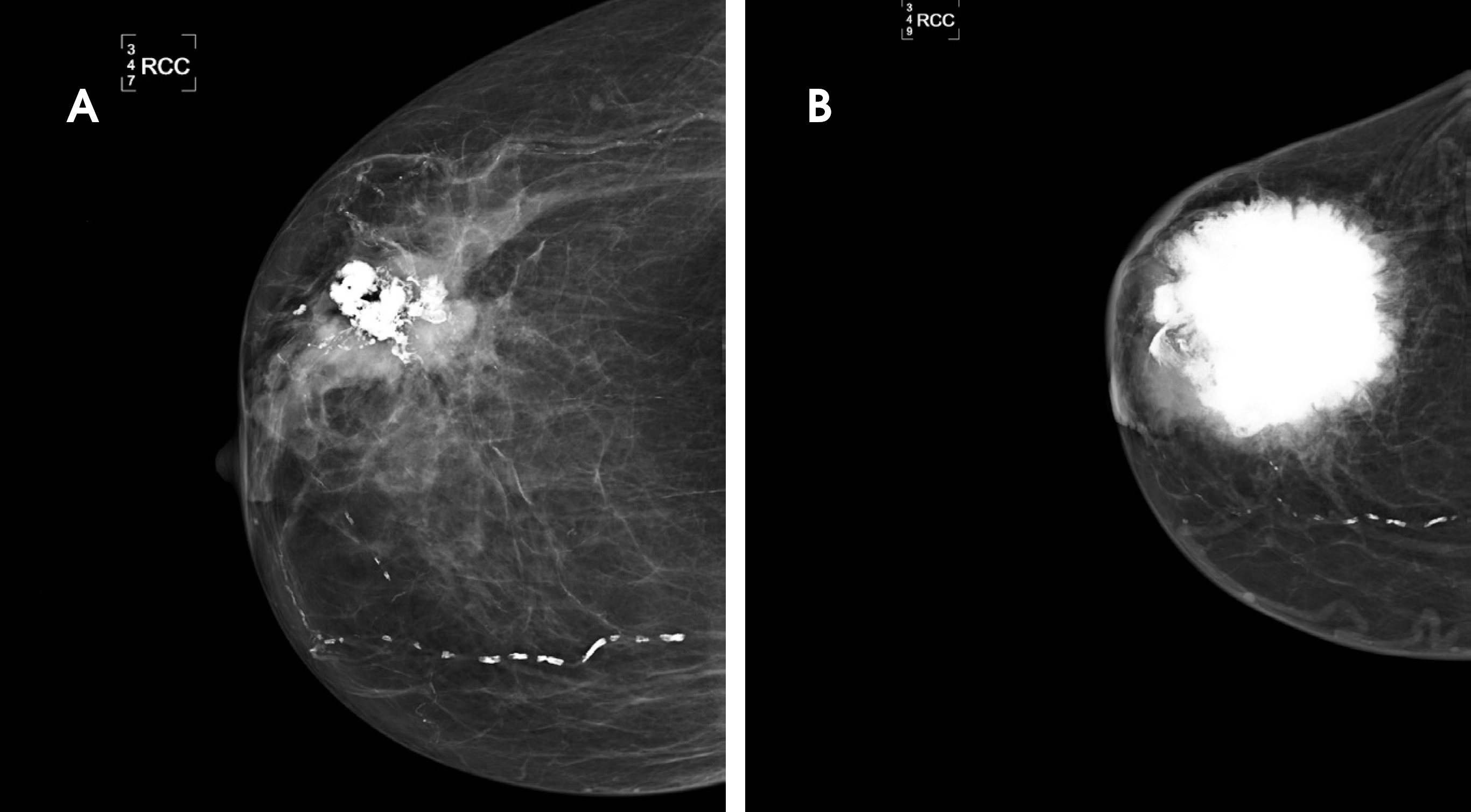
Craniocaudal (CC) mammography of the right breast shows a mass with associated coarse heterogeneous calcifications in the upper outer quadrant (Figure 1A). Figure 1B illustrates an interval increase in size of a large, lobular mass with dystrophic calcification centrally in the upper outer quadrant, measuring 8.7 cm x 7.7 cm. Due to the large size of the mass, manual technique with limited compression was performed to obtain mammographic images.
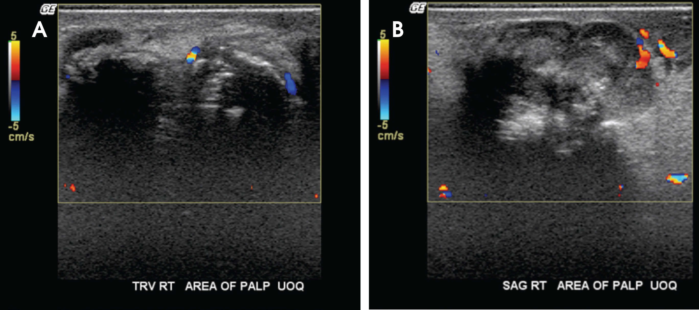
Targeted ultrasound of the palpable mass demonstrates a solid heterogeneous mass with posterior acoustic shadowing located in the upper outer quadrant (Figure 2). This shadowing corresponds to the patient’s palpable mass. There is slight vascularity present at the periphery. Given the indistinct margins of the mass, it could not be accurately measured by ultrasound.
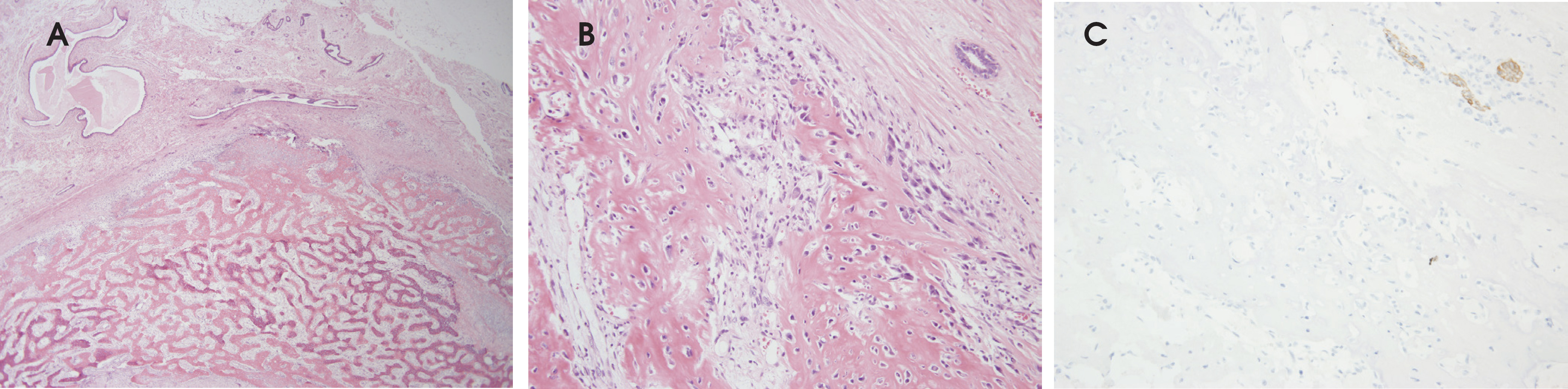
Tissue sections demonstrate a relatively heterogeneous mass composed of trabecular bone, osteoid matrix, cartilage, and spindle cells admixed with epithelioid cells. Regions of osteoid and trabecular bone formation comprise the bulk of the mass, blending indistinctly with regions of cartilaginous and pure spindle cell differentiation (Figure 3). No necrosis is identified.
Cells predominantly demonstrate spindled morphology with bizarre nuclei and prominent nucleoli. A lesser population of admixed epithelioid cells with relatively abundant amphophilic cytoplasm and hyperchromatic eccentric nuclei are also seen. Mitotic activity is brisk.
Multiple tissue sections are stained with a panel of immunostains, including cytokeratins AE1/AE3 (as shown in Figure 3C), CAM 5.2, CK 5/6, and smooth muscle actin (SMA). Malignant cells were negative for all of these markers, differentiating this pathology from metaplastic carcinoma. The high-grade soft tissue osteosarcoma of the breast is a cytologically malignant spindle cell neoplasm dominated by extensive production of osteoid and bone associated with the cytologically atypical cells. There is no evidence of conventional carcinoma or any coexistent phyllodes tumor.
DIAGNOSIS
High-grade soft tissue osteosarcoma of the breast
DISCUSSION
Mammary sarcomas are uncommon cancers that comprise less than 1% of all breast malignancies and less than 5% of all sarcomas.1 Primary osteosarcoma of the breast is extremely rare, representing only 12.5% of mammary sarcomas, or 0.125% of all malignancies of the breast.2 While this specific breast cancer can occur in both young and old patients, it is most commonly observed in older patients, with a mean age of 65 years at presentation.3 The prognosis is relatively poor, with a reported five-year survival rate of 38%.4
Osteosarcomas of the breast may arise either from pre-existing benign or malignant breast neoplasms or from previously healthy parenchyma.5 Some proposed risk factors for extraskeletal osteosarcomas include prior local irradiation, trauma, and foreign body placement, though these are not unique to mammary osteosarcomas.5 The histogenesis of primary osteosarcoma of the breast has not been fully elucidated, though an origin from totipotent mesenchymal cells of the breast stroma or a transformation from a pre-existing fibroadenoma or phyllodes tumor have been suggested.2,6 This cancer is considered highly aggressive and is associated with early recurrence and often hematogenous rather than lymphatic spread, most commonly to the lungs.4,7
Similar to other extraskeletal osteosarcomas, mammary osteosarcoma may exhibit a variety of histologic features. As such, this cancer is characterized as fibroblastic, osteoblastic, or osteoclastic (giant cell-rich). In the osteoblastic type, as seen in our patient, the osteoid is deposited in a fine, branching, lace-like, or coarsely trabecular pattern.4,8
This tumor usually presents mammographically as a well-circumscribed dense lesion within the breast parenchyma with focal or extensive coarse calcifications.4,6,9 These calcifications were evident in our patient, as illustrated in Figure 1. However, it is important to note that the mammographic features of this tumor may be deceptively benign.6 For instance, in an evaluation of several cases of primary osteosarcoma of the breast, Silver and Tavassoli found the mammographic impression to be that of a benign fibroadenoma in 33% of patients.4 In fact, the initial diagnosis in our case was that of a benign fibroadenoma.
Furthermore, metaplastic breast carcinoma should be ruled out before making a diagnosis of primary osteosarcoma of the breast. Mammographically, metaplastic carcinoma generally appears as a high-density mass with circumscribed, obscured, irregular, or spiculated margins.10 This is similar to the mammographic appearance of primary breast osteosarcoma. However, these lesions are often non-calcified,10 which could be useful in differentiating them from primary mammary osteosarcoma.
CONCLUSION
Primary osteosarcoma of the breast is a rare and aggressive malignancy that should be differentiated from two other similar tumors: metaplastic carcinoma and cystosarcoma phyllodes. Due to the rarity and poor prognosis of this disease, it is important that radiologists recognize its mammographic features in order to ensure proper management and optimize patient outcomes.
References
- Voutsadakis I, Zaman K, Leyvraz S. Breast sarcomas: current and future perspectives. Breast. 2011;20((3)):199-204.
- Jernstrom P, Lindberg A, Meland O. Osteogenic sarcoma of the mammary gland. Am J Clin Pathol. 1963;40:521-526.
- Vorobiof G, Hariparsad G, Freinkel W, Said H, Vorobiof D. Primary osteosarcoma of the breast: a case report. Breast J. 2003;9((3)):231-233.
- Silver S, Tavassoli F. Primary osteogenic sarcoma of the breast: a clinicopathologic analysis of 50 cases. Am J Surg Pathol. 1998;22:925-933.
- Rizzi A, Soregaroli A, Zambelli C. Primary osteosarcoma of the breast: a case report. Case Rep Oncol Med.
- Remadi S, Doussis-Anagnostopoulu I, Mac G. Primary osteosarcoma of the breast. Pathol Res Pract. 1995;191:471-474.
- Barnes L, Pietruszka M. Sarcomas of the breast: a clinicopathologic analysis of 10 cases. Cancer. 1977;40:1577-1585.
- Bahrami A, Resetkova E, Ro J, Ibanez J, Ayala A. Primary osteosarcoma of the breast: report of two cases. Arch Pathol Lab Med. 2007;131(5):792.
- Rosen P. Rosen’s Breast Pathology;2001.
- Gunhan-Bilgen I, Memis A, Ustun E, Zekioglu O, Ozdemir N. Metaplastic carcinoma of the breast: Clinical, mammographic, and sonographic findings with histopathologic correlation. AJR Am J Roentgenol. 2002;178:1421-1425.
Citation
. Primary osteosarcoma of the breast. Applied Radiology. 2017;46(12):28-30. doi:10.37549/AR2437.