Pulmonary inflammatory pseudotumor
Applied Radiology — Vol. 37 , Issue 7 , pp. 34 -35
DOI: 10.37549/AR1631
Published: July 1, 2008
Categories
CASE SUMMARY
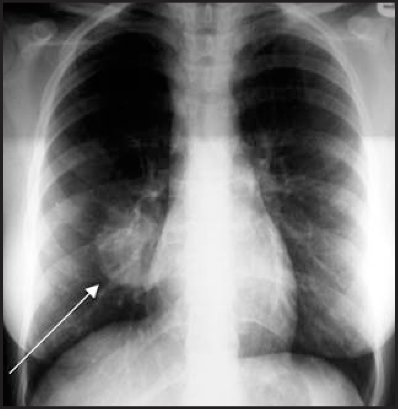
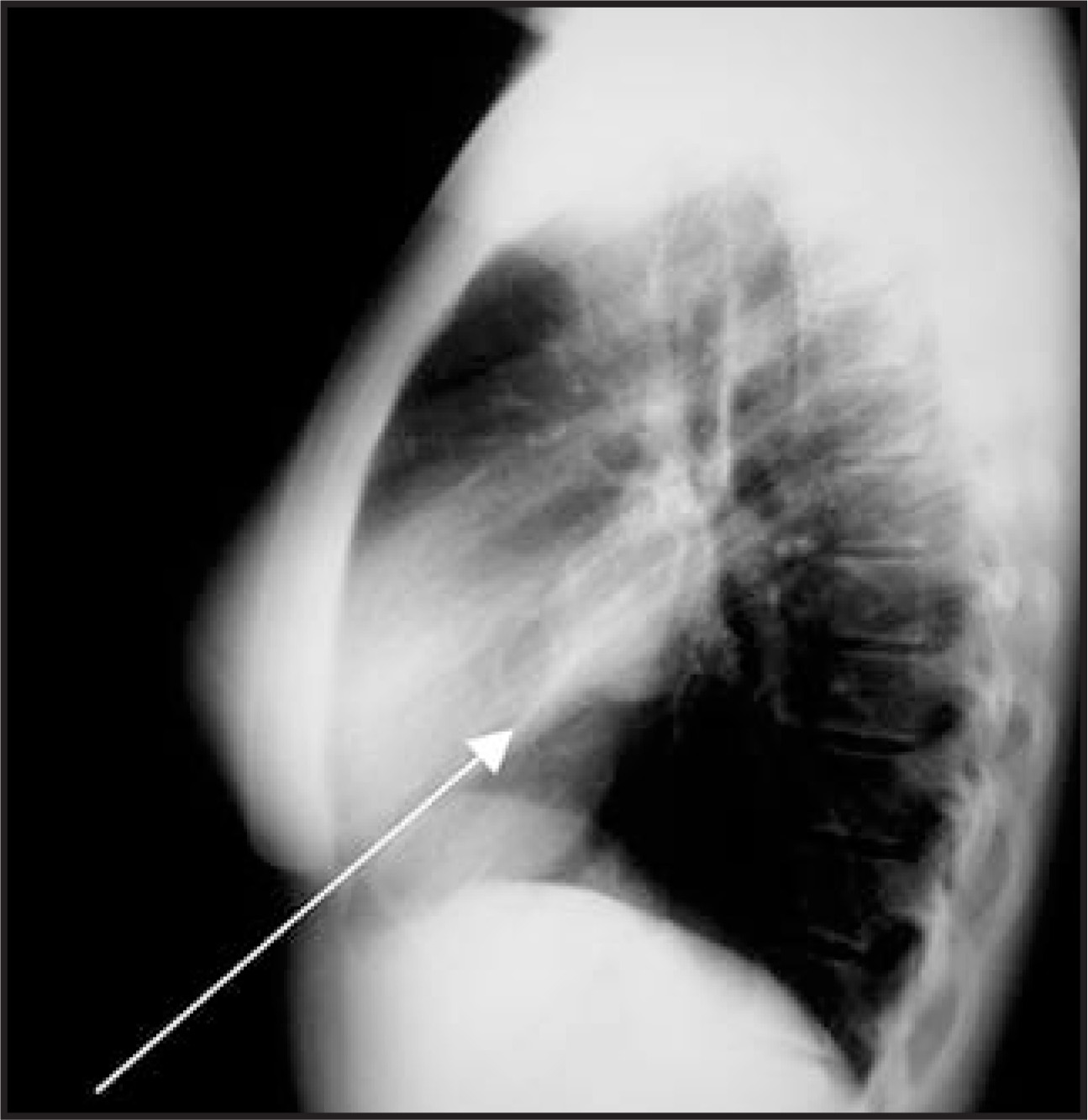
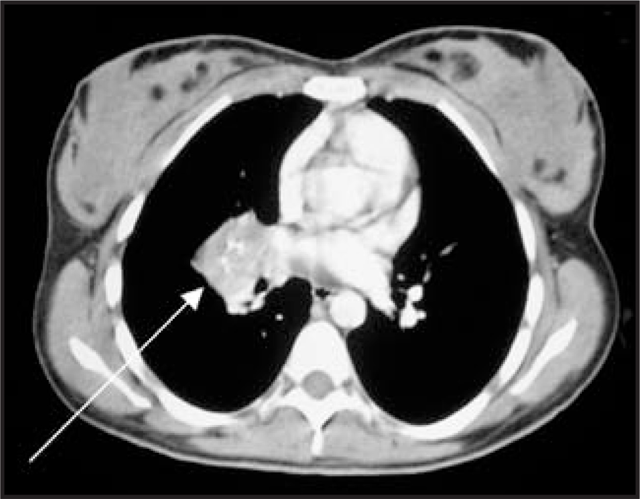
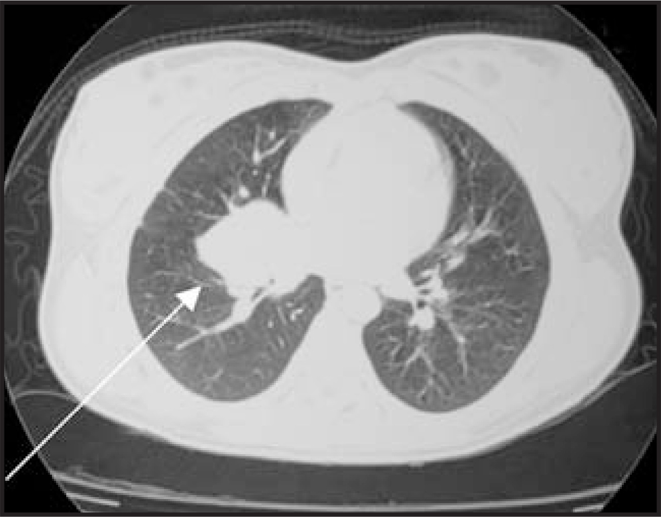
A 16-year-old girl presented to the emergency department with complaints of cough and chest discomfort. She had no significant medical history. On plain-film imaging, she was found to have a large right lung mass (Figures 1 and 2), which was confirmed with computedtomography (CT) (Figures 3 and 4). An antibiotic course did not alter the findings, and she was transferred for surgical evaluation andresection of the mass.
IMAGING FINDINGS
Posteroanterior (Figure 1) and lateral (Figure 2) plain films of the chest show a 4- to 5-cm rounded mass in the right infrahilar region.Contrast-enhanced CT of the chest show a 4.5 × 4-cm heterogenous soft tissue mass in the right infrahilar region (Figures 3 and 4). There areenhancing vessels coursing through it, and it appears to interpose itself between the right middle and lower lobes. The mass narrows and possibly invades the right lower lobe bronchus. The right middlelobe bronchus is not visible, which could be due to invasion or narrowing as well.The differential diagnosis (which varies with mass size and location) included malignant tumor, metastatic disease, pleural pseudotumor, lymphoma, and abscess.
DIAGNOSIS
Pulmonary inflammatory pseudotumor
DISCUSSION
Pulmonary inflammatory pseudo tumor is a relatively unusual benign process that may occur anywhere in the body, but is most commonly found in the lungs.1 It accounts for <1% of all tumors of the lung and airways.2 Alternate terminology includes pulmonary pseudotumor, plasma cell granuloma, inflammatory myofibroblastic tumor, fibroxanthoma, xantho matous pseudotumor, and plasma cell-histiocytoma complex. It has been reported to occur nearly everywhere in the body, but most often it involves the lungs and orbit.1 Brunn first describedit in 19391 and Umiker et al3 termed it inflammatory pseudotumor in 1954 for its proclivity to imitate a malignant process.
Pulmonary inflammatory pseudo tumor typically presents with cough, fever, dyspnea, and hemoptysis, yet some cases are asymptomatic.This lesion usually occurs in the second decade, and it is the most common primary lung mass in children. Males and females are affectedequally.1,4
This benign lesion typically presents on chest radiography as a solitary, peripheral mass, usually >3 cm in size. There is a predilection for thelower lobes, and they are predominantly sharply circumscribed. Rarely, they may calcify, cavitate, invade the mediastinum or hilum, or presentwith pleural effusion.5,6 Five percent of cases are multiple, and 10% are endobronchial.7 On CT, these masses are more often heterogenous thanhomogenous. Although few lesions have been studied with chest MRI, the T1 signal is heterogenous and slightly greater than that of skeletalmuscle. T2 signal is typically high, and heterogenous contrast enhancement with gadolinium is seen.5,6
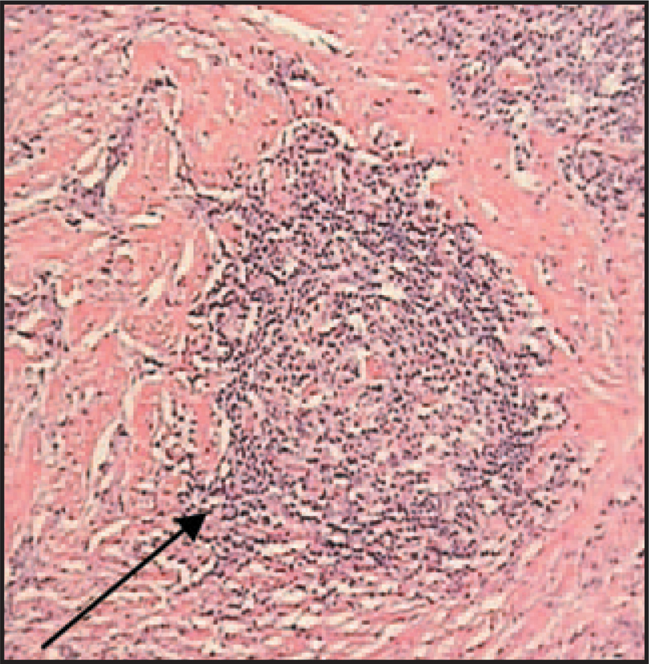
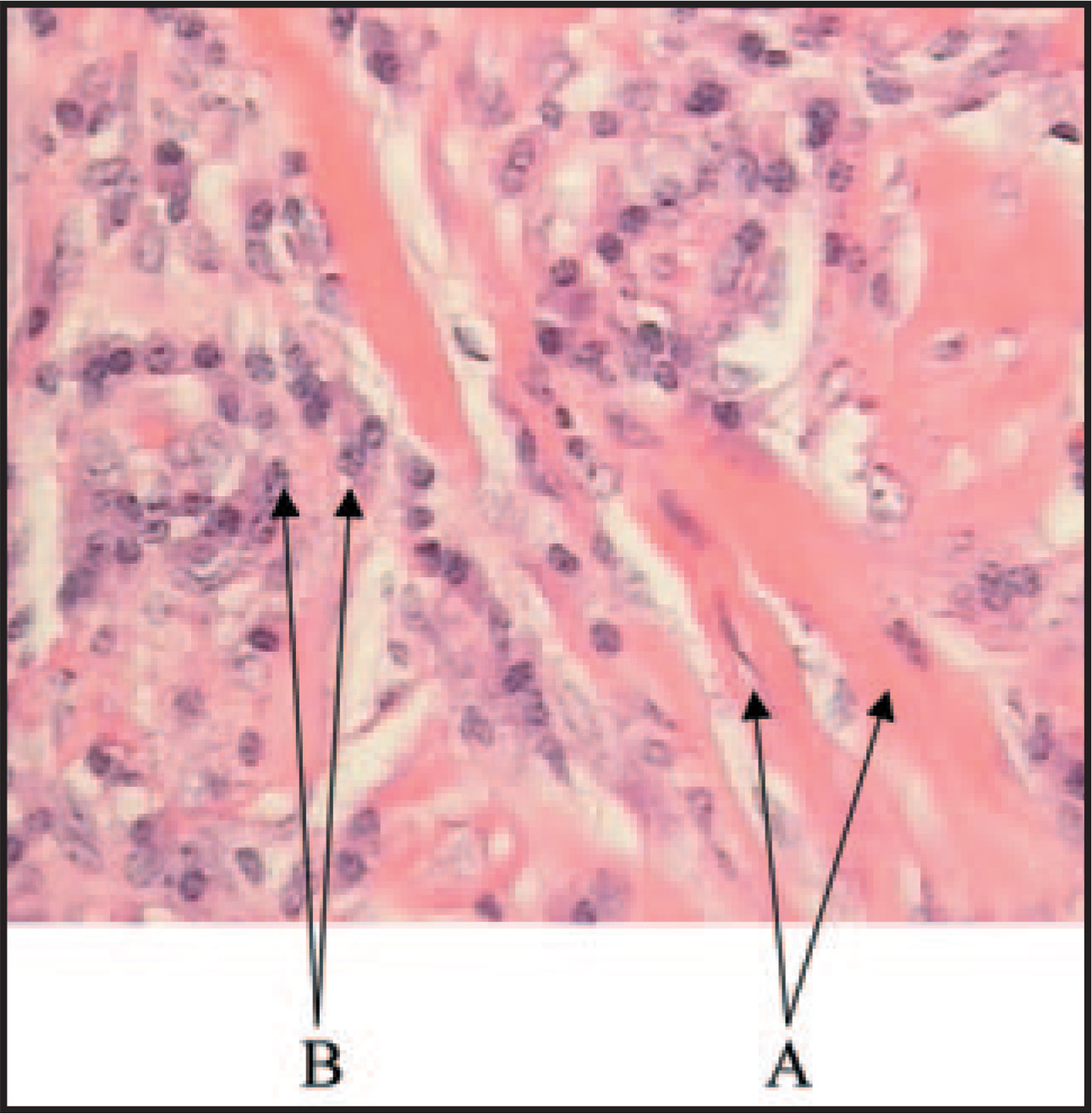
Pathologic evaluation (Figures 5 and 6) of the mass in this case revealed fibrous, hyalinized bands interrupted by irregular patterned sheetsof mononuclear cellularity. These mononuclear cellsinclude lymphocytes (some of which form aggregates), histiocytes, and notable numbers of polyclonal plasma cells.
The treatment of choice for pulmonary pseudotumor is complete surgical resection with local recurrence in 25%. Spontaneous regressionhas been reported. Radiation therapy, chemotherapy, systemic steroids, and antibiotics are less successful or are used as second-line therapiesfor pulmonary lesions.1
CONCLUSION
Pulmonary inflammatory pseudo tumor is an uncommon entity that, although rare, should be considered in the differential diagnosis of a lungmass in a young person. This case showed involvement of the hilum and growth around bronchi, which can lead to further complications. Thedistinction must be made pathologically between this benign entity and other more malignant processes. The preferred treatment is surgicalresection, although there is recurrence in 25% of cases.
References
- Narla L, Newman B, Spottswood S. Inflammatory pseudotumor. RadioGraphics RadioGraphics.
- Dähnert W. Radiology Review Manual. 2002:497.
- Umiker W, Iverson L. Postinflammatory tumors of the lung; report of four cases simulating xanthoma, fibroma, or plasma cell tumor. J Thorac Surg. 1954;28:55-63.
- Hedlund G, Navoy J, Galliani C, Johnson W. Aggressive manifestations of inflammatory pulmonary pseudotumor in children. Pediatric Radiol. 1999;29:112-116.
- Williams S. Pulmonary pseudotumor (plasma cell granuloma or inflammatory myofibroblastic tumor). 2004.
- Agrons G, Rosado-de-Christenson M, Kirejczyk W. Pulmonary inflammatory pseudotumor: Radiologic features. Radiology. 1998;206:511-518.
- Patankar T, Prasad S, Shenoy A, Rathod K. Pulmonary inflammatory pseudotumor in children. Australas Radiol. 2000;44:318-320.
Citation
. Pulmonary inflammatory pseudotumor. Applied Radiology. 2008;37(7):34-35. doi:10.37549/AR1631.