Radiologic-pathologic correlation of papillary lesions of the breast
Applied Radiology — Vol. 34 , Issue 11 , pp. 20 -42
DOI: 10.37549/AR1381
Published: November 1, 2005
Categories
Papillary lesions of the breast occur most frequently in the major lactiferous ducts. Benign intraductal papilloma is a solitary lesion of the duct that typically presents when small and nonpalpable because of the occurrence of spontaneous nipple discharge. Such lesions may be visible on mammography as small masses, dilated ducts, or calcifications, but are more often observed on galactography as intraluminal filling defects. Papillary apocrine metaplasia is a common papillary lesion that is associated with breast cysts. Papillomatosis and adenoma of the nipple are unusual benign papillary lesions. Another benign papillary lesion is radial sclerosing lesion or radial scar. Papillary cancers may develop focally within a cyst wall or may be multicentric, manifesting as multiple small filling defects within numerous ducts. This pictorial essay illustrates radiologic-pathologic correlation of papillary lesions of the breast.
Intraductal papilloma
Clinical presentation
Intraductal papillomas occur most frequently in the sixth decade and are most commonly located in the central or subareolar part of the breast. Papillomas typically occur in major lactiferous ducts and develop within the wall of the duct. In most cases, the presenting symptom is nipple discharge, which may or may not be bloody. Less frequently, a palpable mass is present. The papilloma is a friable, epithelial-covered, pedunculated tumor that may twist on its stalk and infarct, causing the bleeding that is present on clinical examination. This symptom is less common when the papilloma is peripherally located or when the lesion is more solid than frond-forming.1
Imaging
For both solitary and multiple papillomas, it has been found that centrally located lesions are more likely to be detected on galactography as filling defects or duct obstruction. Solitary papillomas, when observed on mammography, may present as a small, circumscribed mass, dilated duct, or cluster of punctate or dystrophic microcalcifications (Figures 1 through 6).1
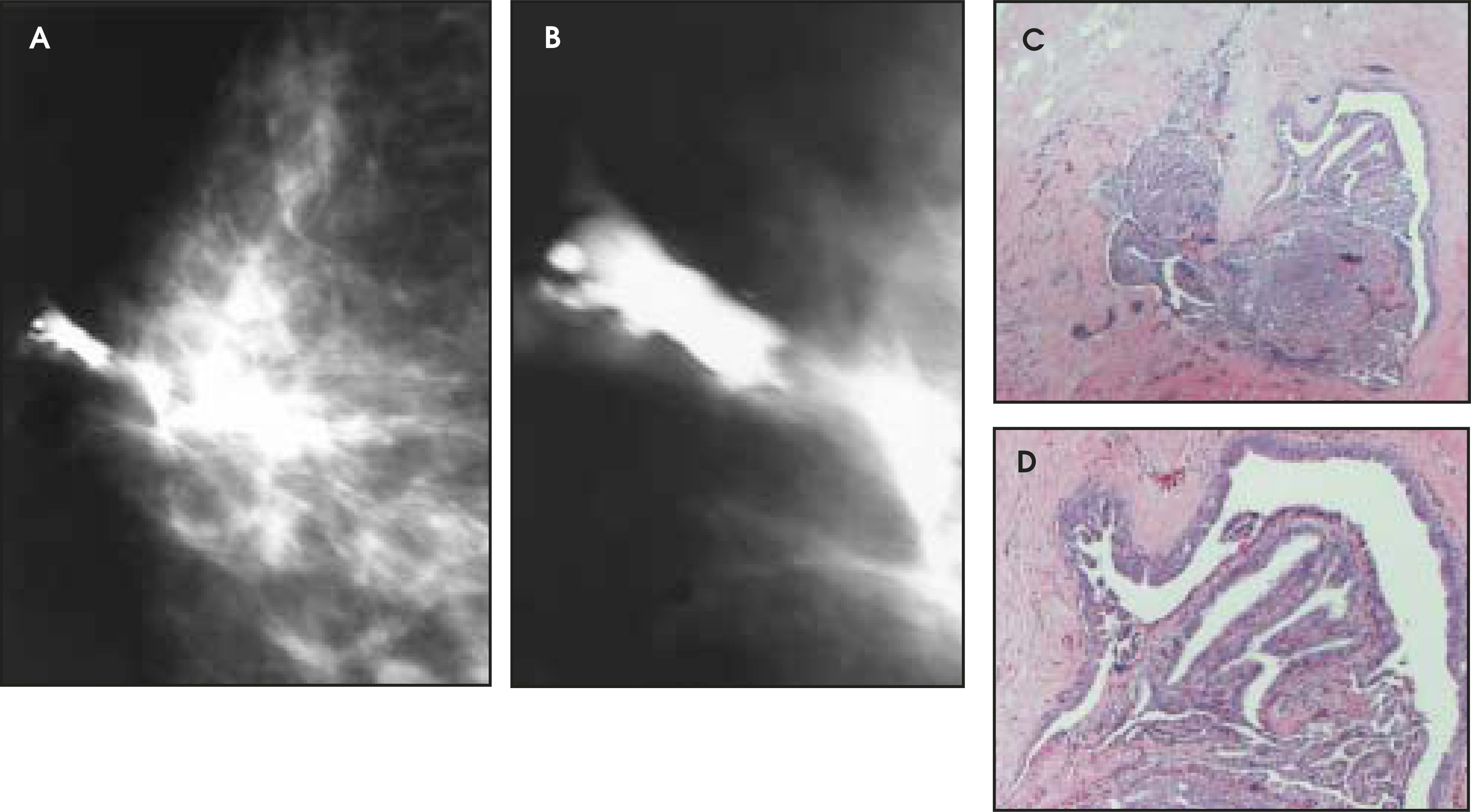
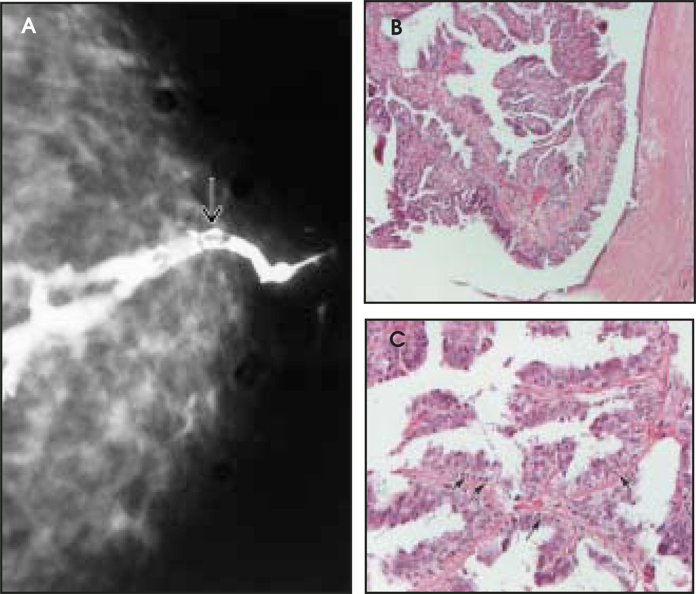
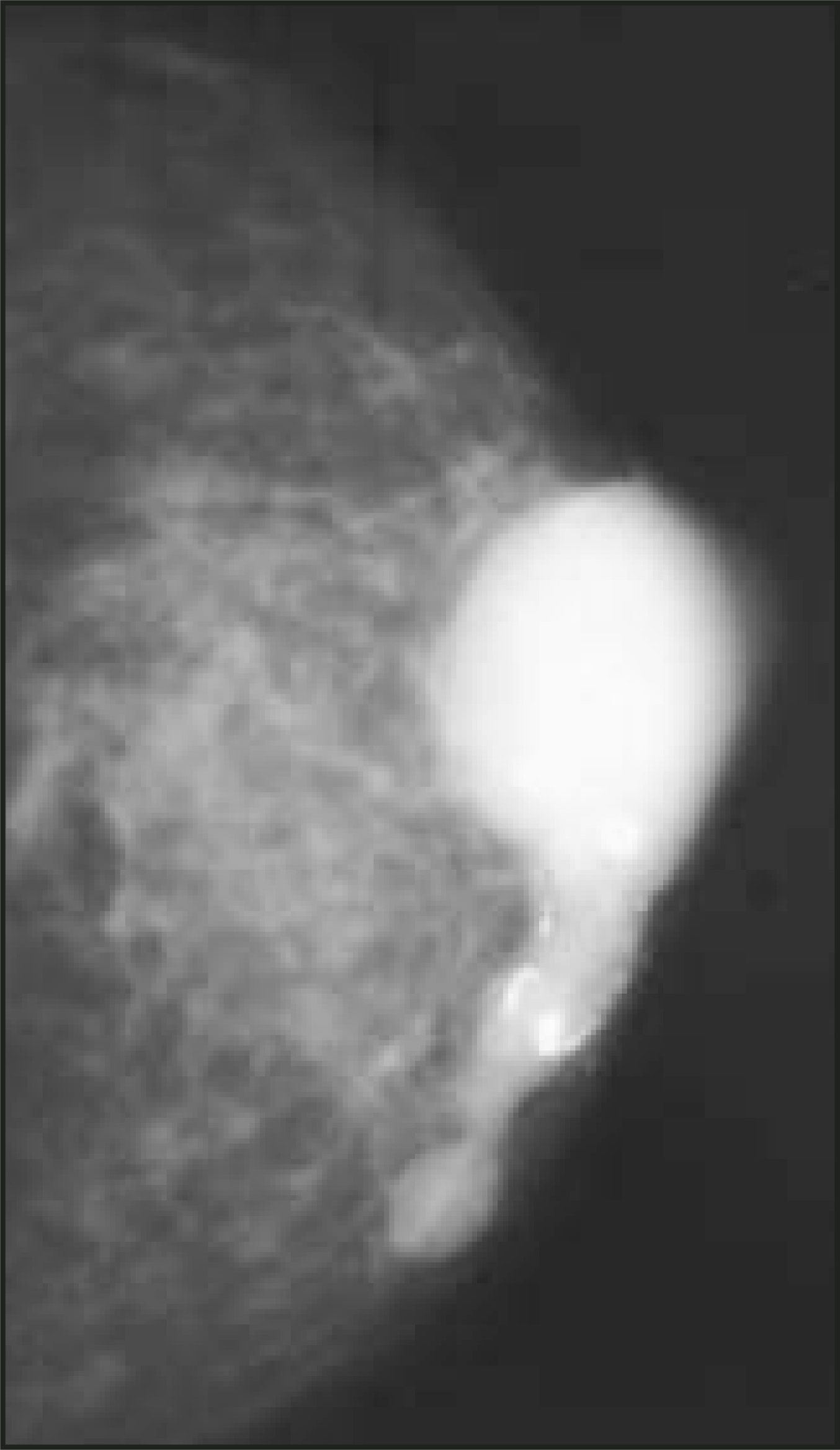
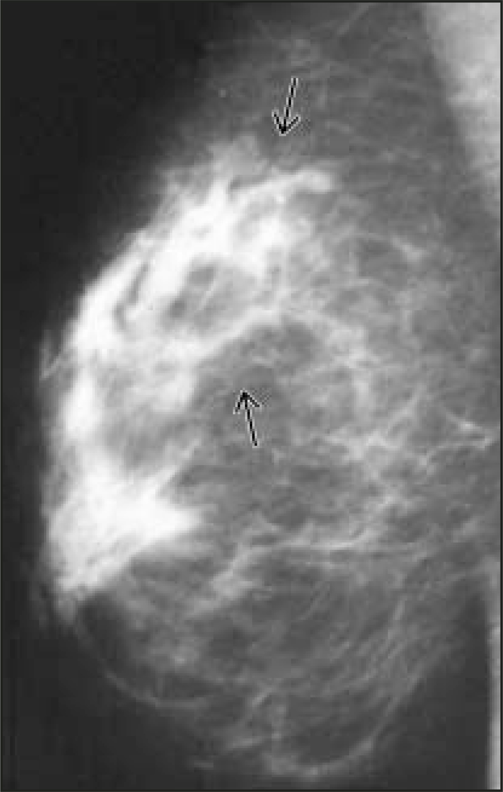
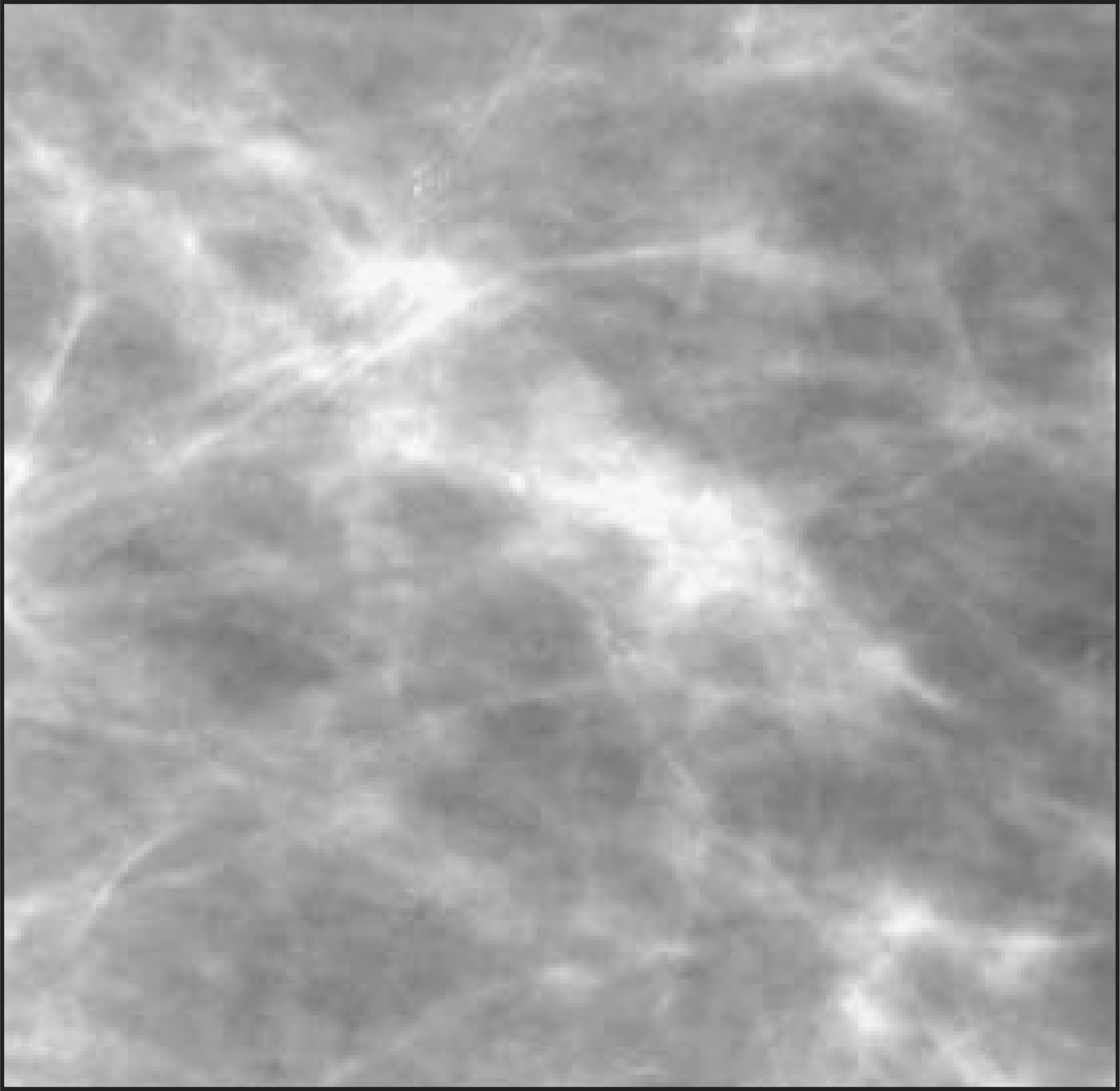
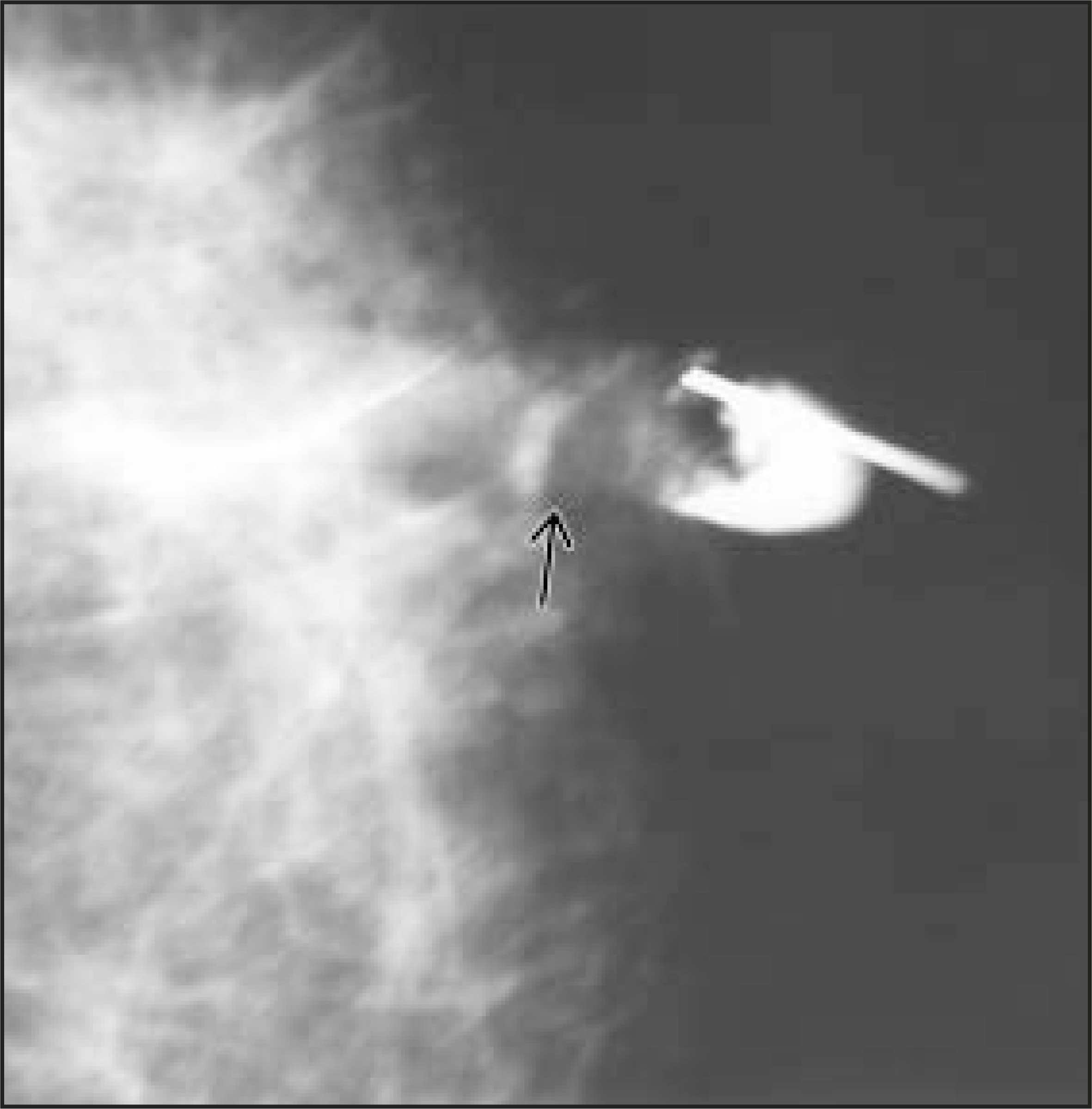
In a review of 24 patients with papillomas, Woods et al2 found that only 2 (8%) presented with an abnormal screening mammogram. Of the 24 patients, 21 (88%) presented with nipple discharge, and 1 patient presented with a palpable mass. Huynh et al3 published a series of 46 patients with unilaterally dilated ducts and found that benign diagnoses predominated. Of these, papilloma was the most common etiology of a unilateral dilated duct pattern.
Papillomas may not be visible using ultrasound; however, when they are seen, they can have a variety of appearances, including a small, solid mass; a dilated duct containing a filling defect; or a cystic lesion with an associated irregular mass or filling defect. When a papilloma presents as a solid mass, it tends to be hypoechoic or of mixed echogenicity, and it often has a thin, echogenic rim, which represents the duct wall.4
Gadolinium-enhanced MRI can also detect solitary papillomas. Daniel et al5 reviewed the MRI findings in 15 patients with pathologically proven papillomas. In this group of patients, 4 of 15 had a negative MRI; 4 had a small, smooth, enhancing mass at the end of an enhancing duct; and 7 had an irregular, enhancing mass. In the patients found to have an irregular mass, enhancement was rapid, and 3 of 7 revealed either spiculated margins or rim enhancement.
Gross and microscopic pathology
Papillomas are discrete benign papillary tumors that develop from the epithelium of the lactiferous ducts. They often have a cystic component, which may contain clear fluid, blood, or clot, which formed as a consequence of ductal dilatation. Most often, the papilloma is an isolated mural nodule, but there may be more than one lining the cyst. When large, a papilloma can obliterate the cyst cavity and grow to several centimeters in size. Microscopically, there is proliferation of the ductal epithelium covering a fibrovascular core. Frequently, there is evidence of repeated hemorrhage and necrosis due to infarction.1
Large, complex papillomas with a cystic component are sometimes called papillary cystadenomas. Large, solid papillomas are called duct adenomas (Figure 7). With multiple, peripheral papillomas, there is an increased association, in the range of 10% to 33%, with occurrence of carcinoma.4 There have also been associations reported between multiple, peripheral papillomas and atypical ductal hyperplasia (ADH), lobular carcinoma in situ (LCIS), and radial scar.6
Papillary apocrine metaplasia
Clinical presentation
Apocrine glands are accessory sex glands that respond to hormonal stimulation. Although apocrine glands do not normally occur in breast tissue, they develop from the same anlage as the breasts. Cells with apocrine features can be found in any proliferative lesion that is benign.1 Apocrine metaplasia can be found in the epithelium lining breast cysts as well as in papillomas and adenomas.1
Imaging
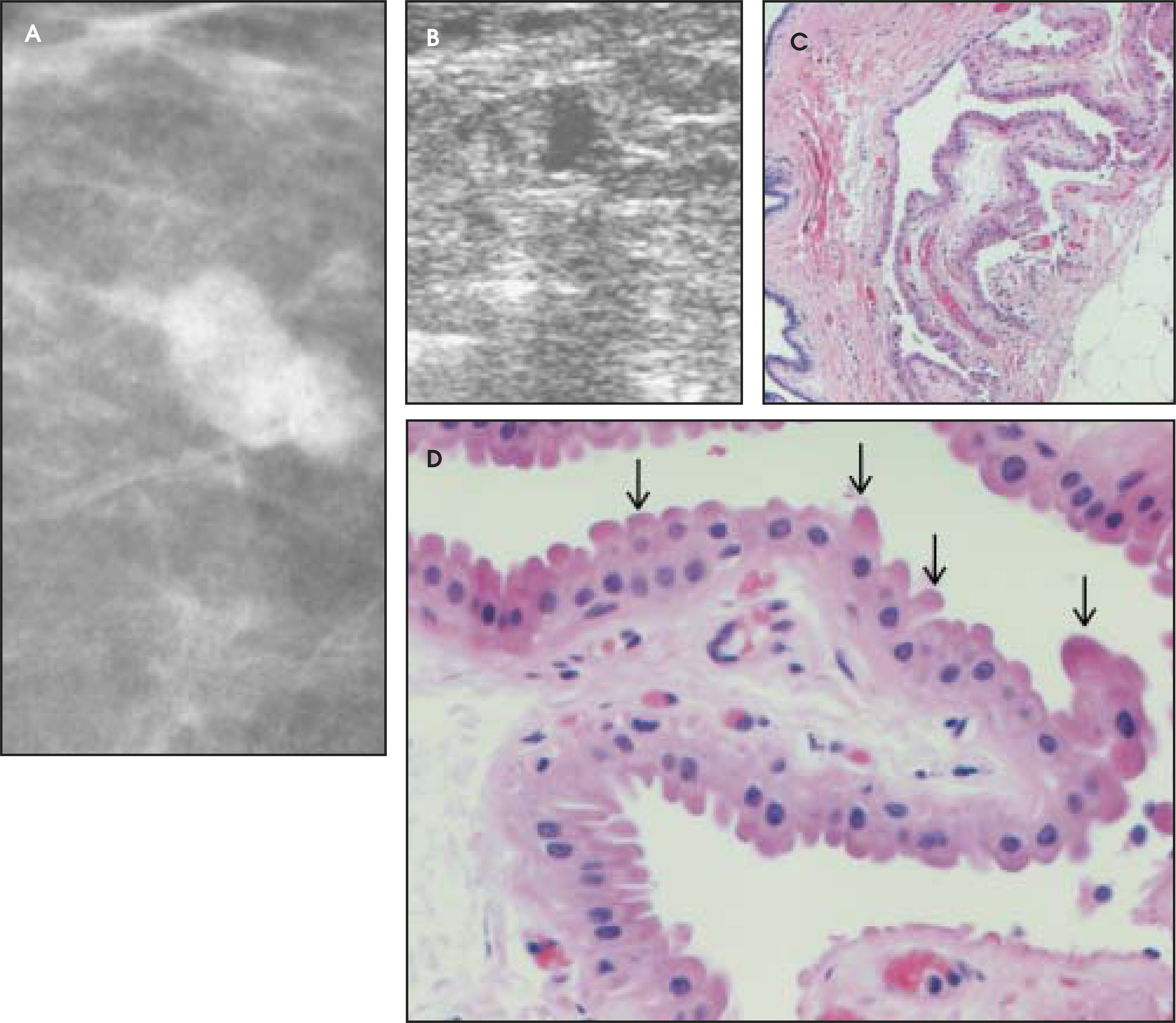
In a retrospective review of 17 cases of apocrine metaplasia, Warner et al7 noted that on mammography, the majority of cases presented as isodense masses averaging 13 mm in size. In approximately 60% of the cases, the borders of the lesion were microlobulated, and in 30% they were macrolobulated. On sonography, 77% were found to be small, anechoic masses with septae (Figures 8 through 11).7
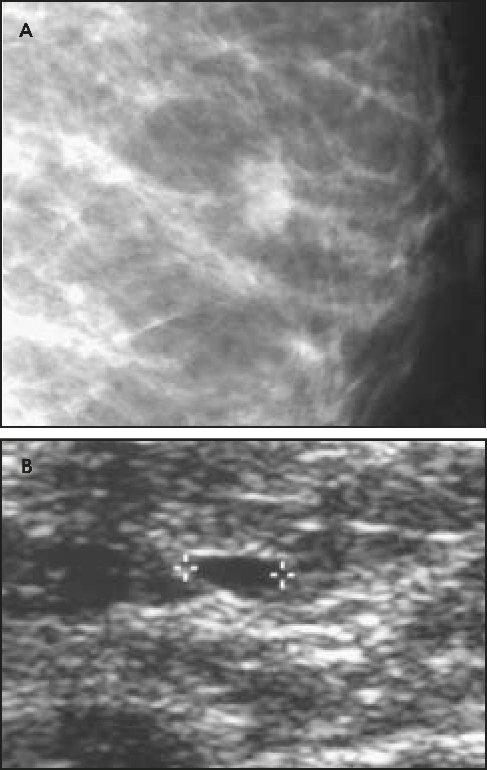
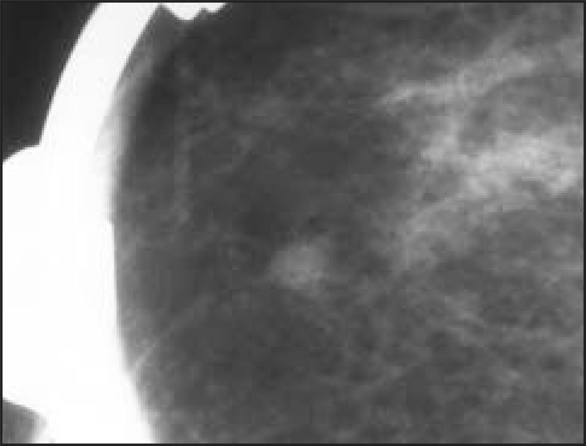
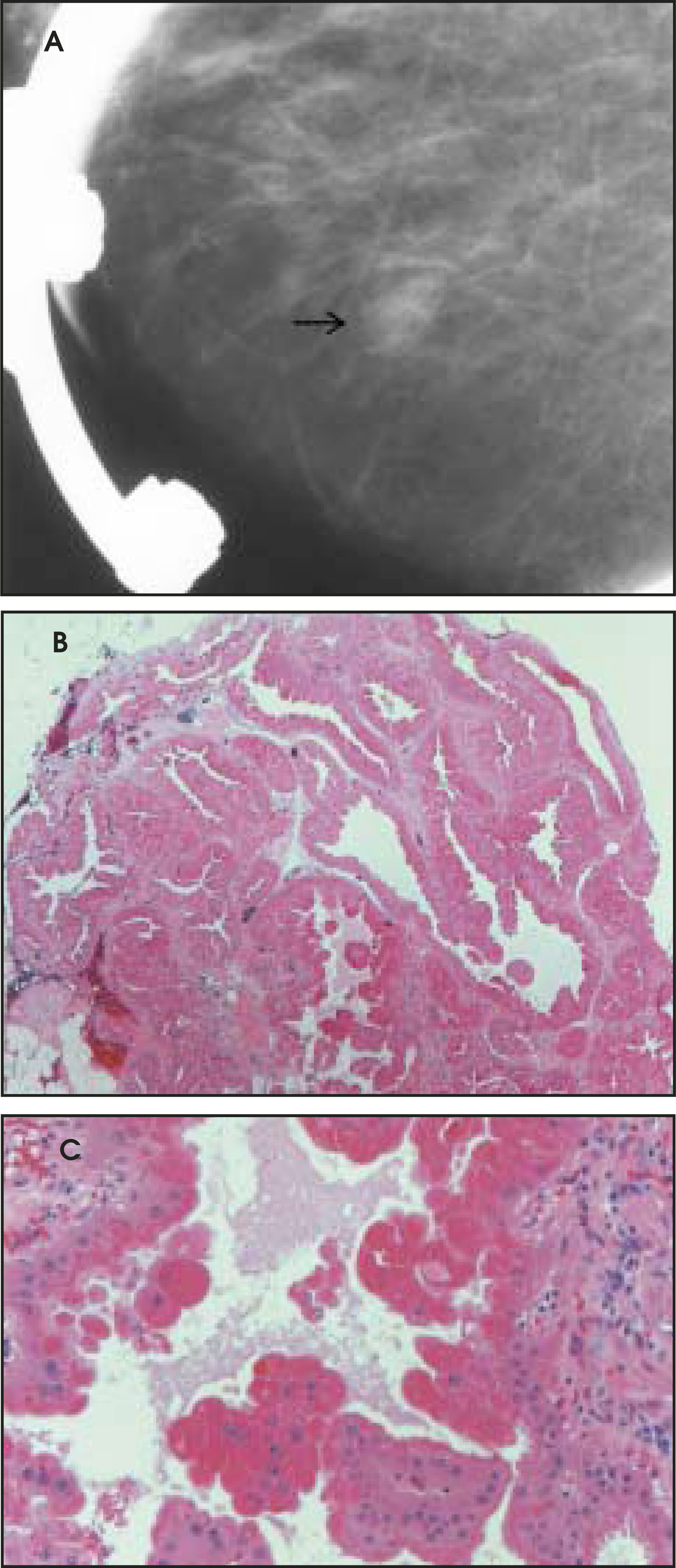
Gross and microscopic pathology
The gross appearance of apocrine metaplasia is nonspecific. Microscopically, apocrine metaplasia is most commonly found in cyst epithelium. Papillary apocrine metaplasia demonstrates a myoepithelial cell layer and micropapillary or branching patterns of hyperplasia.1
Papillomatosis
Clinical presentation
Papillomatosis, or usual duct hyperplasia, does not tend to present clinically, as these are microscopic lesions. When there is a clinical presentation, papillomatosis is an incidental finding coexistent with another palpable lesion, or a large area of papillomatosis presents as an ill-defined, palpable region of breast thickening. The majority of patients with papillomatosis are between ages 35 and 60.1
Imaging
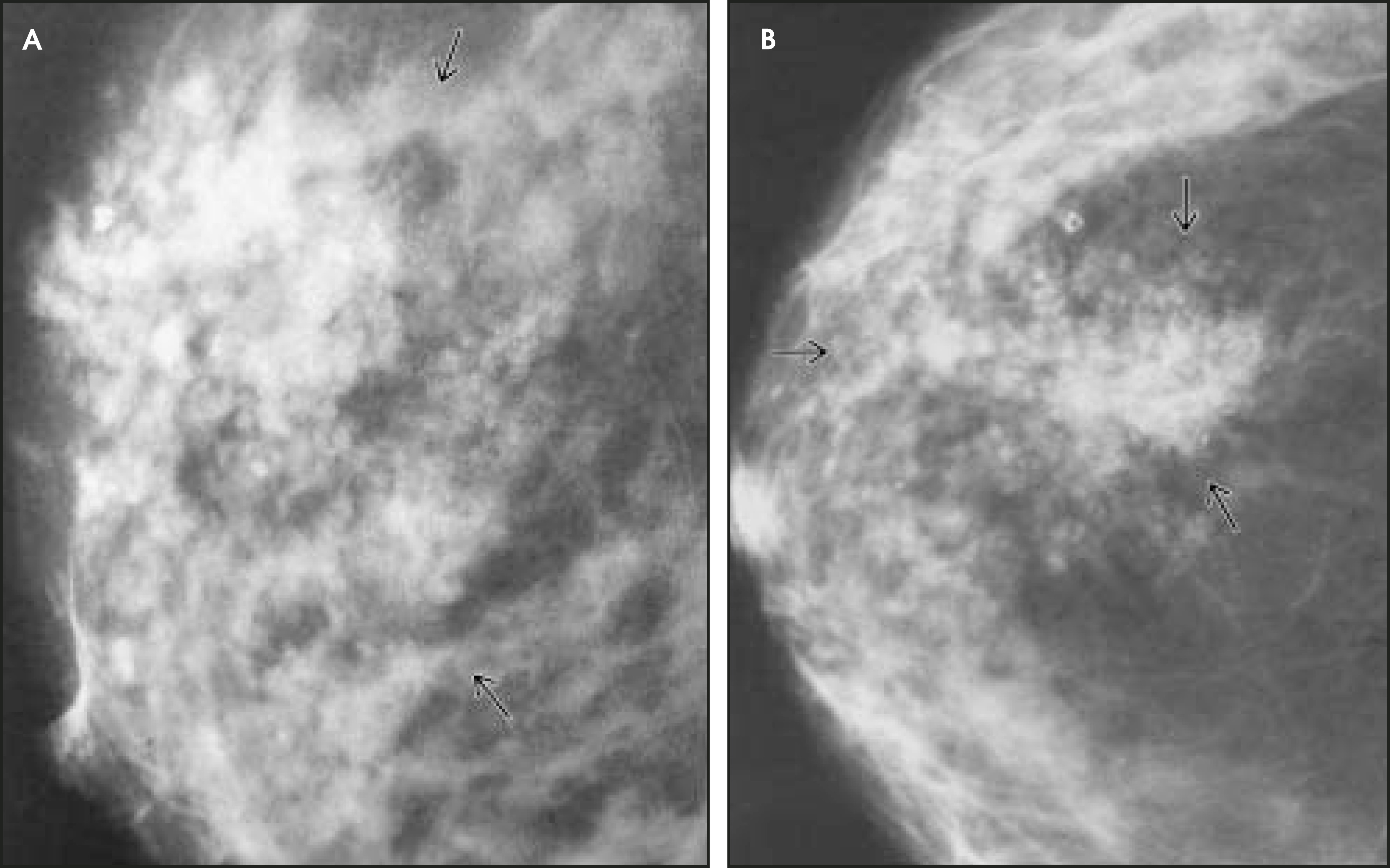
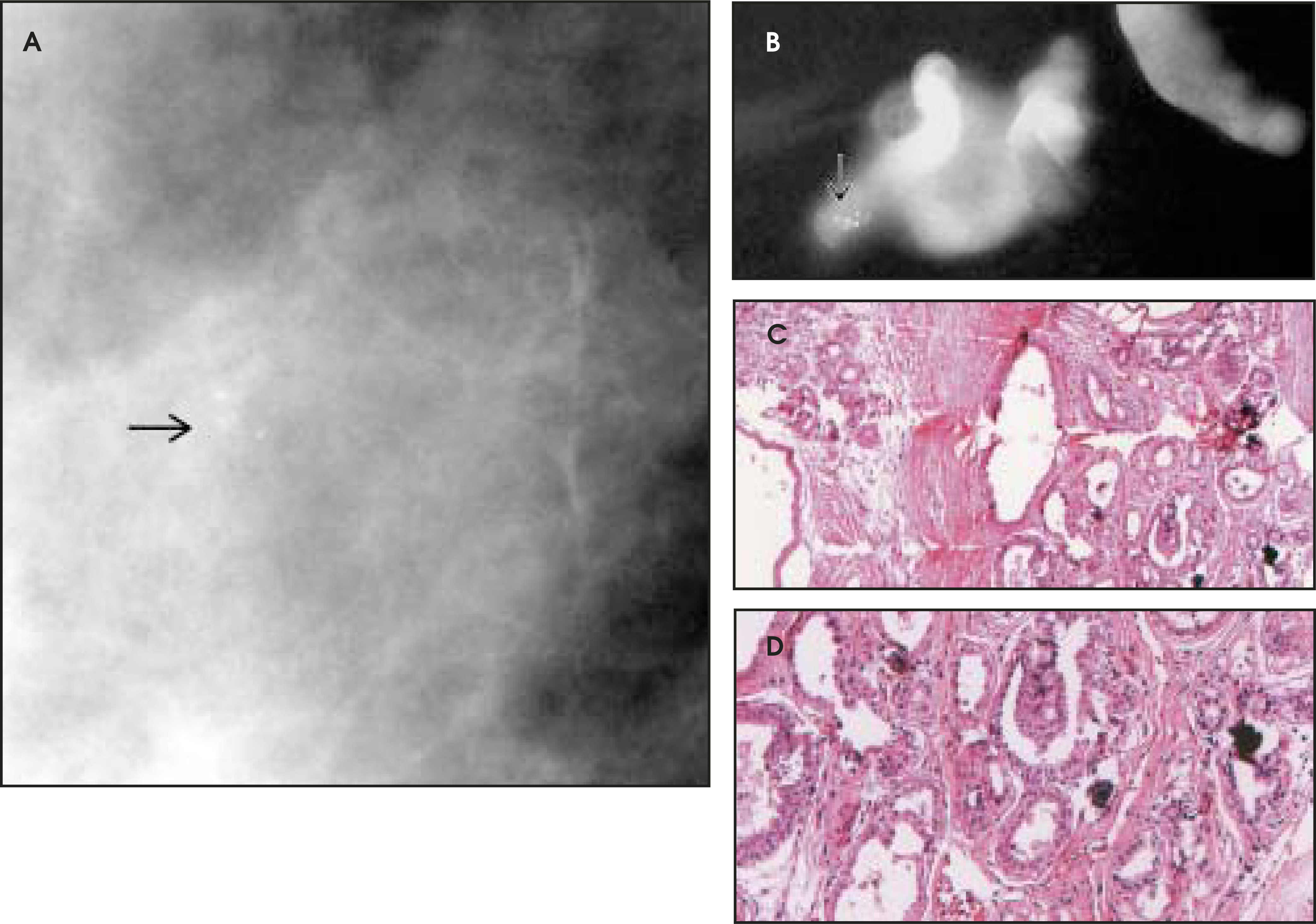
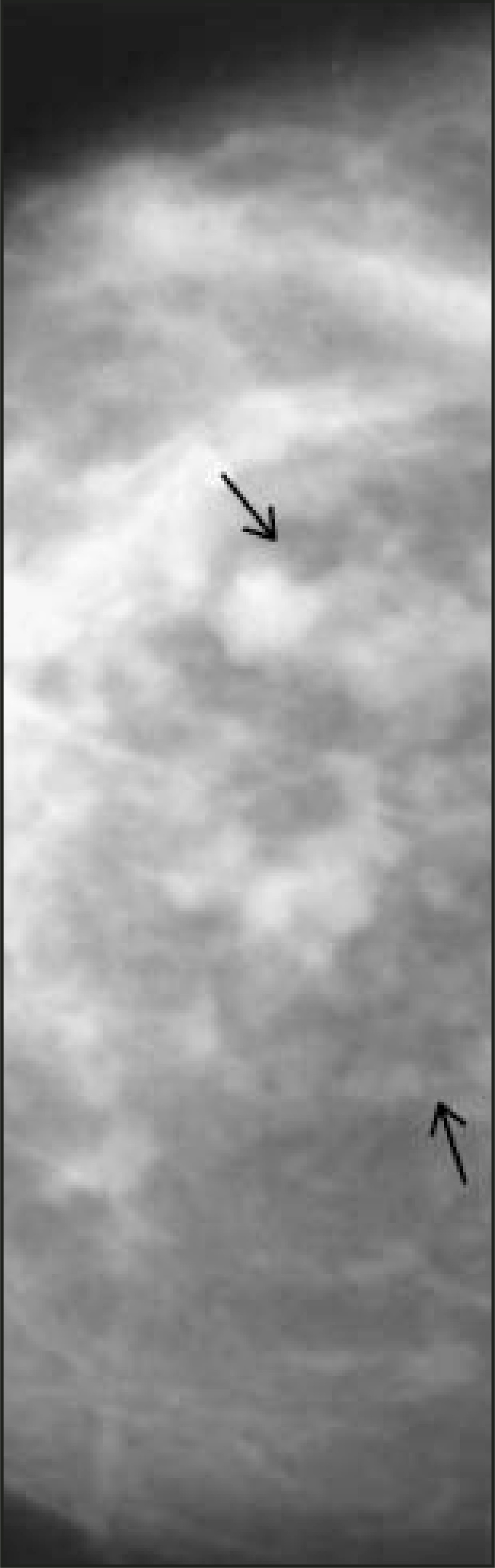
The imaging findings of usual duct hyperplasia, or papillomatosis, are nonspecific. These lesions are often found on percutaneous needle biopsies or surgical biopsies performed for mammographically detected abnormalities. Amorphous or pleomorphic microcalcifications, classified as BIRADS 4 or 5, may occur in areas of duct hyperplasia or papillomatosis. Occasionally, a dilated duct pattern or focal asymmetric density may be the mammographic presentation of papillomatosis (Figures 12 through 14).
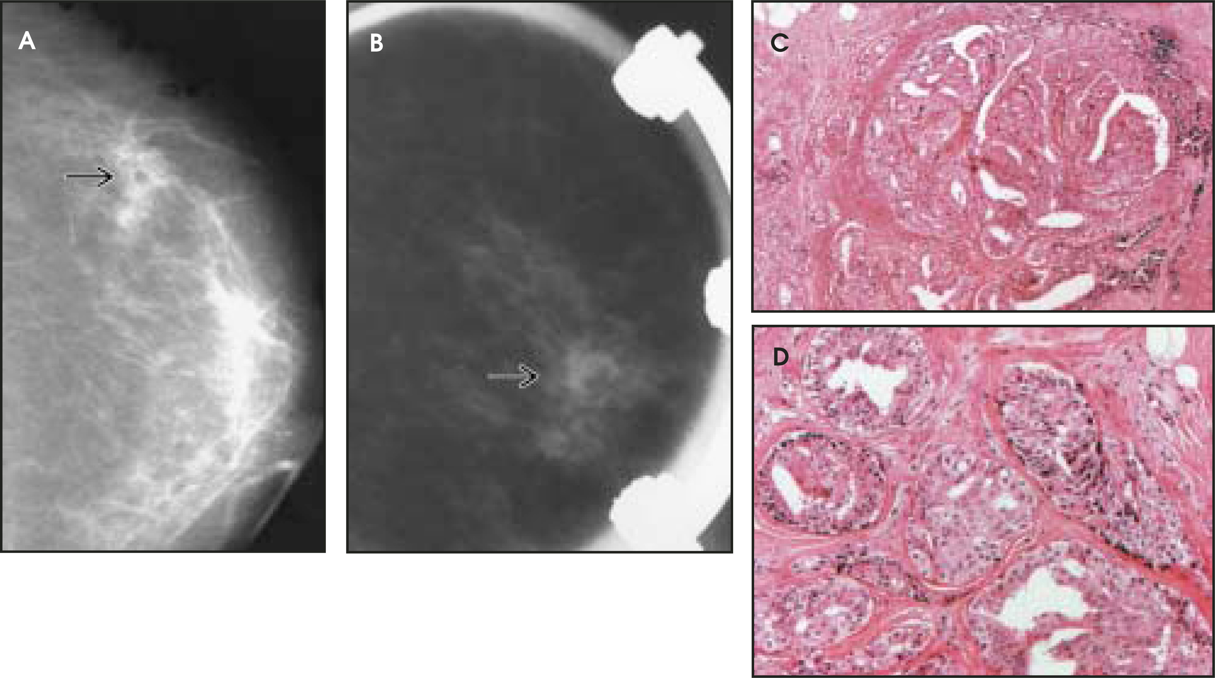
Gross and microscopic pathology
Although the terms papillomatosis and ductal hyperplasia are considered by many to be synonymous, Rosen1 contends that papillomatosis specifically refers to lesions “in which a distinct fibrovascular structure supports papillary epithelial hyperplasia.” Usual ductal hyperplasia can be further defined as mild, moderate, severe, or florid, although sometimes this classification is problematic, given the highly varied structure of this entity.1
Florid papillomatosis of the nipple
Clinical presentation
Florid papillomatosis of the nipple is an entity known by various other names, including adenoma of the nipple. Occasionally, it is confused with syringomatous adenoma, a lesion similar to syringoma of the skin, but these are two distinct pathologic diagnoses.1
Florid papillomatosis of the nipple usually presents as unilateral, bloody discharge from a painful, itchy, ulcerated nipple. The symptoms may worsen late in the menstrual cycle. Most patients with this entity are between the ages of 40 and 50. Nipple enlargement and a palpable mass may be noted, but this is not always the case. Quite often this entity is mistaken for Paget’s disease.1 In fact, based solely on clinical findings, differentiating adenoma of the nipple from Paget’s disease is essentially impossible.8
Imaging
On mammography, florid papillomatosis may mimic carcinoma, since it presents as an indistinct mass.1
Gross and microscopic pathology
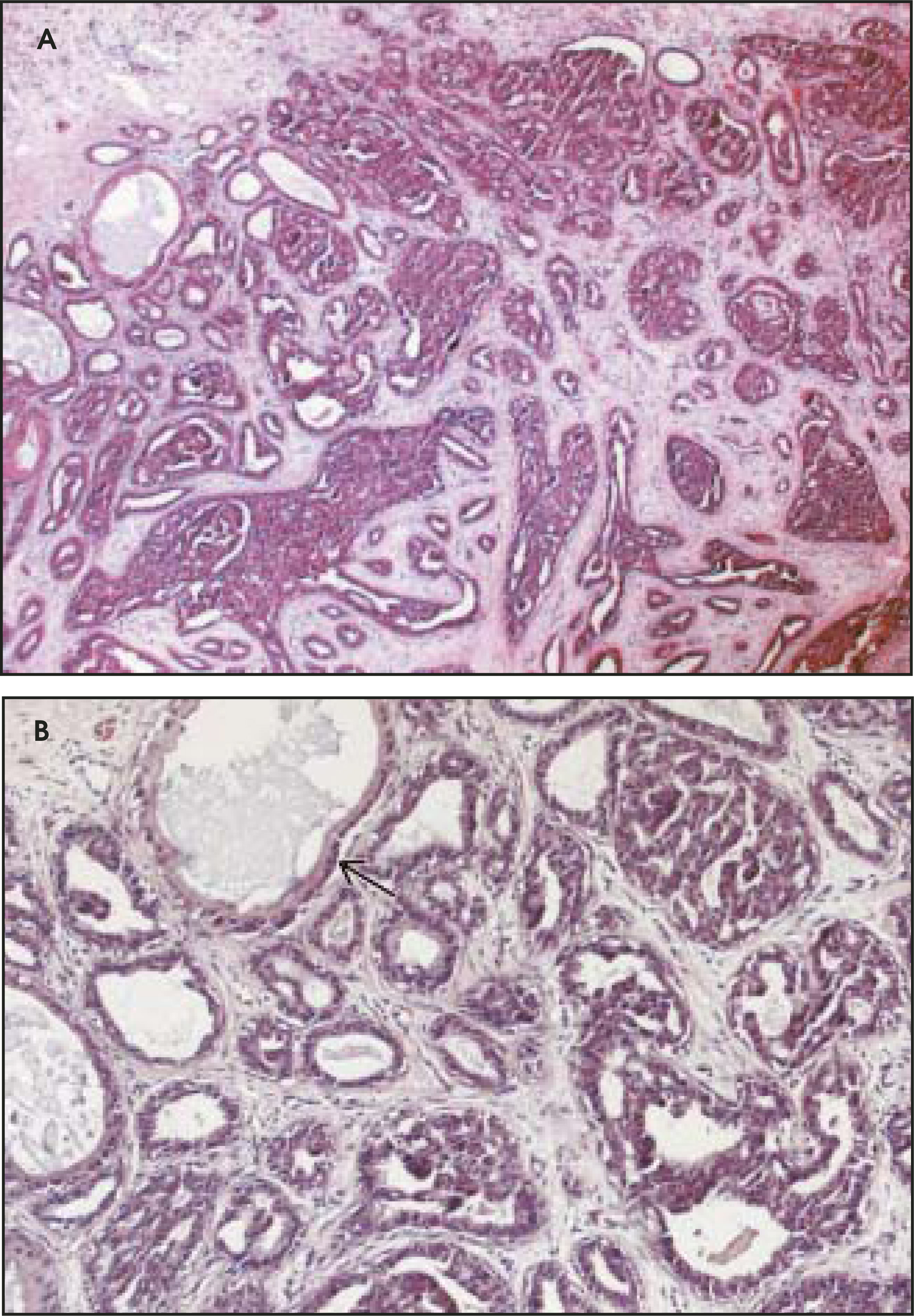
On low-power examination, florid papillomatosis, or adenoma of the nipple, is well circumscribed and unencapsulated; on high-power examination, glands are lined with a 2-cell–thick layer of epithelium and are surrounded by stromal fibrosis and epithelial hyperplasia, resulting in a pseudoinfiltrative appearance (Figure 15).9 There are 4 subtypes of florid papillomatosis, which include the sclerosing papillomatosis, papilloma, adenosis, and mixed proliferation patterns.1
In men, coexistence of florid papillomatosis of the nipple and carcinoma is seen in 50% of cases, suggesting that it may represent a precancerous lesion.1 In women, however, the evidence is less compelling, though it is suggestive of a precancerous nature.1 In a clinicopathologic review of 6 cases of adenoma of the nipple published by Santini et al8 in 1990, the researchers reported 1 case of ductal carcinoma in situ (DCIS) within the adenomatous lesions, as well as another that demonstrated an invasive ductal carcinoma in continuity with the lesions. In 3 other cases, ipsilateral, though independent, infiltrating carcinomas were found. These were classified as apocrine invasive carcinoma, invasive ductal carcinoma (NOS) and invasive papillary carcinoma, respectively. Santini and her colleagues8 concluded that adenoma of the nipple may be a marker for concurrent breast cancer, if not a high-risk lesion with malignant potential itself.
Radial scar
Clinical presentation
Radial scar, or radial sclerosing lesion, is most commonly found in the age group between 40 and 60 and is quite uncommon in patients younger than 30. Most of these lesions are microscopic and escape detection on physical examination. Lesions are ultimately detected by mammography when they are at least 5 mm in size. Lesions <1 cm are called radial scars, while larger ones are often referred to as complex or radial sclerosing lesions. They tend to occur with comparable frequency in breasts that do and do not contain carcinoma.1
Imaging
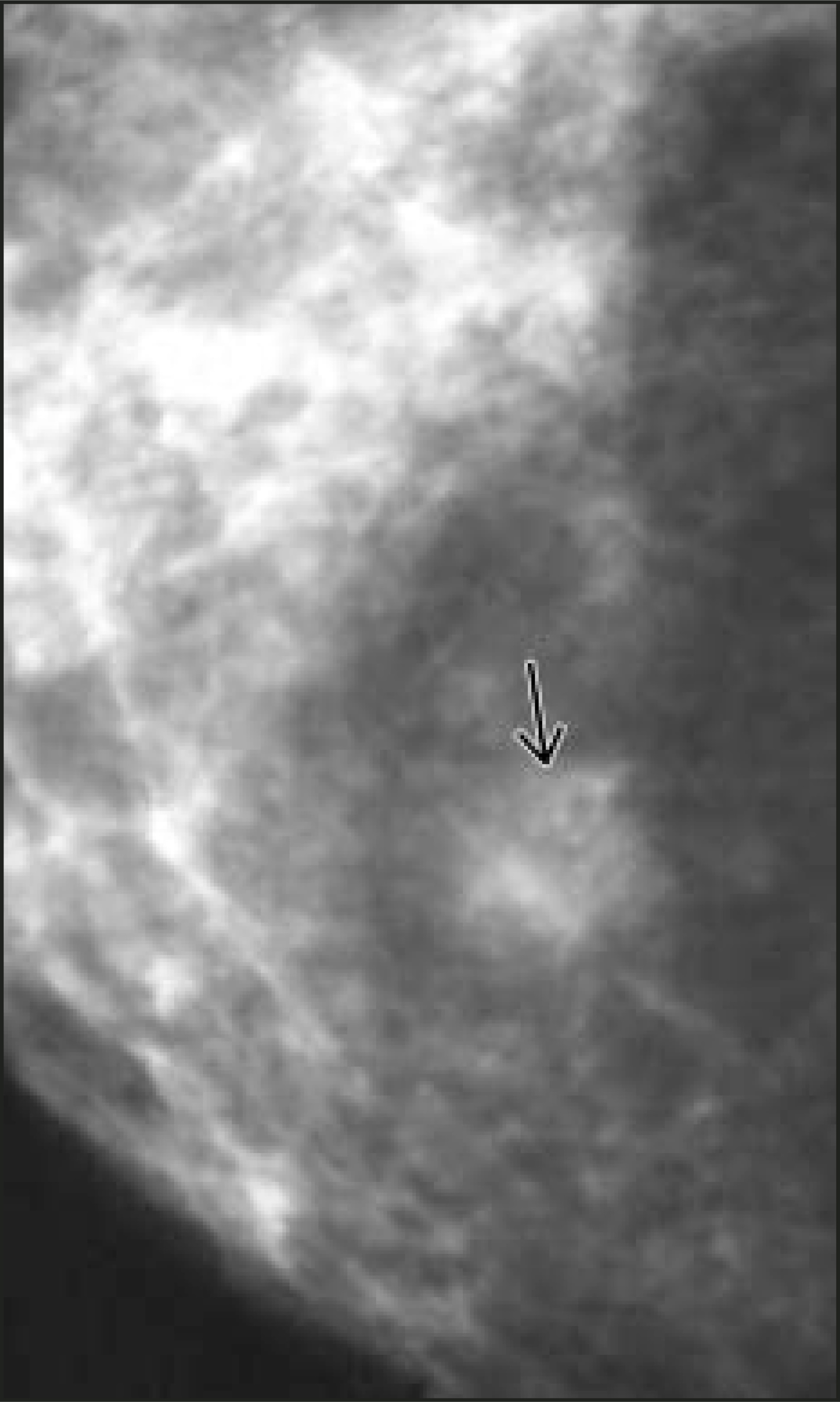
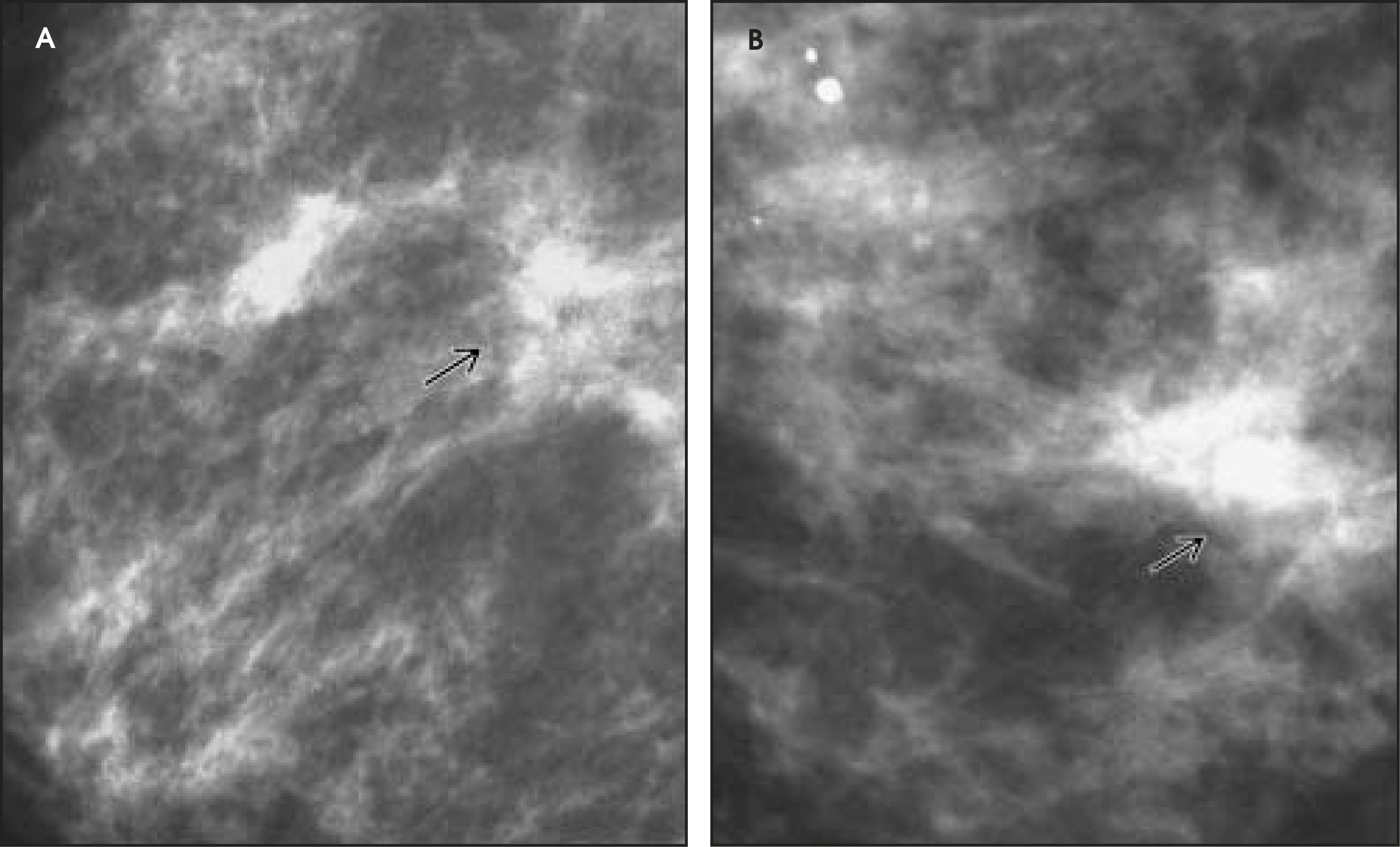
In general, radial scars are indicated by areas of architectural distortion on mammography. Other characteristics include central radiolucency, radiating thin spicules with parallel radiolucent areas, and a variable appearance depending on the projection. This is the so-called “black star” appearance that has been described in conjunction with radial scar.10 None of the aforementioned features are specific to radial scar and they can be seen in carcinomas, scars, or sclerosing adenosis. Amorphous, punctate, or pleomorphic microcalcifications may be seen in radial scars, and occasionally these are the sole mammographic finding. For the most part, these lesions are not palpable and demonstrate no associated skin changes (Figures 16 through 21).11
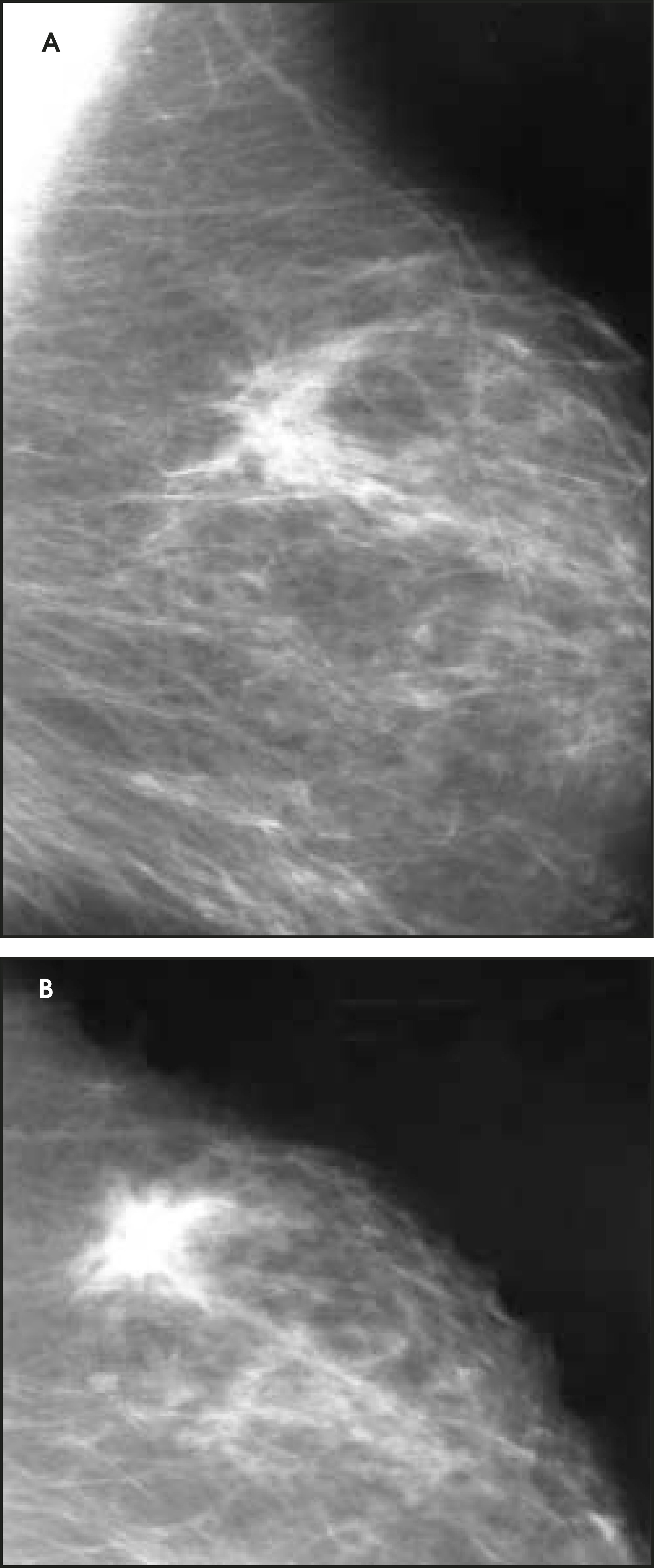
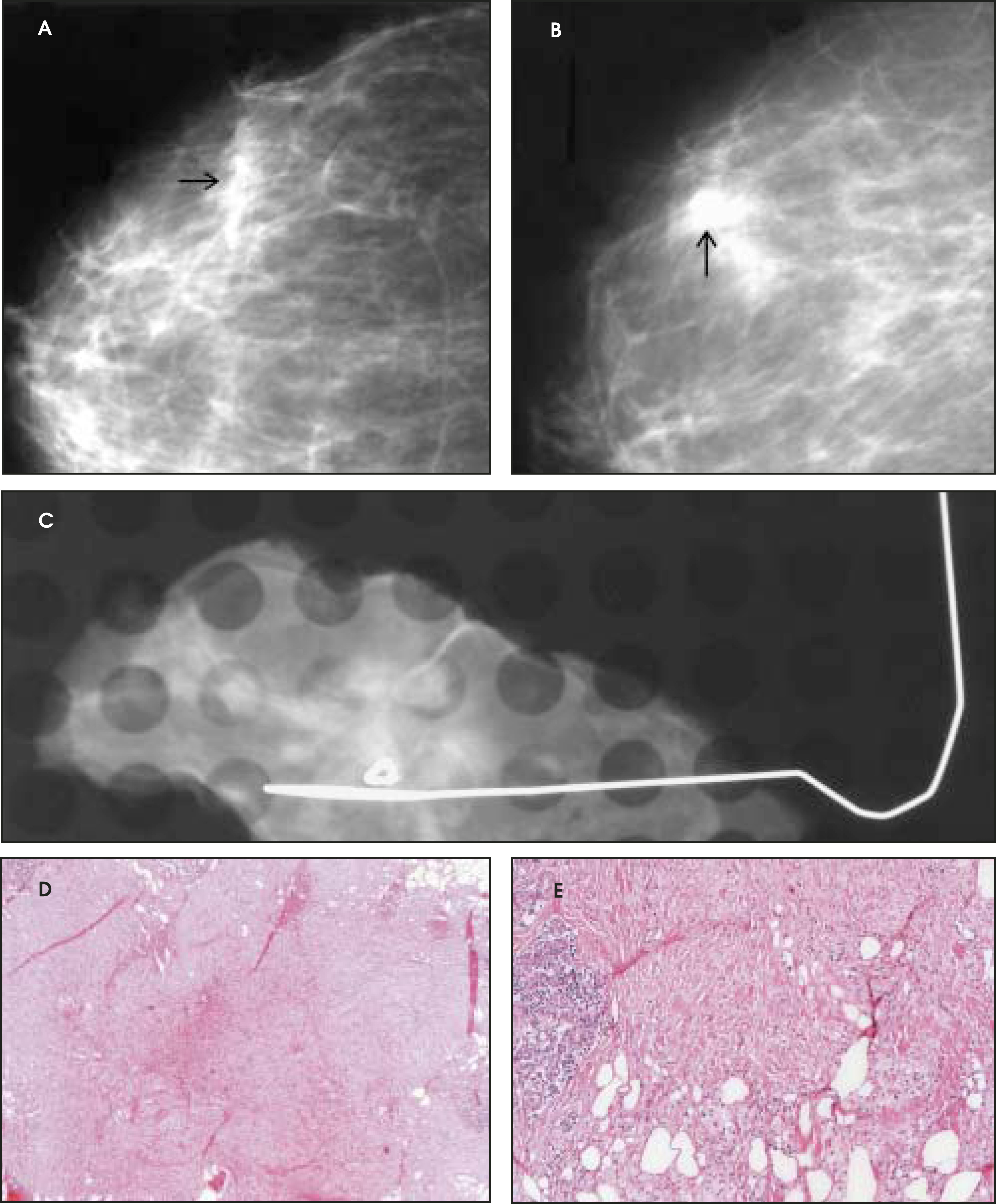
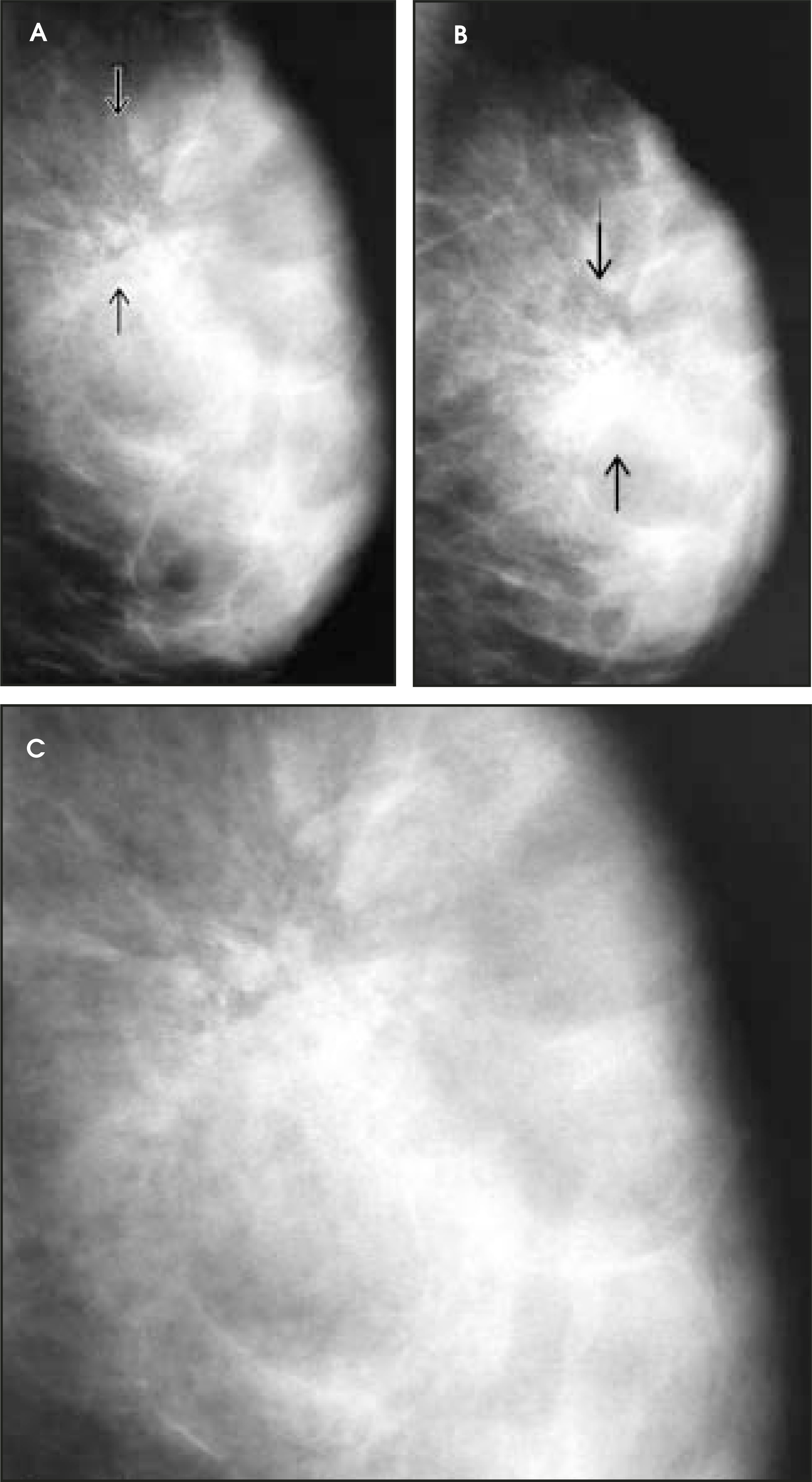
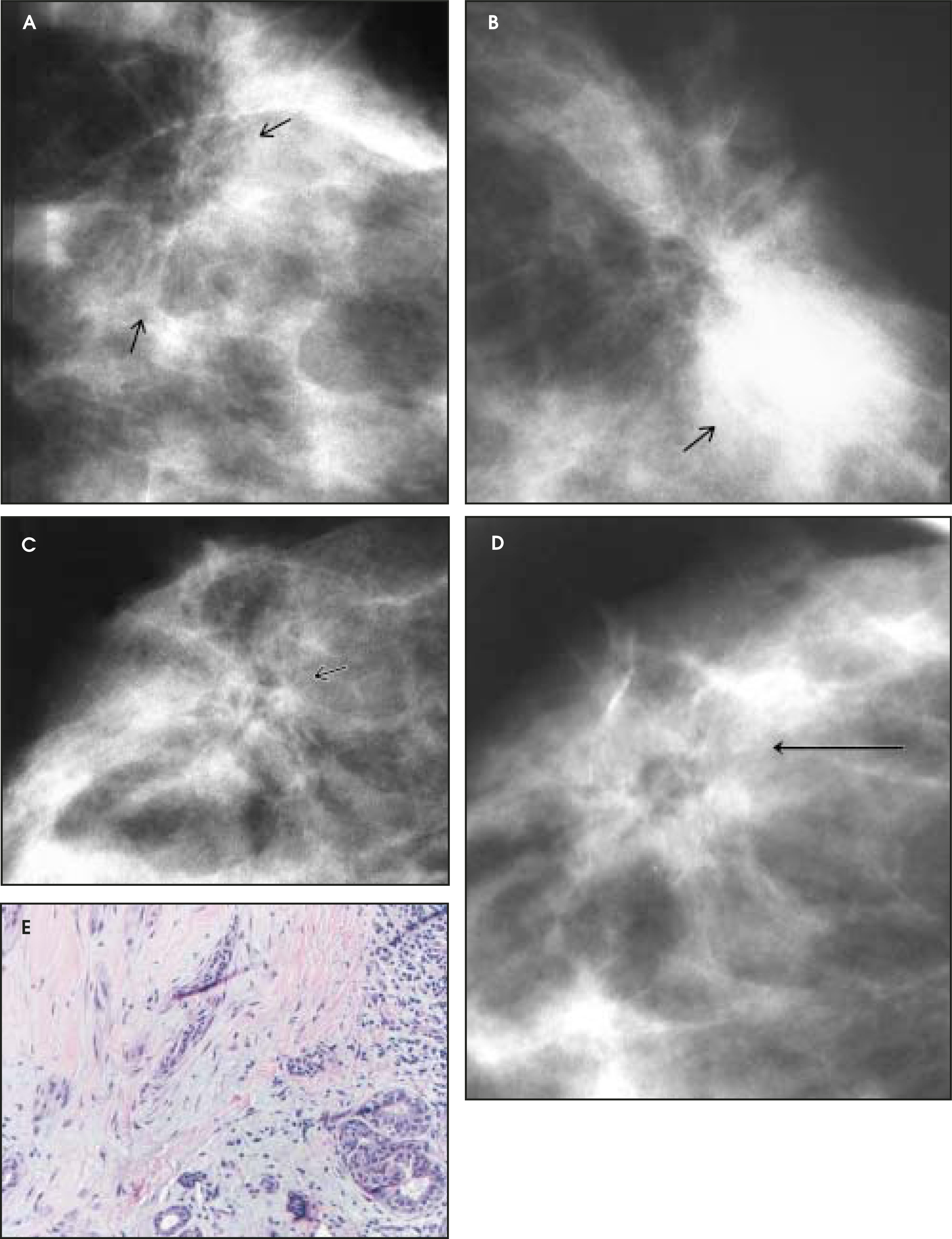
Gross and microscopic pathology
Radial scar typically has a firm, pale center accompanied by radially oriented bands of stroma in a spoke-wheel orientation. The central sclerotic nidus consists of fibrosis and elastosis. In between the radial bands of sclerosis are small ductules and distended lobules. Some radial scars have a “corona” consisting of ducts, lobules, and cysts.
On pathology, the major differential diagnosis with radial scar is tubular carcinoma. Distinguishing features of tubular carcinoma include malignant cells invading tissue as tubular structures, not typically seen in radial scars and lacking a myoepithelial layer.1 Some believe that radial scar is a precursor to tubular carcinoma.12 Interestingly, the mammographic features of radial scar and tubular carcinoma may be identical.
Radial scars can be bilateral or multifocal, and calcification is a common feature. The pseudoinfiltrative appearance of the lesion may make distinguishing it from carcinoma quite difficult, although the presence of the myoepithelial layer seen in benign lesions helps to confirm a diagnosis of radial scar. Additionally, radial scars can be found in conjunction with in situ or infiltrating cancers as well as atypical ductal or lobular hyperplasia.11 Because of this association, the diagnosis of radial scar on core needle biopsy usually prompts excision to exclude more serious pathologies.
Papillary carcinoma
Ductal carcinoma in situ, micropapillary type
Clinical presentation—Most patients with DCIS present asymptomatically with a mammographically detected lesion. Occasionally, serous or bloody nipple discharge or a palpable area of thickening are the clinical presentation of this entity. Micropapillary DCIS has a tendency toward multifocality and often manifests as skip lesions throughout the ductal system. More so than in other forms of DCIS, the patient may present with either palpable thickening or nipple discharge.4
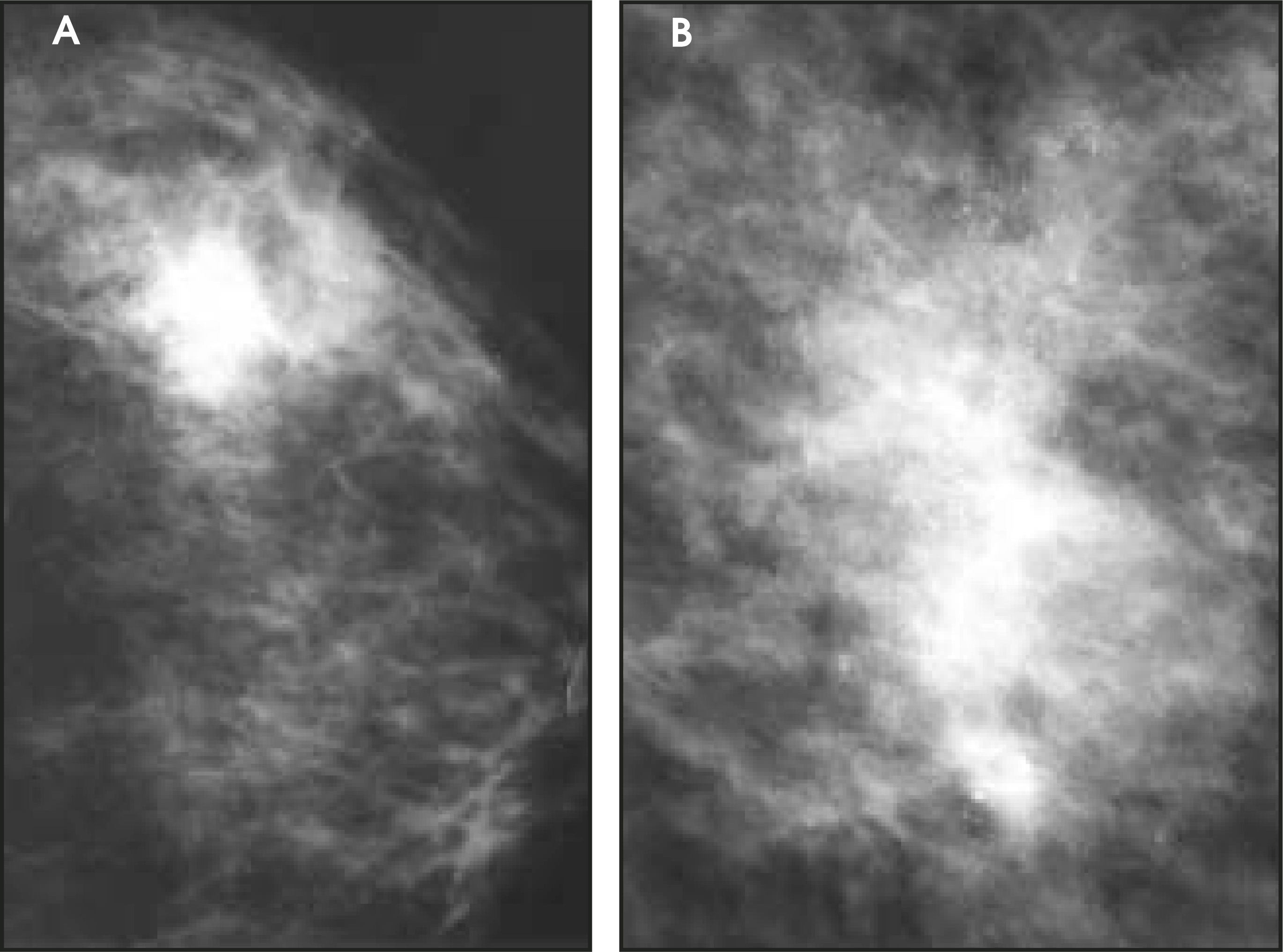
Imaging—On mammography, the majority of cases of DCIS present as microcalcifications that are pleomorphic, amorphous, or fine and linear.13 Occasionally, dilated ducts or an area of asymmetry with or without microcalcifications may be the presenting feature (Figure 22). As many as 65% of cases may be multifocal, and the micropapillary type has a greater tendency toward this.13 In reviewing cases of micropapillary DCIS, Schwartz et al14 found that 86% were multicentric. Multiple clusters of pleomorphic or amorphous calcifications may be the presenting features of micropapillary DCIS. Galactographic findings in patients with nipple discharge and micropapillary DCIS include multiple filling defects and areas of duct narrowing.15 Ultrasound findings include dilated ducts, which may also be associated with thickened walls or intraluminal filling defects.4
Gross and microscopic pathology––In the micropapillary subtype of DCIS, ducts are lined by neoplastic cells with intervening papillary projections. These papillae do not have a fibrovascular core and vary in morphology from thin and long to short and broad-based. No myoepithelial layer is present. A characteristic feature of micropapillary intraductal carcinoma is the presence of “Roman bridge arches.” They result from the creation of microlumens by the coalescence of nearby papillary fronds.1
Invasive papillary carcinoma
Clinical presentation—Papillary carcinoma is a slow-growing lesion that comprises 1% to 2% of breast cancer in women and a minimally higher proportion in men. The range of the mean age of presentation is 63 to 67 years, which is slightly older than for other types of carcinoma. Approximately 50% are located centrally within the breast, and nipple discharge is present in 22% to 34%. Bloody discharge is more common in papillary carcinoma than in papilloma; however, the most common cause of bloody discharge overall is papilloma. The average size of most lesions is 2 to 3 cm; however, they can become large and cystic, involving the entire breast.1
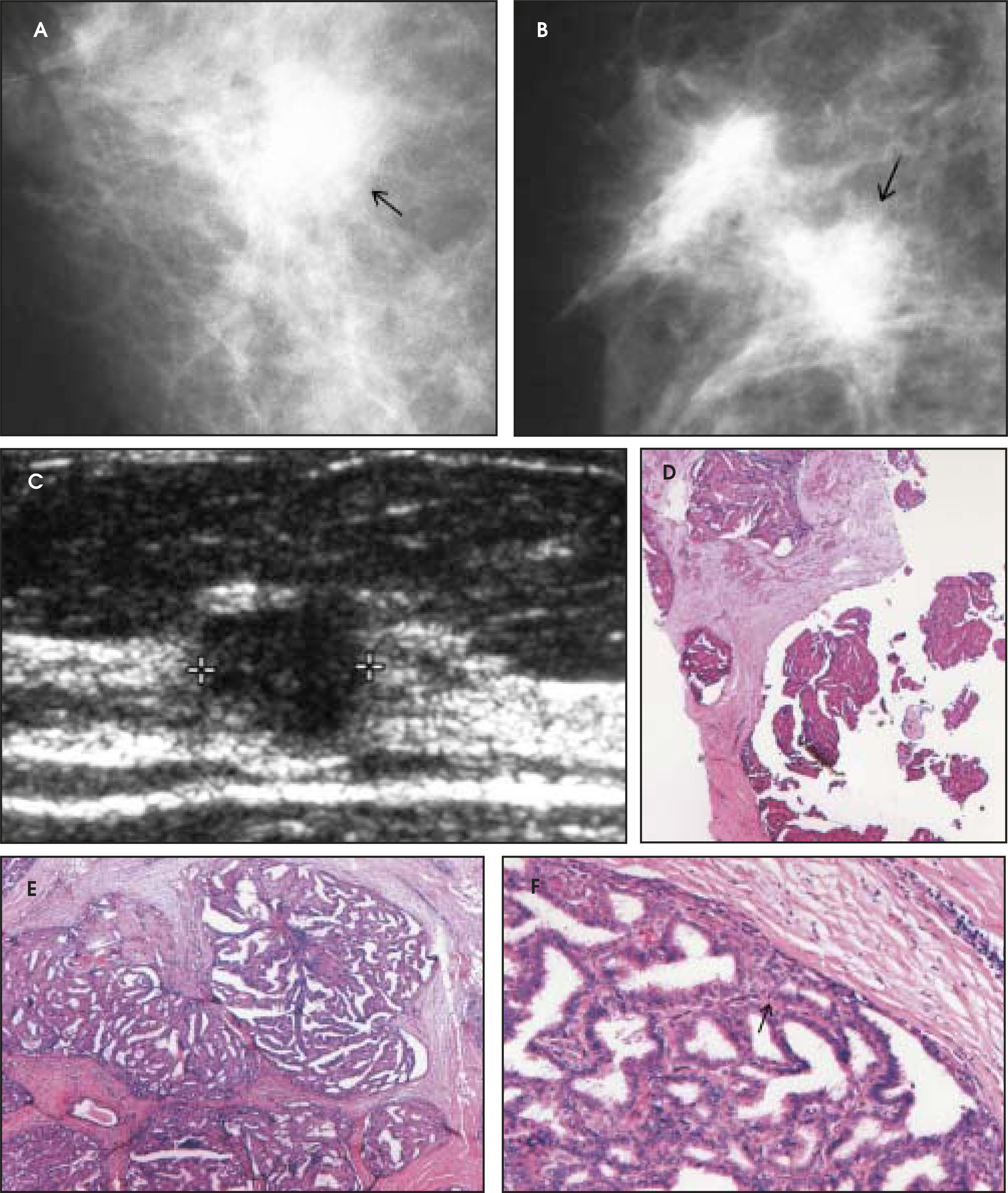
Imaging—Solitary papillary carcinomas tend to be lobulated, rounded lesions, which may be well circumscribed or ill defined on mammography. Invasive papillary carcinoma can also present as several fairly well-defined masses.15 On ultrasound, papillary carcinoma appears as a cystic lesion containing a solid component or as a hypoechoic, solid mass. Another appearance that may be seen is that of prominent ductal extension toward the nipple with a fluid-filled dilated duct between a solid component and the nipple (Figures 23 and 24).16
Gross and microscopic pathology–– Papillary carcinoma is one of the specialized types of invasive ductal carcinoma. Papillary carcinomas tend to be well circumscribed with a variable gross appearance characterized by differing amounts of cystic and solid elements, which may be in part hemorrhagic. Microscopically, branching limbs of fibrovascular stroma and a lack of a myoepithelial layer are noted.1
Conclusion
Papillary lesions of the breast include the spectrum of benign, high-risk, and malignant pathologies and, thus, may involve major or smaller lactiferous ducts. An understanding of the range of pathologic entities will aid the radiologist in considering these lesions on imaging studies.
Acknowledgments
The authors gratefully acknowledge the assistance of Daniel C. Mathews, Medical Student, Medical College of Virginia, Virginia Commonwealth University Health System, Richmond, VA, with case collection and Ms. Louise Logan with manuscript preparation.
References
- Rosen P. Rosen’s Breast Pathology. 2001:77-385.
- Woods E, Helvie M, Ikeda D. Solitary breast papilloma: Comparison of mammographic, galactographic, and pathologic findings. AJR Am J Roentgenol. 1992;159:487-491.
- Huynh P, Parellada J, Shaw de Paredes E. Dilated duct pattern at mammography. Radiology. 1997;204:137-141.
- Shaw de Paredes E, Hijaz T, Trivedi A. Assessment of the abnormal lactiferous duct. Semin Breast Dis. 2004;7:21-48.
- Daniel B, Gardner R, Birdwell R. Magnetic resonance imaging of intraductal papilloma of the breast. Magn Reson Imaging. 2003;21:887-892.
- Cardenosa G, Eklund G. Benign papillary neoplasms of the breast: Mammographic findings. Radiology. 1991;181:751-755.
- Warner J, Kumar D, Berg W. Apocrine metaplasia: Mammographic and sonographic appearances. AJR Am J Roentgenol. 1998;170:1375-1379.
- Santini D, Taffurelli M, Gelli M. Adenoma of the nipple: A clinico-pathologic study and its relation with carcinoma. Semin Breast Dis. 1990;3:153-163.
- Vianna L, Millis R, Fentiman I. Adenoma of the nipple: A diagnostic dilemma. Br J Hosp Med. 1993;50:639-642.
- Tabar L, Dean P. Teaching Atlas of Mammography. 1985:89-90.
- Kennedy M, Masterson A, Kerin M, Flanagan F. Pathology and clinical relevance of radial scars: A review. J Clin Pathol. 2003;56:721-724.
- Vega A, Garijo F. Radial scar and tubular carcinoma. Acta Radiology. 1993;34:43-47.
- Dershaw D, Abramson A, Kinne D. Ductal carcinoma in situ: Mammographic findings and clinical implications. Radiology. 1989;170:411-415.
- Schwartz G, Patchefsky A, Finklestein S. Nonpalpable in situ ductal carcinoma of the breast. Predictors of multicentricity and microinvasion and implications for treatment. Arch Surg. 1989;124:29-32.
- Shaw de Paredes E. Atlas of Film-Screen Mammography. . 1992:133-517.
- Stavros A. Breast Ultrasound. 2004:179-180.
Citation
. Radiologic-pathologic correlation of papillary lesions of the breast. Applied Radiology. 2005;34(11):20-42. doi:10.37549/AR1381.