Radiological Case: Metastatic meningioma
Applied Radiology — Vol. 44 , Issue 11 , pp. 68 -71
DOI: 10.37549/AR2230
Published: November 1, 2015
Categories
CASE SUMMARY
A 41-year-old male with recurrent intracranial meningioma with extensive metastatic spread to the lungs. We present radiologic imaging of the indolent natural progression of the lung metastases over a span of 7 years with no systemic treatment. The uniqueness of the case presentation will further the understanding of this rare entity.
IMAGING FINDINGS
Meningiomas constitute approximately 15% of primary brain tumors.1 Most meningiomas are benign with only a minor percentage of cases exhibiting malignant or metastatic behavior. Metastases have a predilection to pulmonary, bone and liver locations.2 The majority of pulmonary metastases are solitary, with only a few case reports of extensive lung involvement.3 Correct diagnosis requires histologic sampling of lung lesions.
A 41-year-old male with a history of intracranial meningioma, World Health Organization (WHO) grade II was initially diagnosed in 1999 and initially resected at another facility. At his first presentation to our institution in 2007, the patient complained of headaches and seizures. Work-up revealed locally recurrent intracranial meningioma on brain MRI (Figure 1), as well as pulmonary nodules on chest radiographs (Figure 2). Subsequent CT-guided fine needle aspiration biopsy of a lung nodule confirmed metastatic meningioma (CK5/6, TTF1 and progesterone receptors negative; EMA focally positive)
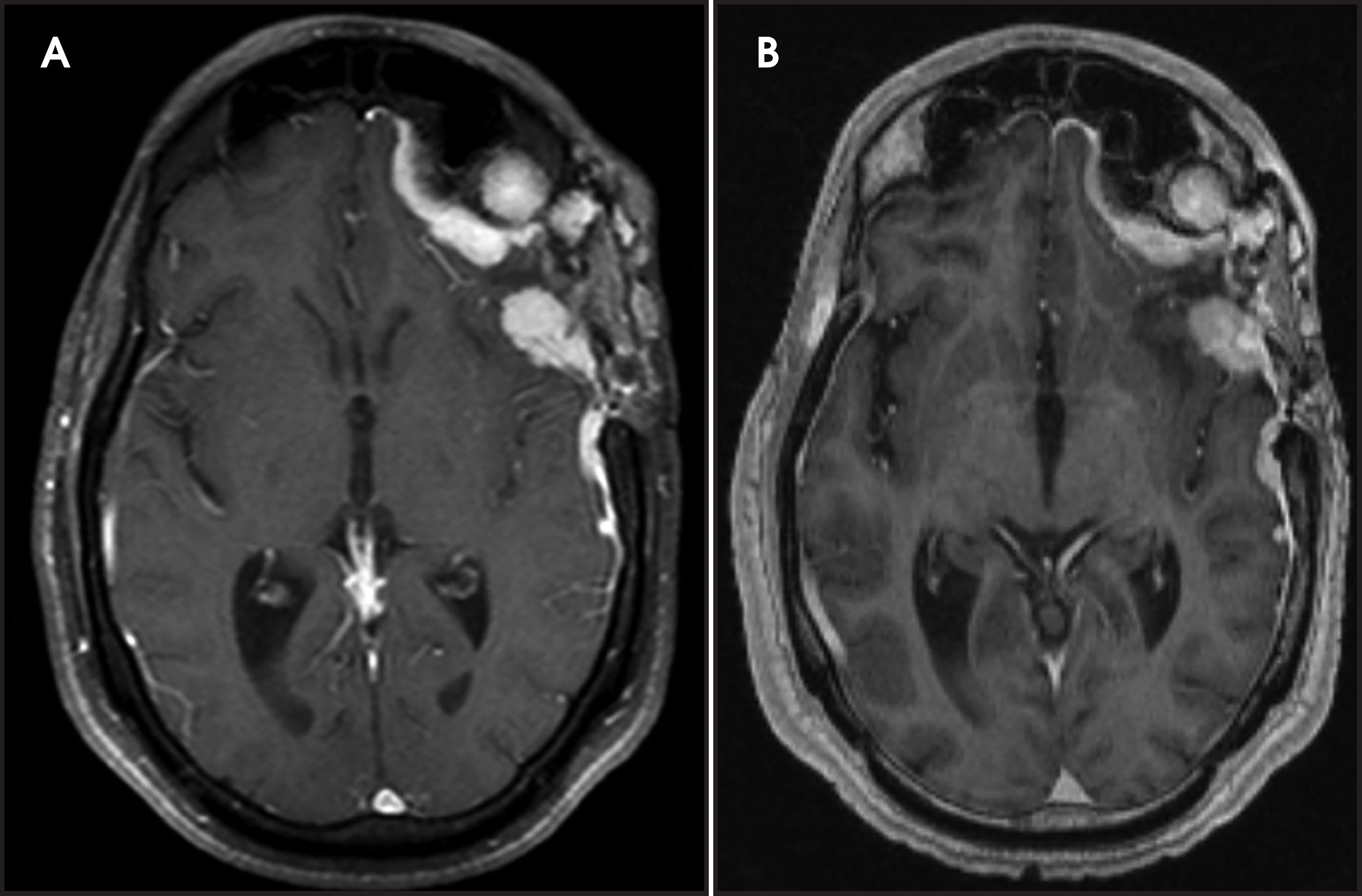
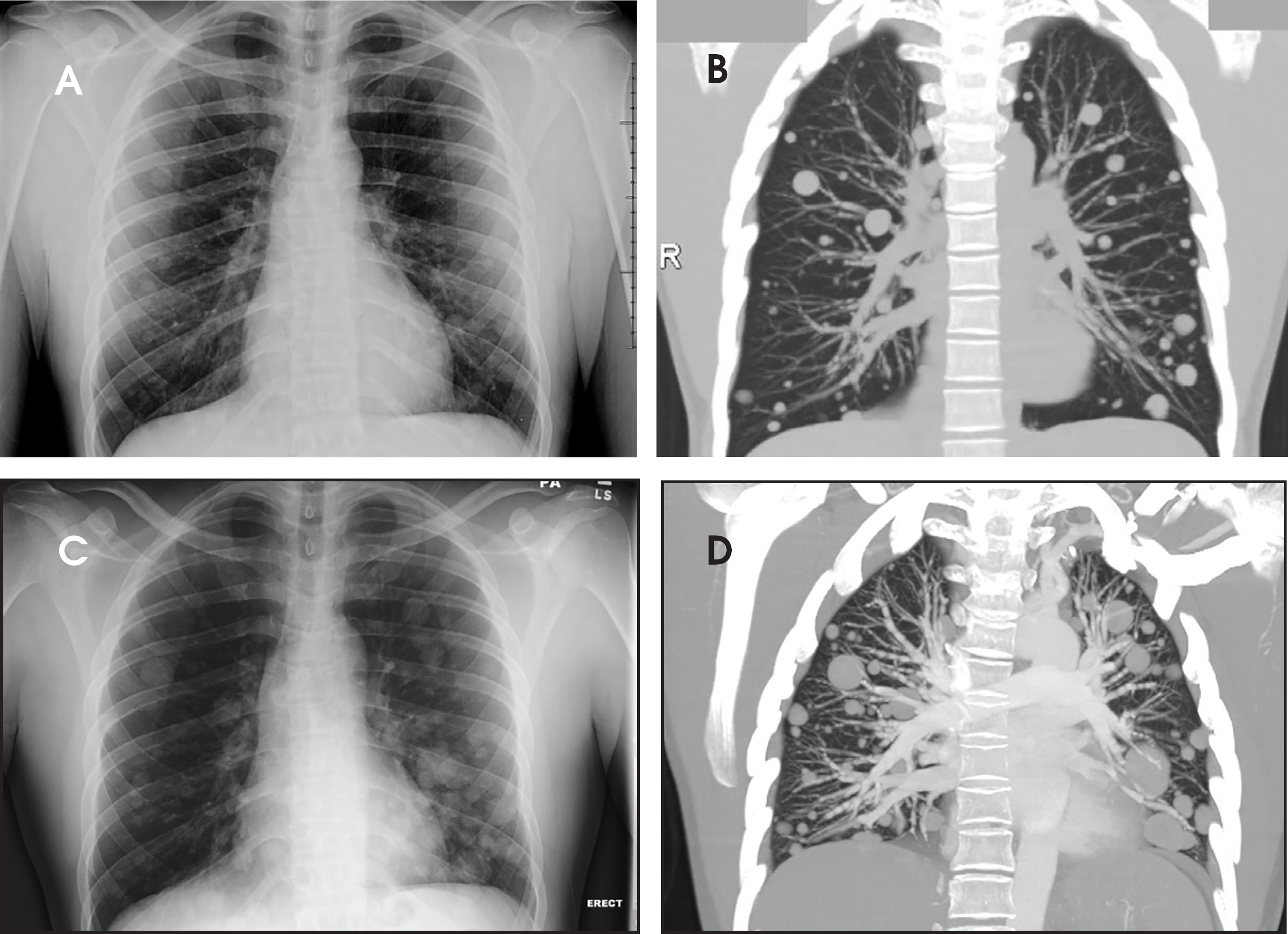
The patient received CyberKnife radiotherapy for the brain tumor with partial clinical response of headaches and seizures However, he repeatedly declined systemic chemotherapy for the metastatic pulmonary involvement.
Upon later readmissions for recurrent seizures, follow-up serial chest imaging revealed slow but persistent growth of these untreated pulmonary nodules (Figure 2) On his most recent hospitalization, he experienced hemoptysis, which was attributed to bronchial invasion of the metastatic disease.
DIAGNOSIS
Metastatic meningioma
DISCUSSION
Central nervous system meningiomas are classified by the WHO into four grades of malignancy: WHO I - benign meningioma (88% to 95% of all cases), WHO II - atypical meningioma (5% to 6%), WHO III - malignant meningioma (1%), and WHO IV - meningioma with sarcomatous degeneration (extremely rare).1,10 Although metastatic spread of meningioma is more likely to occur in WHO grades II - IV, grade I lesions can also metastasize.11,12 Due to the much higher relative incidence of grade I meningiomas, 34% of metastatic meningiomas in one series originated from grade I lesions.2 Recent attempts to define histological parameters that would predict metastatic potential of meningioma have been inconclusive.13
Less than 1% of meningiomas exhibit malignant behavior, with over 130 cases of metastatic meningioma described in the literature.1-9 Metastatic lesions in a series by Surov et al. were most frequently found in the lung (37%), bones (17%), spine (15%), and liver (9%), with only 13% of cases having multiple metastases.2 Another series described lung metastases in 61% of metastatic meningiomas.14 Two other cases of extensive pleural metastases have been published.8,15
The case presented here describes the very slow progression of multifocal pulmonary metastases. Lung metastases are typically diagnosed incidentally and as asymptomatic solitary pulmonary nodules.13,16 Several case reports described positive response to systemic chemotherapy treatment or resection,18,19,20 and only a few case reports described any progression of extensive pulmonary disease.24 In contrast, our case documents the indolent growth of lung metastases over a long period of time. The clinically aggressive behavior of the recurrent tumor coupled with the pulmonary disease prompted the suspicion of this rare diagnosis, although biopsy was still necessary.
Metastatic disease is not typically considered in the setting of very slow-growing lung nodules. Other reported causes of slow-growing lung metastases include adenoid cystic carcinoma30, pleomorphic adenoma3, metastasizing uterine leiomyoma22, and less commonly, thymoma and renal cell carcinoma.23,24 Epithelioid hemangioendothelioma is an additional uncommon cause of chronic slow growing lung lesions.25 Mengingioma is now added to this uncommon, but distinct list.
CONCLUSION
This case demonstrates the slow imaging and clinical progression of metastatic spread of meningioma to the lungs. Metastatic meningioma to lung has been well described; however, this case documents the very slow course of untreated metastases over 7 years.
References
- Perry A. Meningeal tumours WHO classification of tumours of the central nervous system. 2007.
- Surov A, Gottschling S. Distant metastases in meningioma: An underestimated problem.. J Neuroonco. 2013;112((3)):323-327.
- Yeung T, Wong C. Prolonged survival with multiple pulmonary metastases from adenoid cystic carcinoma after conservative management.. J HK Coll Radiol.. 2009;12:68-71.
- Murrah C, Ferguson E, Jannelle R. Resection of multiple pulmonary metastases from a recurrent intracranial meningioma.. Ann Thorac Surg.. 1996;61:1823-1824.
- Stoller J, Kavuru M, Mehta A. Intracranial meningioma metastatic to the lung.. Cleve Clin J Med.. 1987;54:521-527.
- Baisden B, Hamper U, Ali S. Metastatic meningioma in fine-needle aspiration of the lung.. Diagn Cytopathol.. 1999;20:291-294.
- Slavin M. Metastatic malignant meningioma.. J Clin Neuroophthalmol.. 1989;9:55-59.
- Tao L. Pulmonary metastasis from intracranial meningioma diagnosed by aspiration biopsy cytology.. Acta Cytol.. 1991;35:524-528.
- Rawat B, Franchetto A, Elavathil J. Extracranial metastasis of meningioma.. Neuroradiology.. 1995;37:38-41.
- Jääskeläinen J, Haltia M, Servo A. Atypical and anaplastic meningiomas: Radiology, surgery, radiotherapy, and outcome.. Surg Neurol.. 1986;25((3)):233-242.
- Miller D, Ojemann R. Benign metastasizing meningioma: Case report.. J Neurosurg.. 1985;62:763-766.
- Fulkerson D, Horner T, Hattab E. Histologically benign intraventricular meningioma with concurrent pulmonary metastasis: case report and review of the literature.. Clin Neurol Neurosurg.. 2008;110:416-419.
- Hishima T, Fukayama M, Funata N. Intracranial meningioma masquerading as a primary pleuropulmonary tumor.. Pathol Int.. 1995;45:617-621.
- Kaminski J, Movsas B, King E. Metastatic meningioma to the lung with multiple pleural metastases.. Am J Clin Oncol.. 2001;24:579-582.
- Allen I, McClure J, McCormick D. LDH isoenzyme pattern in a meningioma with pulmonary metastasis.. J Pathol.. 1977;123:187-191.
- Weber C. Primary pulmonary malignant meningioma with lymph node and liver metastasis in a centenary woman: An autopsy case.. Virchows Arch.. 2013;462((4)).
- Nakayama Y, Horio H, Horiguchi S, Hato T. Pulmonary and pleural metastases from benign meningeal meningioma: A case report.. Ann Thorac Cardiovasc Surg. 2014;20((5)):410-413.
- Kanzaki R, Higashiyama M, Fujiwara A. Surgical resection of pulmonary metastases from meningioma: Report of a case.. Surg Today.. 2011;41((7)):995-998.
- Erman T, Hanta I, Haciyakupoğlu S, Zorludemir S, Zeren H, Göçer A. Huge bilateral pulmonary and pleural metastasis from intracranial meningioma: A case report and review of the literature.. J Neurooncol.. 2005;74((2)):179-181.
- D’Aiuto M, Veronesi G, Pelosi G. Two-year survival after multiple bilateral lung metastasectomies for cranial meningioma.. Ann Thorac Surg.. 2005;80:1129-1130.
- Stoller J, Kavuru M, Mehta A, Weinstein C, Estes M, Gephardt G. Intracranial meningioma metastatic to the lung.. Cleve Clin J Med.. 1987.
- Londero A, Perego P, Mangioni C, Lellé R, Londero F, Marchesoni D. Locally relapsed and metastatic uterine leiomyoma: A case report.. J Med Case Reports.. 2008;2:308.
- Nomori H, Watanabe K, Ohtsuka T. Pulmonary metastasis 12 years after resection of thymoma with microscopic capsule invasion.. Jpn J Clin Oncol.. 2004;34:630-633.
- Tatsuta M, Shiozaki K, Masutani S. Splenic and pulmonary metastases from renal cell carcinoma: Report of a case.. Surg Today.. 2001;31:463-465.
- Cronin P, Arenberg D. Pulmonary epithelioid hemangioendothelioma: An unusual case and a review of the literature.. Chest.. 2004;125:789-793.
- Adlakha A, Rao K, Adlakha H. Meningioma metastatic to the lung.. Mayo Clin Proc.. 1999;74:1129-1133.
- Incarbone M. Primary pulmonary meningioma: Report of a case and review of the literature.. Lung Cancer.. 2008;62:401-407.
- Fulkerson D, Horner T, Hattab E. Histologically benign intraventricular meningioma with concurrent pulmonary metastasis: Case report and review of the literature.. Clin Neurol Neurosurg.. 2008;110:416-441.
Citation
. Radiological Case: Metastatic meningioma. Applied Radiology. 2015;44(11):68-71. doi:10.37549/AR2230.