Rapidly enlarging fibromatosis-like spindle cell carcinoma of the breast
Applied Radiology — Vol. 46 , Issue 7 , pp. 36 -39
DOI: 10.37549/AR2398
Published: July 1, 2017
1 Radiology Fellow at Beth Israel Deaconess Medical Center, Boston, MA
2 Medical Student at the Baylor College of Medicine, Houston, TX
3 Radiologist at the University of Texas Medical Branch, Galveston, TX
4 Radiologist at the Baylor College of Medicine, Houston, TX
5 Department of Radiology at the Baylor College of Medicine, Houston, TX
Categories
CASE SUMMARY
A 48-year-old woman presented to the emergency center with a rapidly enlarging, painful right breast mass. Upon physical examination, the right breast was asymmetrically enlarged (Figure 1) compared to the left breast. A firm, slightly mobile, non-tender, non-erythematous right breast mass was palpated without expression of discharge, skin changes, or visible nipple inversion. Laboratory studies showed leukocytosis. Imaging and biopsy of the mass resulted in a diagnosis of fibromatosis-like spindle cell carcinoma - a rare metaplastic carcinoma morphologically resembling pure fibromatosis and that is locally aggressive with a tendency to locally recur. Radical mastectomy and pathological evaluation confirmed the diagnosis of triple negative, fibromatosis-like spindle cell carcinoma.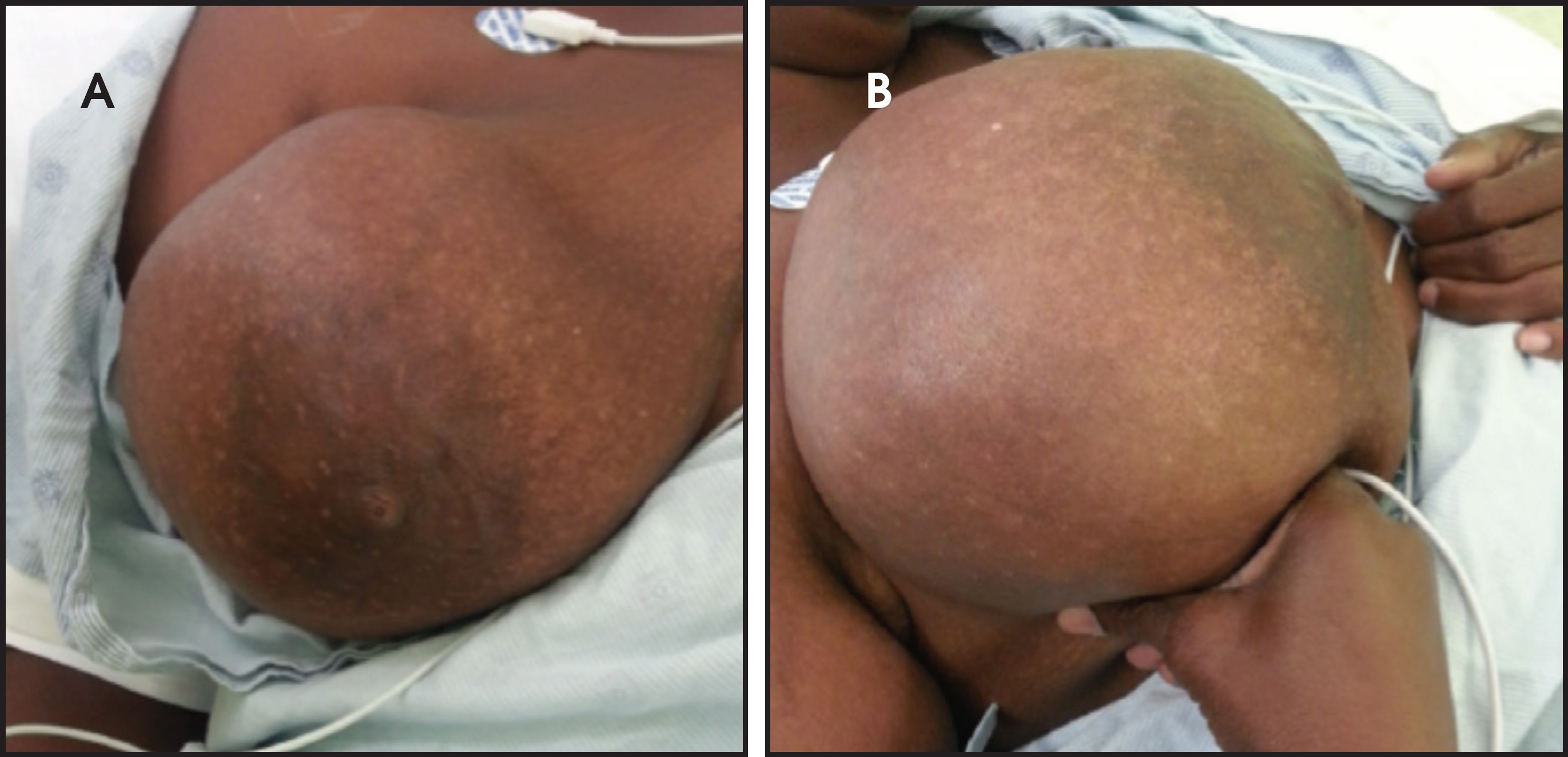
IMAGING FINDINGS
Initial emergent ultrasound of the palpable abnormality (Figure 2A) revealed an approximately 16.4 cm oval, indistinct complex cystic mass with solid components occupying the majority of the right breast. The solid mural component with papillary projections and peripheral vascularity measured up to 8.5 cm (Figure 2B). Follow-up ultrasound revealed a heterogeneously hypoechoic solid component positioned within the upper outer breast (Figure 2C). Benign appearing nodes were identified on survey of the axilla (Figure 2D).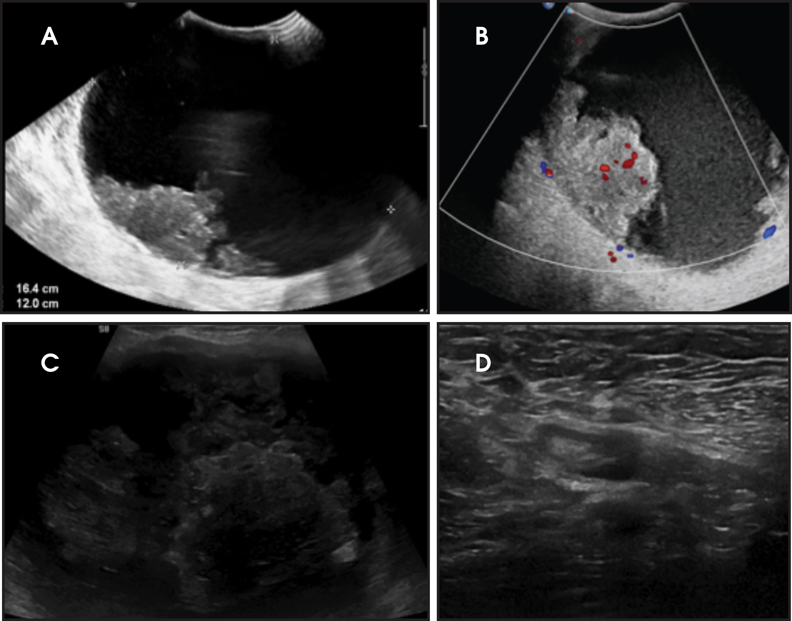
Complete diagnostic evaluation with mammography identified a solitary, large, round, right breast mass with indistinct margins measuring up to 18.3 cm in the greatest dimension without associated calcifications, skin thickening or visualized axillary adenopathy (Figure 3).
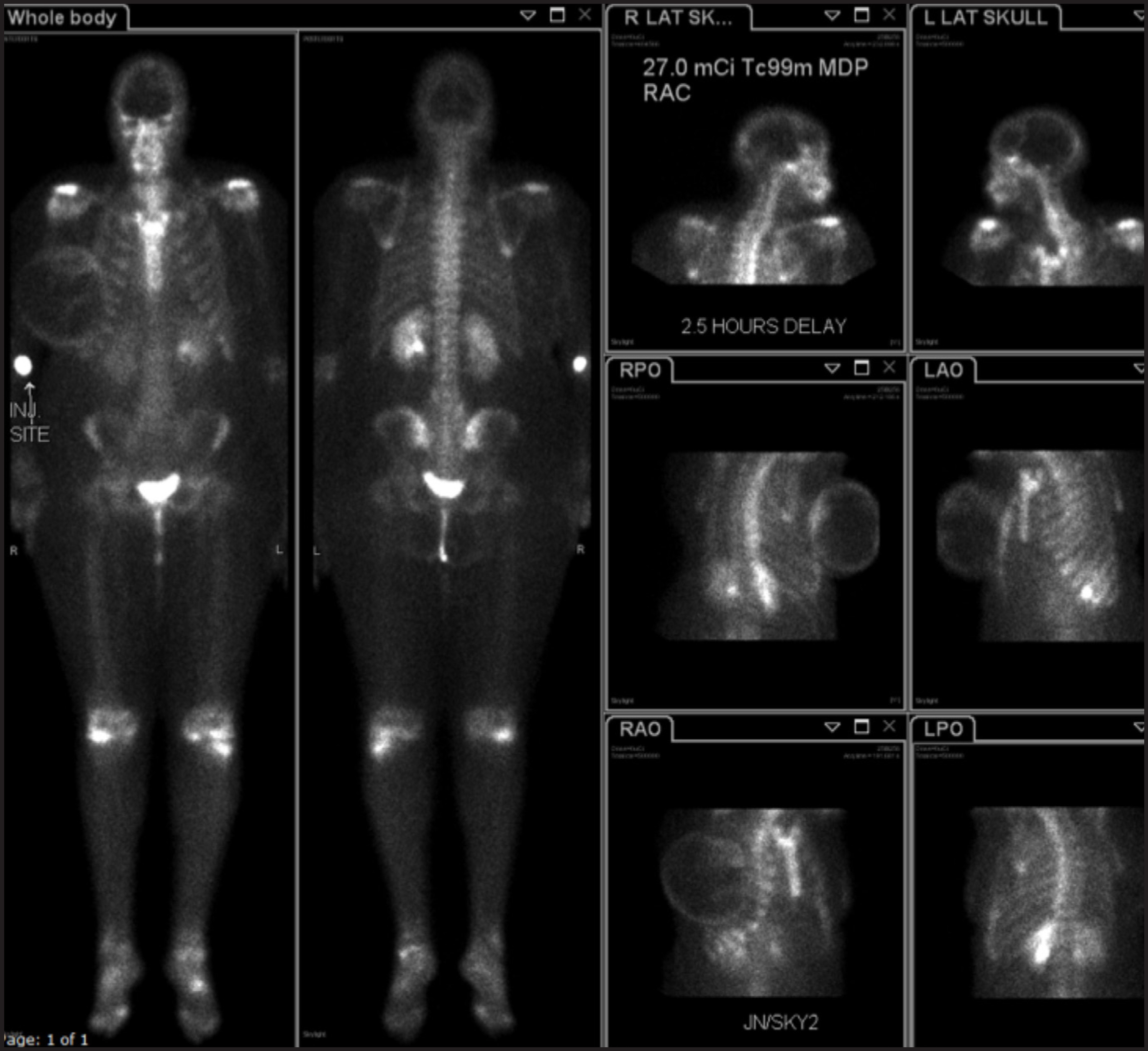
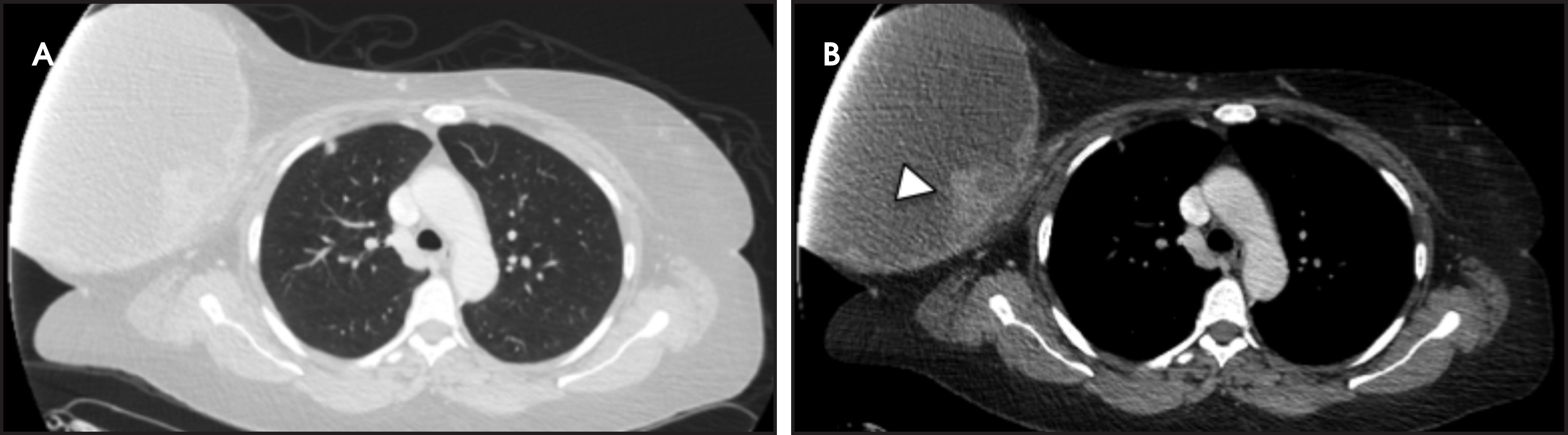
Delayed total body images from bone scan (Figure 4) revealed age-appropriate physiologic distribution of tracer activity throughout the skeletal system; however, a rim of increased tracer activity outlined the right breast mass, consistent with inflammatory process. The CT of the chest, abdomen and pelvis with contrast (Figure 5) discovered multiple hyperdense and cavitary nodules within the lungs, findings suspicious for metastatic disease. Subsequent pulmonary nodule fine needle aspiration confirmed metastatic disease. An MRI was not performed secondary to the large size of the breast mass.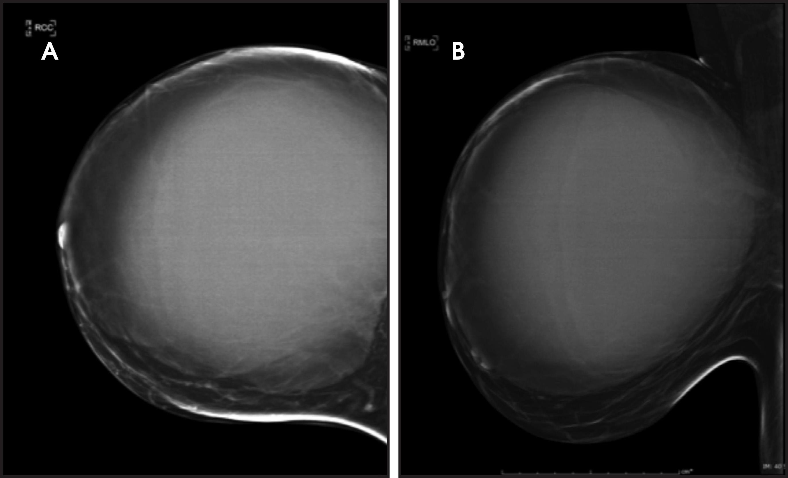
DIAGNOSIS
Fibromatosis-like spindle cell carcinoma
DISCUSSION
Fibromatosis-like metaplastic carcinomas are an uncommon tumor of the breast, being a subtype of spindle cell carcinomas, which compose only 0.3% of invasive breast carcinomas.1 These tumors can span a wide morphological spectrum, from admixed conventional carcinomas to fibromatosis-like carcinomas with primarily spindle cells. In a recent study of metaplastic spindle cell breast tumors 16 of the 33 lesions were defined as fibromatosis-like based on <5% of the tumor cells showing epithelial traits.2,3
These lesions primarily resemble fibromatosis histologically with a proliferation of bland spindle cells, but also contain clusters of epithelial cells which represent the likely metaplastic origin. The presence of an epitheliod component is critical for diagnosis, by distinguishing pure fibromatosis from carcinoma with predominant fibromatosis.3 Immunohistochemistry with antibodies against myoepithelial and cytokeratin markers should assist in establishing a diagnosis. In this patient, core needle biopsy revealed spindled neoplastic cells and nests of epitheliod cells that were positive for pancytokeratin, vimentin, and SMA immunostains. This histopathological distribution is infrequent as only five of the 24 patients in a case series of fibromatosis-like spindle cell carcinomas had neoplastic cells that co-expressed pancytokeratin and SMA.4 This lesion was also negative for estrogen receptors, progesterone receptors, and HER2 over-expression, typical for spindle cell carcinomas and limiting treatment options.5
Clinically, these lesions typically present as a palpable, painless sometimes rapidly enlarging breast mass in women with a mean age in the seventh decade of life.3,4 The large, painful, and cystic presentation of our tumor is unique, as a case series identified pain and swelling, cystic appearance of the mass, and the large size to be uncommon.3 Typically, these tumors have well-circumscribed but microscopically infiltrative borders, no cystic component or encapsulation, and have an average size of 2.5-3 cm in the greatest dimension.3,4
In general, metaplastic carcinomas of the breast such as spindle cell carcinoma show more benign features on imaging and less axillary nodal involvement than intraductal carcinomas in mammography and sonography studies.6 Compared to intraductal carcinomas, these malignancies are more likely to have round or oval shape with circumscribed margins and less likely to have any combination of irregular shape, spiculated margins, segmentally distributed pleomorphic calcifications, and posterior acoustic shadowing.7 In addition to these imaging findings, spindle cell carcinomas can have cystic change on breast ultrasound. This component is due to necrosis from rapid tumor proliferation.8 On MRI, metaplastic carcinomas are more likely to have increased signal intensity on T2-weighted MRI compared to IDC.6 While mammography, ultrasound, and MRI can help differentiate metaplastic carcinomas from IDC, immunohistochemistry is necessary to differentiate types and subtypes of metaplastic carcinomas.5
Fibromatosis-like carcinomas have a higher chance of local recurrence than lymph node metastasis. The risk of nodal metastasis is possibly related to the amount and distribution of epitheliod cells in the tumor. Due to this decreased risk of metastases, a sentinel node biopsy is recommended before axillary clearance.3,4 Distant metastases most often localize to the bone and lung and have the same histology as the primary tumor.1,3,4 In recently recorded cases, two patients have died from the metastatic disease burden, indicating its potential for aggressive behavior.9 Our patient was found to have biopsy proven metastasis to the lungs without locally metastatic disease to the axillary nodes. Nuclear medicine bone scan can be performed to evaluate for osseous metastasis given the tumor’s propensity for bony spread. While our patient had no findings to suggest osseous metastasis, inflammatory changes associated with the breast lesion were identified.
Surgical management is currently the mainstay of treatment for spindle cell carcinomas.10 Early-stage lesions are the ideal candidate for partial mastectomy and radiation therapy has been shown to slightly augment survival. Incomplete removal carries a high risk of aggressive local recurrence for spindle cell carcinomas.3 In later stages of disease, complete mastectomy is recommended, though survival is poor. Radiation therapy in these later stages has not been shown to provide survival benefit.
CONCLUSION
While as a group spindle cell carcinomas have a poor prognosis compared to other invasive mammary carcinomas, the fibromatosis-like subtype of these carcinomas typically have a more favorable outcome, behaving more like a fibromatosis than a carcinoma with the extent of fibromatosis portending a more favorable prognosis relative to other metaplastic carcinomas.
References
- Khan H, Wyld L, Dunne B. Spindle cell carcinoma of the breast: a case series of a rare histological subtype. Eur J Surg Oncol. 2003;29:600-603.
- Gobbi H, Simpson J, Jensen R, Olson S, Page D. Metaplastic spindle cell breast tumors arising within papillomas, complex sclerosing lesions, and nipple adenomas. Mod Pathol. 2003;16:893-901.
- Gobbi H, Simpson J, Borowsky A, Jensen R, Page D. Metaplastic breast tumors with a dominant fibromatosis-like phenotype have a high risk of local recurrence. Cancer. 1999;85:2170-2182.
- Sneige N, Yaziji H, Mandavilli S. Low-grade (fibromatosis-like) spindle cell carcinoma of the breast. Am J Surg Pathol. 2001;25:1009-1016.
- Tse G, Tan P, Putti T, Lui P, Chaiwun B, Law B. Metaplastic carcinoma of the breast: a clinicopathological review. J Clin Pathol. 2006;59:1079-1083.
- Choi B, Shu K. Metaplastic carcinoma of the breast: multimodality imaging and histopathologic assessment. Acta Radiol. 2012;53:5-11.
- Yang W, Hennessy B, Broglio K. Imaging differences in metaplastic and invasive ductal carcinomas of the breast. AJR Am J Roentgenol. 2007;189:1288-1293.
- Kitada M, Hayashi S, Matsuda Y, Ishibashi K, Oikawa K, Miyokawa N. Spindle cell carcinoma of the breast as complex cystic lesion: a case report. Cancer Biol Med. 2014;11:130-133.
- Kinkor Z, Svitakova I, Ryska A, Kodet R, Hrabal P. Metaplastic spindle-cell (fibromatosis-like) carcinoma of the breast – report of 4 cases. Cesk Patol. 2002;38:164-168.
- Moten A, Jayarajan S, Willis A. Spindle cell carcinoma of the breast: a comprehensive analysis. Am J Surg. 2016;16:30004-30006.
Citation
. Rapidly enlarging fibromatosis-like spindle cell carcinoma of the breast. Applied Radiology. 2017;46(7):36-39. doi:10.37549/AR2398.