Rare case of angiosarcoma of the adrenal gland seen with multiple modalities
Applied Radiology — Vol. 46 , Issue 10 , pp. 30 -32
DOI: 10.37549/AR2423
Published: October 1, 2017
Categories
Case Summary
A 72-year-old female with a prior history of breast cancer presented with mild acute kidney injury. She underwent renal ultrasonography which identified a mass and prompted further imaging. An MRI demonstrated a nonspecific large left suprarenal mass which was subsequently resected. Pathology demonstrated the mass to be angiosarcoma. A six-month postoperative FDG PET-CT was preformed which showed new hypermetabolic focus in the left prevascular mediastinum. Subsequent CT-guided biopsy revealed metastatic disease.
Imaging Findings
Initial abnormalities on the renal ultrasound prompted further investigations and due to the patient's AKI a MRI was performed. Renal protocol MRI examination showed a large, complex mass appearing to arise from the left adrenal gland (Figure 1), which correlated to the suprarenal lesion on the sonogram. This mass was markedly heterogeneous on the T1 and T2-weighted image, with areas of T1 hyperintensity suspected to represent hemorrhagic material, as well as central T2 hyperintensity likely related to necrosis, and exhibited irregular internal and nodular peripheral enhancement. The patient underwent resection of the suprarenal mass which was consistent with epithelioid angiosarcoma with extensive infarction.
Subsequent to the pathologic diagnosis, the patient underwent an 18-F fluorodeoxyglucose positron emission tomography-computed tomography (FDG PET-CT) scan to assess for metastatic disease, which showed expected post-surgical and reactive changes with no definitive metabolic evidence of surgical bed residual tumor or regional or distant metastasis.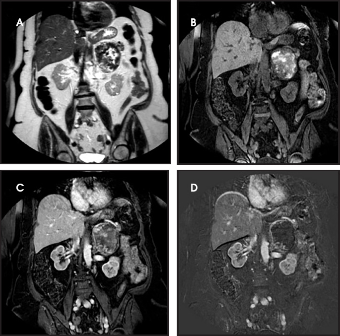
A follow-up PET-CT scan 6 months later demonstrated regional recurrence as well as a new hypermetabolic focus in the left prevascular mediastinum (Figure 2). A CT-guided biopsy of the anterior mediastinal mass revealed metastatic disease from primary angiosarcoma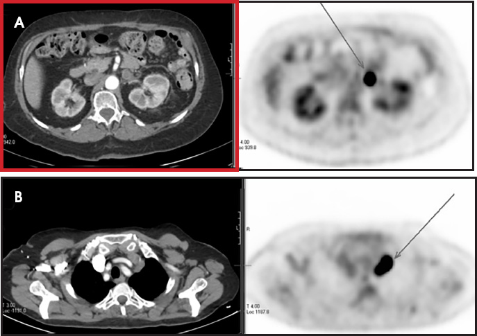
Diagnosis
Adrenal epithelioid angiosarcoma.
Differential diagnoses: hemorrhagic and or necrotic metastases, adrenal cortical carcinoma or pheochromocytoma.
Discussion
The adrenal, or suprarenal, glands are a unique, duplex endocrine organ comprised of a cortex formed from the mesoderm and central medulla of neural crest origin. As a result, their endocrine function comprises both glucocorticoid and catecholamine production. The adrenal glands represent a common site of disease and are well evaluated by cross-sectional imaging, particularly on CT and MRI examinations of the abdomen. The frequency of adrenal abnormalities in the general population is 9%.1
For purposes of differential diagnosis, masses of the adrenal gland are commonly grouped into those that are fat containing, cystic, hypervascular and overtly malignant. Primary cortical carcinomas of the adrenal gland are rare tumors, found in approximately two patients per million.2 Malignant mesenchymal primary tumors of the adrenal gland are extremely uncommon; the few reported cases of soft-tissue sarcoma have included tumors of vascular and smooth muscle origin.3
Adrenal angiosarcoma represents less than 1% of malignancies.4 There have been few reported cases of primary adrenal angiosarcoma in the literature.5 Most have been found incidentally or with nonspecific symptoms, such as abdominal pain. While most of these lesions have been solitary, there has been a single reported instance with a coexistent, functional adrenal adenoma.6
The described MRI appearance of adrenal angiosarcoma is that of punctate calcifications, intense peripheral enhancement and a necrotic center.7 It can also have loss of signal on out-of phase imaging secondary to foci of intracytoplasmic lipid.8 In our case, the MRI appearance was heterogeneous in signal intensity on all sequences, containing foci of T1-weighted hyperintensity, T2-weighted hypointensity related to hemorrhagic byproducts, T2-hyperintensity in regions of necrosis seen at pathology, and non-uniform, peripheral contrast enhancement. This nonspecific appearance leads to difficulty making a definitive diagnosis based on MRI.
Review of the literature demonstrates challenges in the pathologic diagnosis as well. One obstacle is a common microscopic cystic appearance that can be confused with adrenal cortical carcinoma. Also, most cases of reported adrenal angiosarcoma demonstrate an epithelioid histologic pattern as opposed to a vasoformative pattern. Histologic testing for vascular markers, such as CD31 and factor VIII, is a key part in the pathologic evaluation.5
The clinical outcomes of patients with adrenal angiosarcomas are not well studied. The five-year overall survival rates of angiosarcoma is 24% to 31%.9 A review of 17 reported cases showed five patients who died with disease after surgical removal of the primary tumor (including from postoperative complications).5 Patients with soft tissue sarcoma typically undergo a combination of surgery, chemotherapy and radiation therapy. In our case, the patient developed metastatic disease after surgery, detected on surveillance PET-CT. A search of the literature revealed reports of adrenal angiosarcoma with hypermetabolic metastatic disease on FDG PET.10,11 However, there is no broader literature to suggest that angiosarcoma is consistently hypermetabolic on PET. Soft tissue sarcomas, in general, exhibit variable FDG hypermetabolism, typically in relationship to their histologic grade.12
Conclusion
Angiosarcomas commonly occur in the skin, soft tissue, breast, bone, liver, and spleen. The MRI appearance is variable owing to the various stages of hemorrhage, necrosis, cystic degeneration, and viable tissue present within the lesion. Although rare, angiosarcoma can also arise in the adrenal glands.
Our case report shows that adrenal gland angiosarcoma can have a similar appearance to adrenal cortical carcinoma. Adrenocortical carcinomas are usually large at diagnosis and appear heterogeneous on both T1- and T2-weighted images owing to the presence of internal hemorrhage and necrosis.
The behavior of adrenal angiosarcoma on FDG PET is not well studied. In our case, as well as in two additional reports, the surgical bed recurrence and metastasis were hypermetabolic.
References
- Glazer H, Weyman P, Sagel S, Levitt R, McClennan B. Nonfunctioning adrenal masses: incidental discovery on computed tomography.. AJR Am J Roentgenol.. 1982;139:81-85.
- Hedican S, Marshall F. Adrenocortical carcinoma with intracaval extension.. J Urol.. 1997;158:2056-2061.
- Travis W, Oertel J, Lack E, Lack E. Miscellaneous tumors and tumefactive lesions of the adrenal gland.. Pathology of the adrenal glands.. 1990:351-378.
- Weiss S, Goldblum J. Enzinger and Weiss's soft tissue tumors.. . 2008.
- Sung J, Ahn S, Kim S, Park Y, Choi Y. Angiosarcoma arising within a long-standing cystic lesion of the adrenal gland: a case report.. J Clin Oncol.. 2013;31.
- Hayashi T, Gucer H, Mete O. A mimic of sarcomatoid adrenal cortical carcinoma: epithelioid angiosarcoma occurring in adrenal cortical adenoma.. Endocr Pathol.. 2014;25:404-409.
- Otal P, Escourrou G, Mazerolles C, Janne d'Othee B, Mezghani S, Musso S. Imaging features of uncommon adrenal masses with histopathologic correlation.. Radiographics. 1999;19:569-581.
- Schlund J, Kenney P, Brown E, Ascher S, Brown J, Semelka R. Adrenocortical carcinoma: MR imaging appearance with current techniques.. J Magn Reson Imaging.. 1995;5:171-174.
- Fury M, Antonescu C, Van Zee K, Brennan M, Maki R. A 14-year retrospective review of angiosarcoma: clinical characteristics, prognostic factors, and treatment outcomes with surgery and chemotherapy.. Cancer J.. 2005;11:241-247.
- Derlin T, Clauditz T, Habermann C. Adrenal epithelioid angiosarcoma metastatic to the epicardium: diagnosis by 18F-FDG PET/CT.. Clin Nucl Med.. 2012;37:914-915.
- Lepoutre-Lussey C, Rousseau A, Al Ghuzlan A, Amar L, Hignette C, Cioffi A. Primary adrenal angiosarcoma and functioning adrenocortical adenoma: an exceptional combined tumor.. Eur J Endocrinol.. 2012;166:131-135.
- Nieweg O, Pruim J, van Ginkel R, Hoekstra H, Paans A, Molenaar W. Fluorine-18-fluorodeoxyglucose PET imaging of soft-tissue sarcoma.. J Nucl Med. 1996;37:257-261.
Citation
. Rare case of angiosarcoma of the adrenal gland seen with multiple modalities. Applied Radiology. 2017;46(10):30-32. doi:10.37549/AR2423.