RCOM RADIOLOGICAL
Applied Radiology — Vol. 36 , Issue 1 , pp. 39 -41
DOI: 10.37549/AR1482
Published: January 1, 2007
Categories
CASE SUMMARY
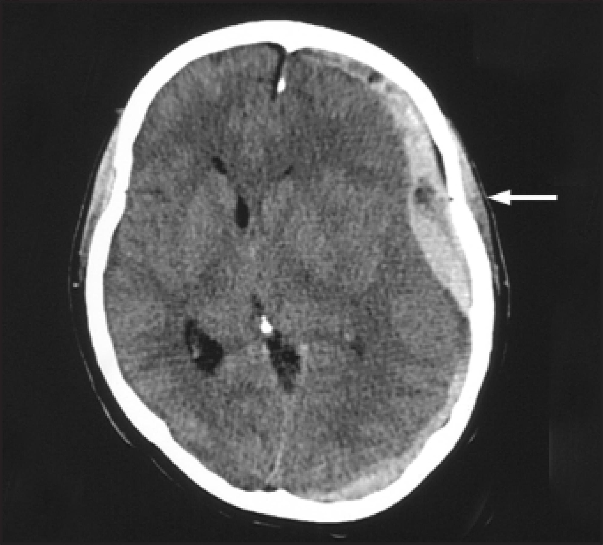
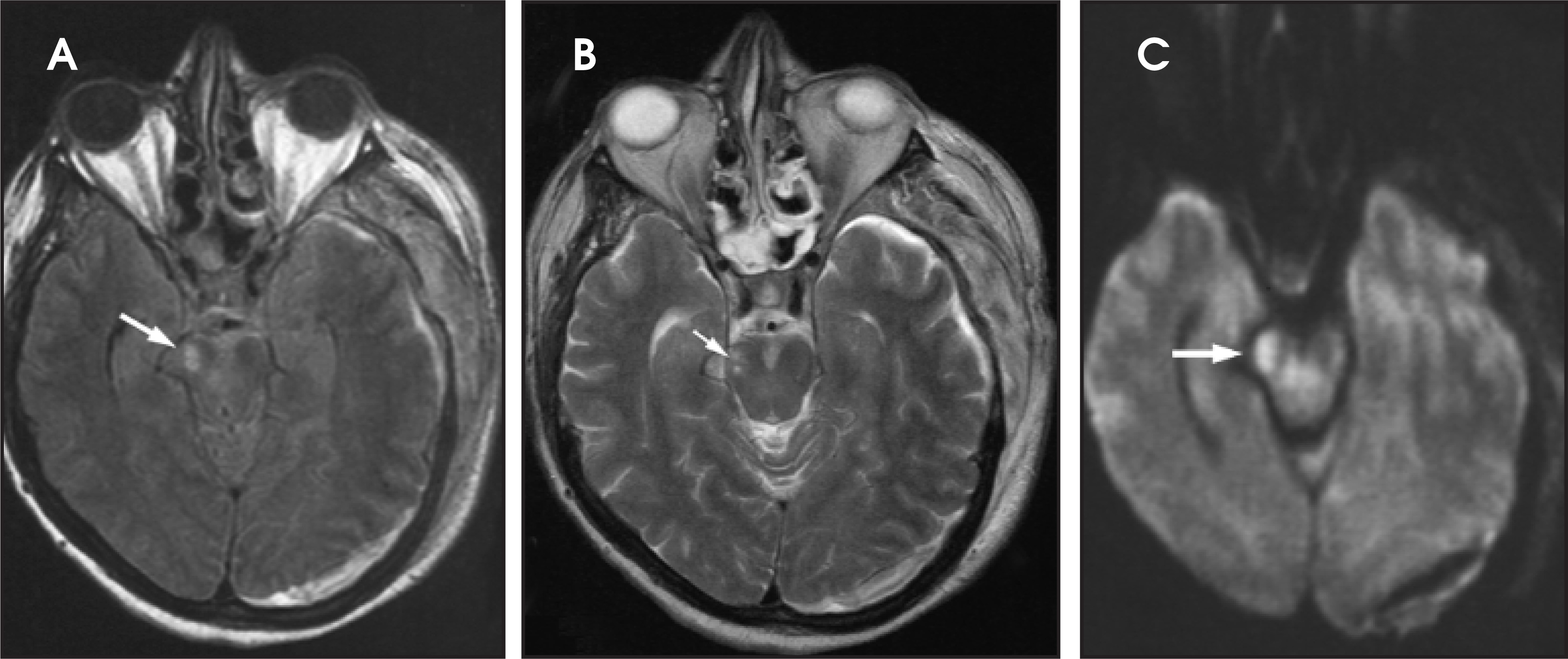
A 50-year-old woman with myasthenia gravis became lethargic after falling from her bed. A computed tomography (CT) scan of the head was performed, which showed a left acute subdural hematoma with significant mass effect and shift (Figure 1). She underwent an emergency craniotomy for hematoma evacuation. Postoperatively, her physical examination revealed a left dilated pupil, a left oculomotor nerve palsy with ptosis, and a dense hemiparesis on the left side. Magnetic resonance imaging (MRI) (Figure 2) was performed to evaluate the deficit.
IMAGING FINDINGS
CT of the brain revealed a large acute left convexity subdural hematoma with mass effect and left-to-right midline shift (Figure 1, arrow). Postoperative MRI of the brain revealed an area of high signal intensity in the right crus cerebri on the fluid-attenuated inversion recovery, T2-weighted, and diffusion-weighted imaging sequences (Figure 2, arrows).
DIAGNOSIS
Kernohan’s phenomenon, resulting from a traumatic left acute subdural hematoma
DISCUSSION
Transtentorial, or uncal, herniation is a well-described neurologic phenomenon caused by an ex-panding supratentorial mass. The medial aspect of the temporal lobe is forced downward over the tentorium, compressing the neighboring oculomotor nerve and resulting in ipsilateral pupillary dilatation, which is often followed by oculomotor ophthalmoplegia. With time, the integrity of the crus cerebri and its descending corticospinal tracts is disturbed, and a contralateral motor deficit is produced with a deteriorating level of consciousness. Lateral pressure on the midbrain can, however, compress the opposite crus cerebri against the free edge of the tentorium, forming an indentation in the crus known as Kernohan’s notch. This may produce a hemiparesis ipsilateral to the expanding mass known as Kernohan’s phenomenon, which is a false localizing sign. This was first described by Kernohan et al1 in 1929 after an autopsy study revealed a notched cerebral peduncle from a contralateral herniation syndrome. Because of streak artifacts from the petrous bone, CT is usually unable to detect these changes. MRI can be useful in revealing a deformity or injury of the cerebral peduncle that results from ongoing or past transtentorial herniation.2-6
CONCLUSION
Kernohan’s phenomenon should be suspected when a motor deficit develops ipsilateral to a space-occupying supratentorial mass. MRI is the study of choice to confirm injury to the contralateral crus cerebri.
References
- Kernohan J, Woltman H. Incisura of the crus due to contralateral brain tumor Arch Neurol Psychiatry. 1929;21:274-287.
- Hamada H, Kuwayama N, Endo S, Takaku A. Detection of Kernohan’s notch on magnetic resonance imaging in a patient with acute subdural haematoma. Br J Neurosurg. 2000;14:498-499.
- Kole M, Hysell S. MRI correlate of Kernohan’s notch. Neurology. 2000;55:1751.
- Wolf R, ter Weeme C, Krikke A. Kernohan’s notch and misdiagnosis. Lancet. 1995;345:259-260.
- Iwama T, Kuroda T, Sugimoto S. MRI demonstration of Kernohan’s notch: Case report. Neuroradiology. 1992;34:225-226.
- Cohen A, Wilson J. Magnetic resonance imaging of Kernohan’s notch. Neurosurgery. 1990;27:205-207.
Citation
. RCOM RADIOLOGICAL. Applied Radiology. 2007;36(1):39-41. doi:10.37549/AR1482.