RCOM RADIOLOGICAL
Applied Radiology — Vol. 36 , Issue 5 , pp. 40 -41
DOI: 10.37549/AR1507
Published: May 1, 2007
Categories
CASE SUMMARY
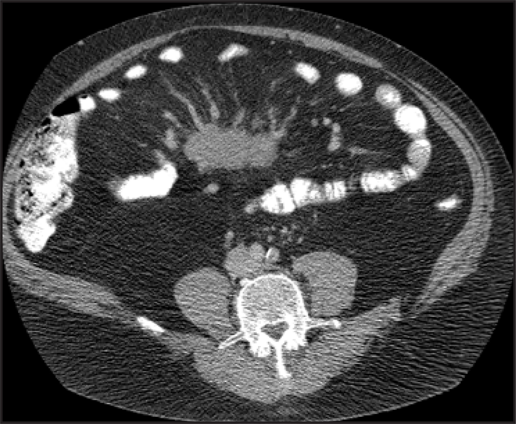
A 69-year-old man presented with persistent abdominal pain, bloating, and gas. The patient experienced mild tenderness to palpation over the midabdomen but had unremarkable laboratory studies. The patient denied any recent trauma, infection, or history of previous abdominal surgery. An abdominal computed tomography (CT) study was performed (Figure 1).
IMAGING FINDINGS
CT scans of the abdomen and pelvis were obtained and revealed a 3.75 × 3.70 × 7.60-cm mesenteric mass of soft tissue density with an associated prominence of the adjacent mesenteric vasculature (Figure 1). The stomach, small intestine, and large intestine were normal in appearance. The remaining vasculature and lymphatics were also normal in appearance. A subsequent upper gastrointestinal study with a small bowel follow-through fluoroscopic study showed mass effect in the midabdomen with no intraluminal filling defect or primary tumor. Based on the CT appearance of the mesenteric mass in this patient, reasonable differential diagnoses included carcinoid, dermoid, lymphoma, mesenteric edema, carcinomatosis, and metastases.
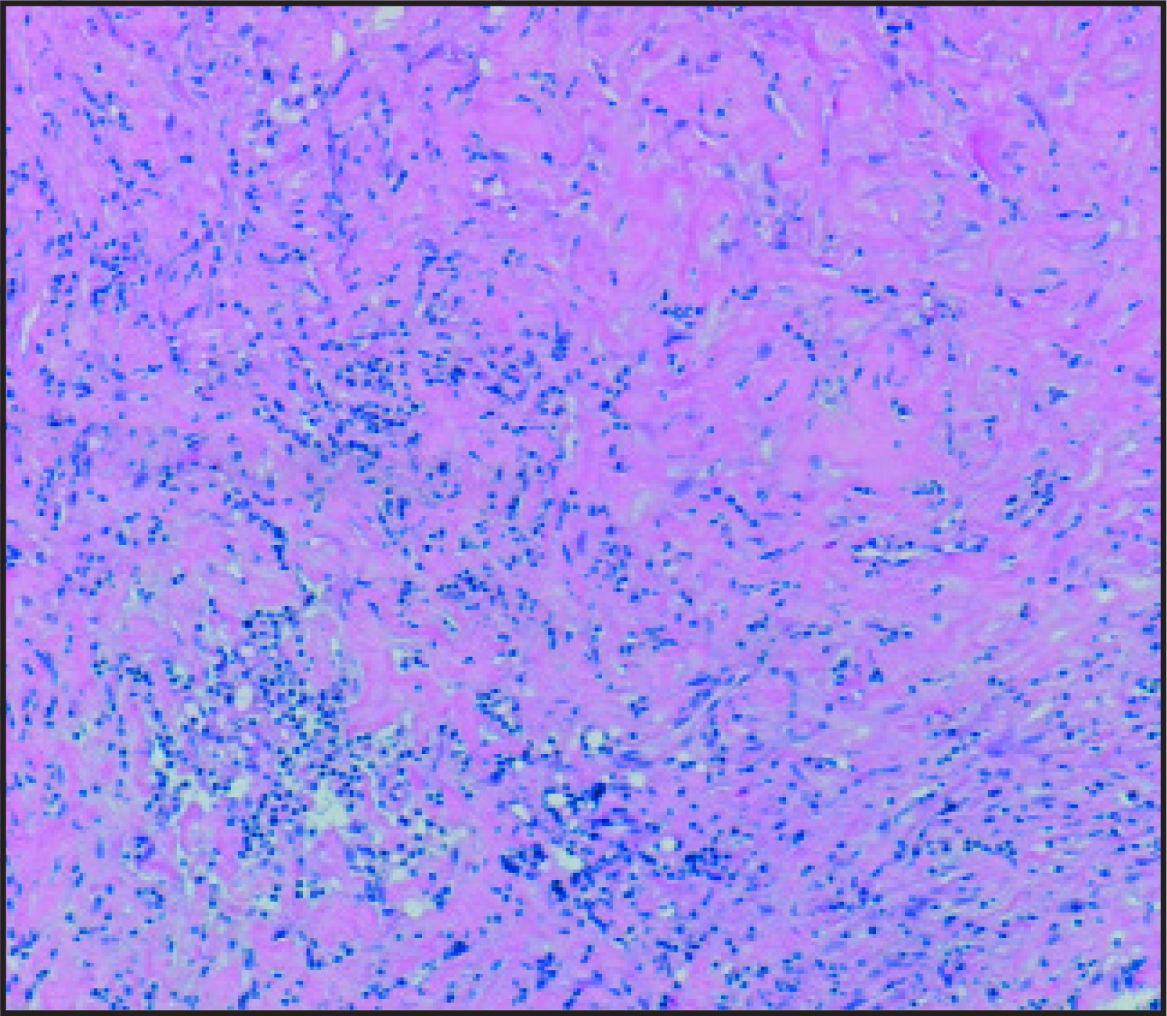
Laparoscopic surgical biopsy and pathologic analysis determined the final diagnosis of sclerosing mesenteritis (Figure 2).
DIAGNOSIS
Sclerosing mesenteritis
DISCUSSION
Sclerosing mesenteritis is a rare diagnosis of exclusion that is characterized by chronic inflammation of the mesentery, usually along the small bowel. Also described as retractile mesenteritis, liposclerotic mesenteritis, xanthogranulomatous mesenteritis, systemic nodule panniculitis, and mesenteric Weber-Christian disease, sclerosing mesenteritis appears to have an increased incidence in men with a mean age of 60 years.1 Possible etiologies currently proposed include autoimmunity in the setting of collagen vascular disease, trauma, infection, and ischemia. This condition has also been associated with such disorders as Riedel thyroiditis, orbital pseudotumor, sclerosing cholangitis, and retroperitoneal fibrosis.2 One series reported a coexisting malignancy in up to 69% of patients.3
The clinical presentation of sclerosing mesenteritis varies, but the patient can present with abdominal pain, diarrhea, intestinal obstruction or ischemia, and a mass.2 Laboratory values can show an elevation of the erythrocyte sedimentation rate but may be normal.
Histologic analysis displays a mixture of sclerosis, fat necrosis, and chronic inflammation, as seen in this patient (Figure 2). Given that similar pathologic findings can occur in the mesentery adjacent to such things as lymphomas, carcinomas, abscesses, and leaking aortic aneurysms, cytologic analysis is advisable. In this patient, cytokeratin and BerEP4 reaction for carcinoma were negative.
CT attenuation can vary from slightly increased attenuation above mesenteric fat, the often described “misty mesentery” to soft tissue mass. As seen in this patient, preservation of the mesenteric fat around prominent vessels, known as the “fat-ring sign” may be seen and can help to differentiate mesenteric sclerosis from other neoplastic diagnoses.2 A tumoral pseudocapsule may also be identified as observed in 50% of patients in one series.4 Calcifications, cystic components, and mesenteric or retroperitoneal lymphadenopathy may also exist.2 Of note, correlative imaging with 18-fluorine fluorodeoxyglucose (FDG) positron emission tomography has shown a doughnut-shaped pattern of FDG uptake, which likely represents the peripheral mesenteric inflammation and central sclerosis or fibrosis.5
Treatment options may include surgical resection or debulking and medical management with steroids, colchicines, oral progesterone, or immunosuppressive agents.2
CONCLUSION
Sclerosing mesenteritis is a rare pathologic diagnosis characterized by inflammation of the mesentery that should be considered in CT evaluation of a patient with a soft tissue abdominal mass while ensuring appropriate work-up to rule out diagnoses that may have a similar appearance with a worse prognosis.
References
- Durst A, Yarom R, Luttwak E. Malignant fibromatous peritoneal mesothelioma associated with liposclerotic mesenteritis. Am J Gastroenterol. 1971;55:477-481.
- Horton K, Lawler L, Fishman E. CT findings in sclerosing mesenteritis (panniculitis): Spectrum of disease. RadioGraphics. 2003;23:1561-1567.
- Daskalogiannaki M, Voloudaki A, Prassopoulos P. CT evaluation of mesenteric panniculitis: Prevalence and associated diseases. AJR Am J Roentgenol. 2000;174:427-431.
- Sabate J, Torrubia S, Maideu J. Sclerosing mesenteritis: Imaging findings in 17 patients. AJR Am J Roentgenol AJR Am J Roentgenol.
- Nguyen B. F-18 FDG PET demonstration of sclerosing mesenteritis. Clin Nucl Med. 2003;28:670-67.
Citation
. RCOM RADIOLOGICAL. Applied Radiology. 2007;36(5):40-41. doi:10.37549/AR1507.