RCOM RADIOLOGICAL
Applied Radiology — Vol. 36 , Issue 5 , pp. 46 -47
DOI: 10.37549/AR1508
Published: May 1, 2007
Categories
CASE SUMMARY
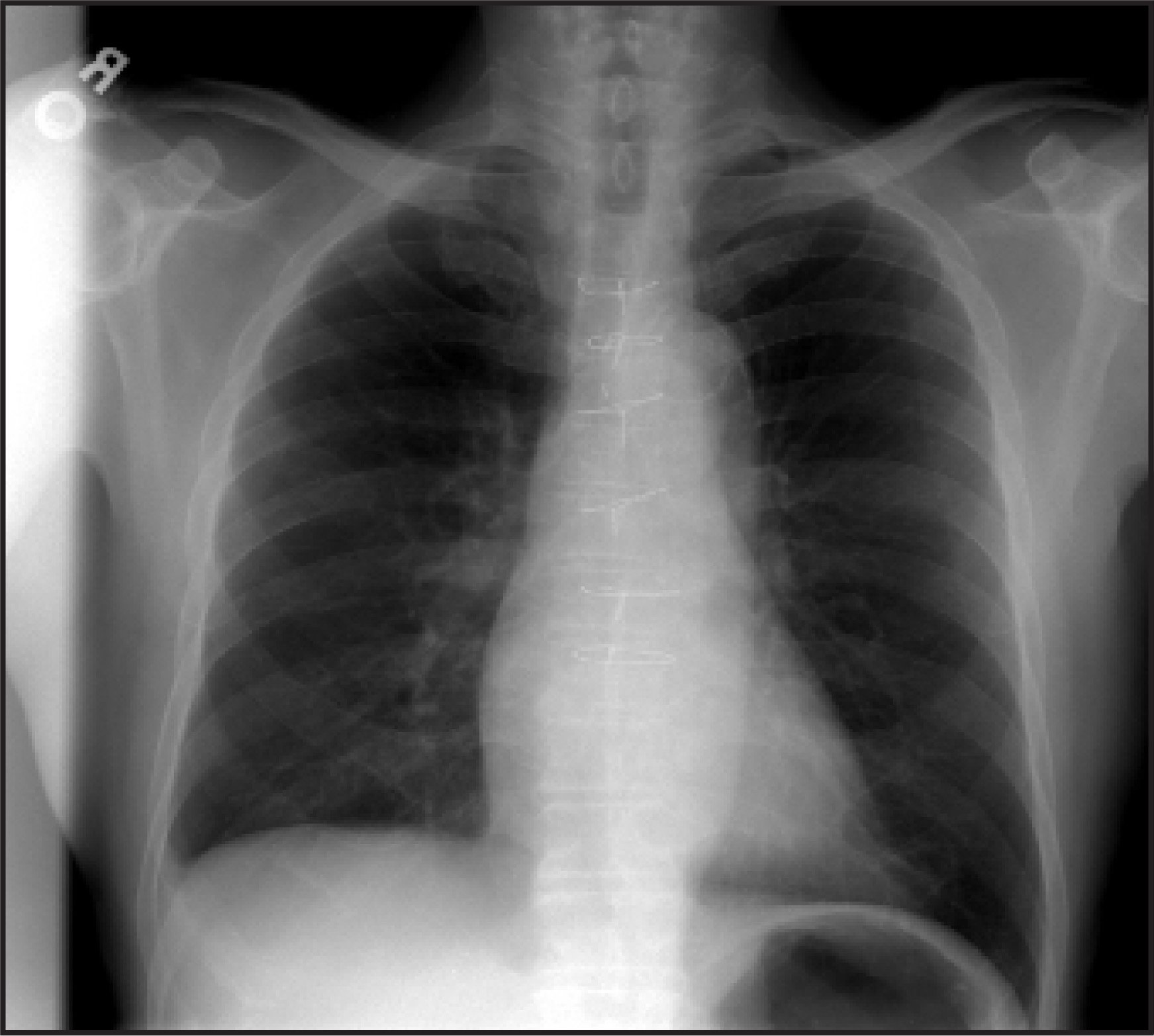
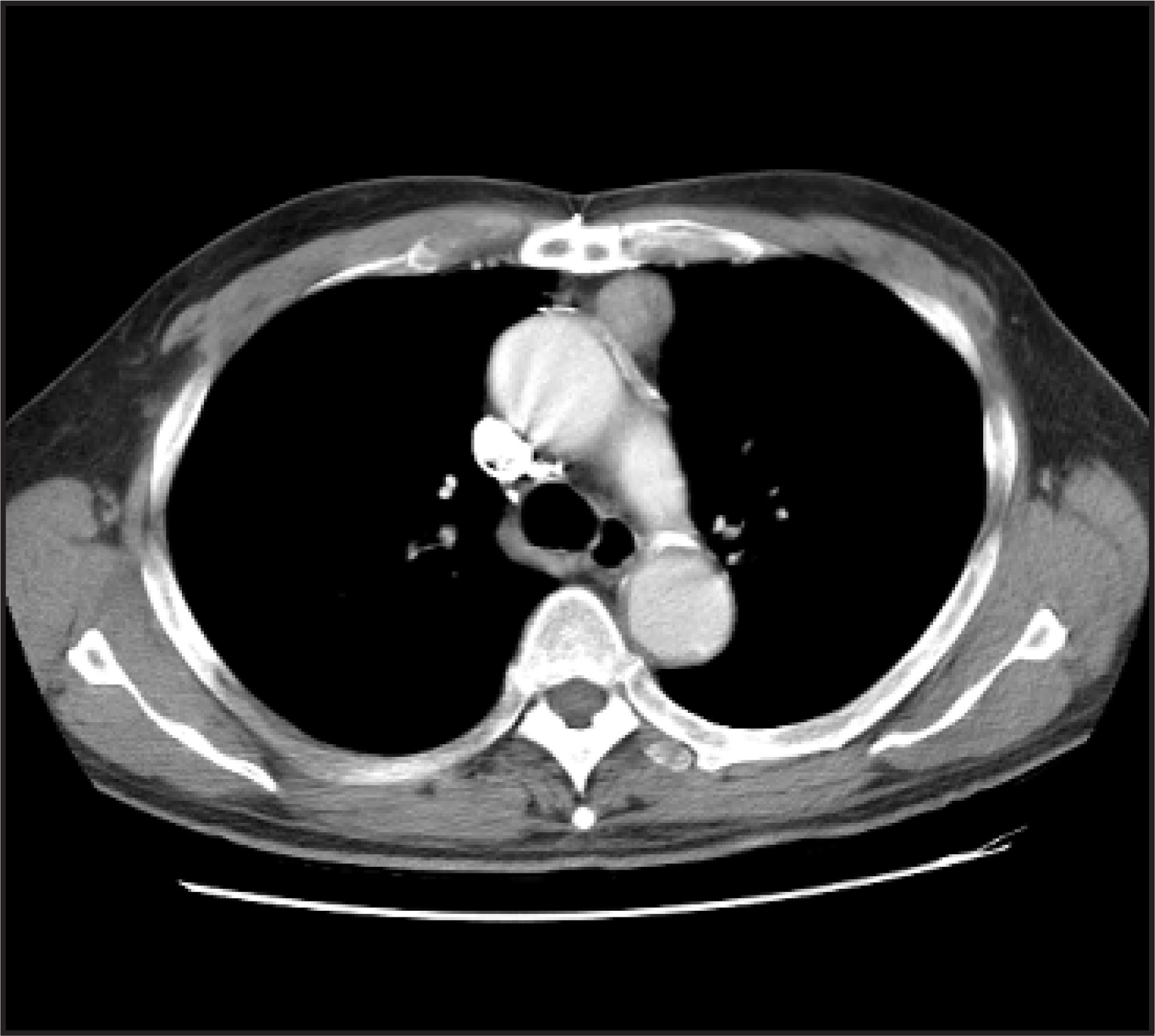
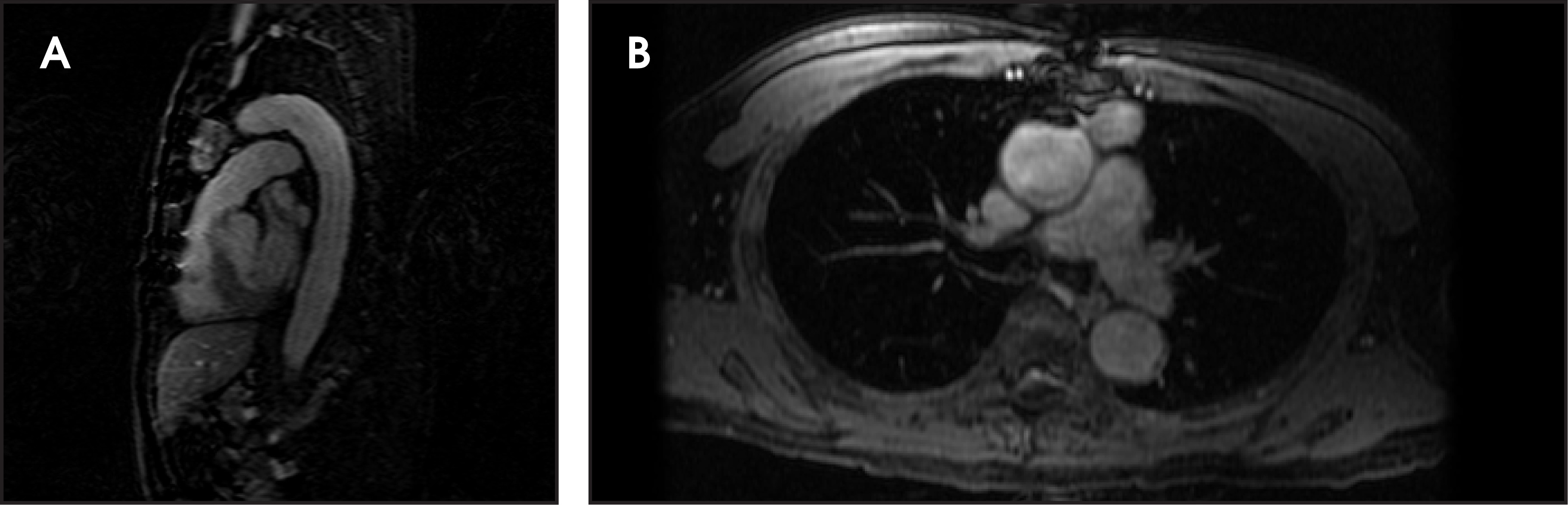
A 72-year-old man presented with an incidental anterior mediastinal mass that had been identified on chest radiography while he was undergoing a workup for laryngitis. Significant physical examination findings included a pulse of 120 bpm, a blood pressure of 155/90 mm Hg, and a well-healed sternotomy scar. Laboratory data were unremarkable. Chest X-ray (Figure 1), chest computed tomography (CT) (Figure 2), and chest magnetic resonance imaging (MRI) (Figure 3) were performed.
IMAGING FINDINGS
Chest X-ray revealed a round opacity within the anterior mediastinum (Figure 1). On subsequent CT, a 2.8-cm round homogeneous dense mass without calcification (Figure 2) can be seen. An electrocardiographic-gated MRI of the aortic arch and 3-dimensional angiogram reconstruction showed a 4.0- × 2.5- × 2.5-cm anterior mediastinal mass near the origin of the ascending aorta adjacent to the coronary artery native bypass graft origin; on postgadolinium MRI, there was evidence of near homogeneous uptake equaling the adjacent aorta (Figure 3). The identification of the exact origin of this mass was limited because of blooming artifact from an overlying sternotomy wire. Small filling defects within the lesion may represent thrombus.
DIAGNOSIS
Saphenous vein coronary artery bypass graft (CABG) pseudoaneurysm
DISCUSSION
Saphenous vein graft aneurysms are rare complications of CABG surgery. Since 1975, approximately 75 cases have been published in the literature. These aneurysms are typically classified as true aneurysms or (false) pseudoaneurysms. True aneurysms usually appear late in the postoperative course and are associated with atherosclerosis.1 True aneurysms, which are defined as >3 cm diameter dilatations, involve expansion of all the layers of the wall and are most likely to be identified in the body of the graft.2
Pseudoaneurysms occur in the weeks or months following the operation and can be related to anastomotic site weakness, wound infection, and iatrogenic or intrinsic wall weakness, possibly related to bifurcation points or valve sites.2 These false aneurysms involve dilatation of limited layers of the graft wall and can result in life-threatening hemorrhage.3 Given such life-threatening complications, treatment options may include thrombectomy, embolization, resection, or exclusion of the aneurysm.2
Regarding this patient’s case, prior to MRI evaluation, reasonable differential diagnoses may also have included lymphoma, thymoma or other thymic lesions, and germ cell tumors. Despite the patient being asymptomatic and 19 years after CABG, this aneurysm was classified as a pseudoaneurysm, given its proximity to the origin of the native graft origin.
CONCLUSION
Pseudoaneurysm of the aorta or saphenous vein graft following CABG surgery is a rare complication that can occur many years postoperatively and should be considered in the setting of a new anterior mediastinal mass.
References
- Le Breton H, Pavin D, Langanay T. Aneurysms and pseudo-aneurysms of saphenous vein coronary artery bypass grafts. Heart. 1998;79:505-508.
- Trop I, Samson L, Cordeau M. Anterior mediastinal mass in a patient with prior saphenous vein coronary artery bypass grafting. Chest. 1999;115:572-576.
- Walsh G, Glynn A, Slavotinek J. Giant coronary artery bypass graft pseudoaneurysm presenting as a haemothorax. Clin Radiol. 2001;56(1):74-75.
Citation
. RCOM RADIOLOGICAL. Applied Radiology. 2007;36(5):46-47. doi:10.37549/AR1508.