RCOM RADIOLOGICAL
Applied Radiology — Vol. 36 , Issue 5 , pp. 36 -37
DOI: 10.37549/AR1510
Published: May 1, 2007
Categories
CASE SUMMARY
A 68-year-old man with a long history of uncontrolled hypertension presented with a single 4-minute episode of a generalized tonic-clonic seizure, and 2 days of fluctuating headache, visual disturbance, nausea, and vomiting. On neurologic examination, he was drowsy but able to follow simple commands. He had no focal neurologic deficit. The physical examination revealed a blood pressure of 230/75 mm Hg and no pedal edema. The fundoscopic examination was normal.
IMAGING FINDINGS
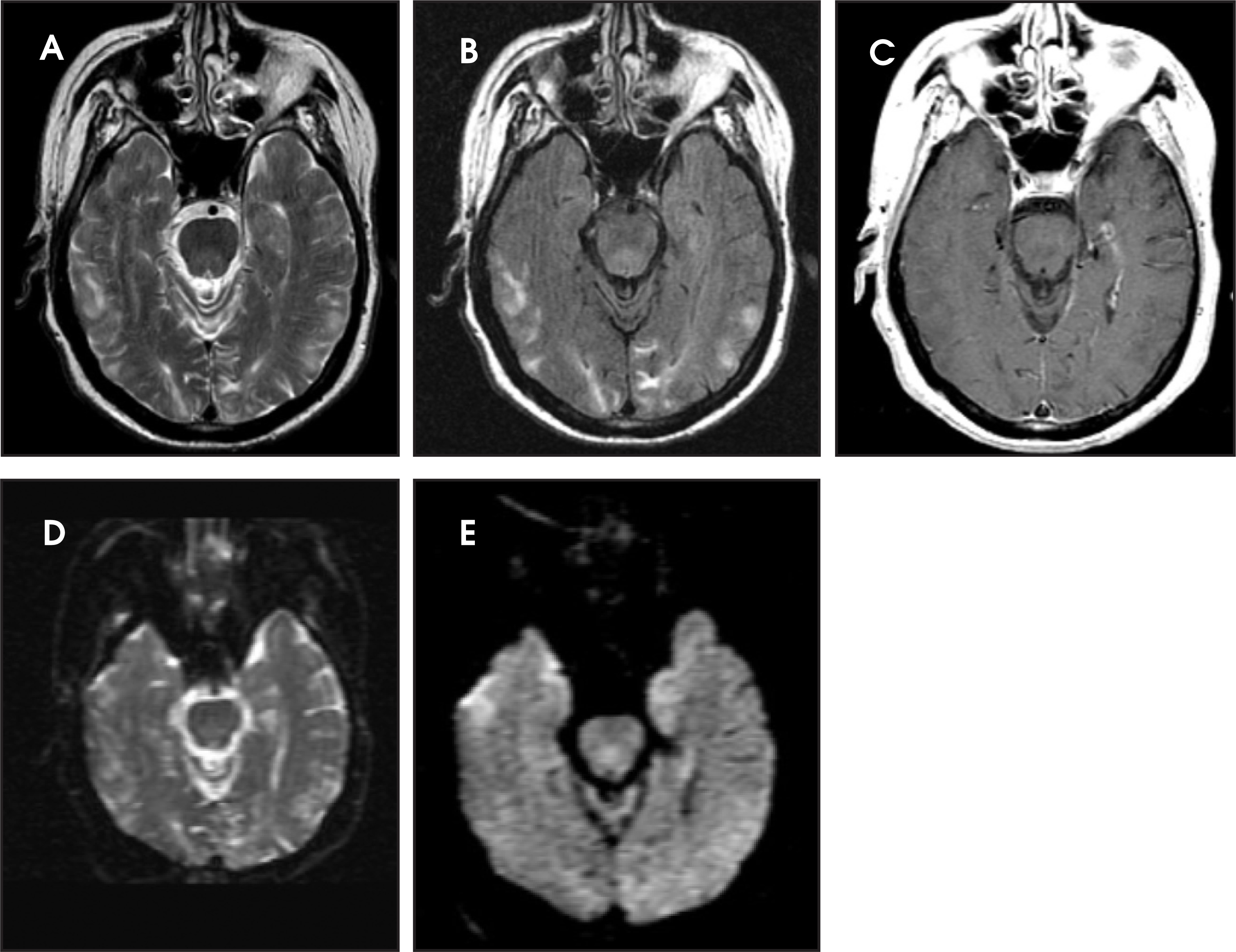
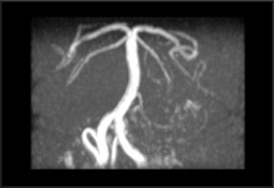
Magnetic resonance imaging (MRI) and magnetic resonance angiography (MRA) of the brain were performed with a GE Signa LX 1.5T MRI scanner (GE Healthcare, Waukesha, WI). Contrast media (Magnevist, gadopentetate dimeglumine, Berlex Laboratories, Montville, NJ) was injected intravenously (0.1 mmol/kg body weight). The MRI showed bilateral symmetric vasogenic edema mainly involving the subcortical white matter in the parieto-occipital, posterior temporal, and posterior frontal lobes (Figure 1). The MRA of the vertebrobasilar system was unremarkable without any area of stenosis or vasospasm (Figure 2).
DIAGNOSIS
Posterior reversible encephalopathy syndrome
DISCUSSION
Reversible posterior leukoencephalopathy syndrome is a clinicoradiologic entity characterized by headaches, altered mental status, seizures, and visual loss and is associated with white matter vasogenic edema predominantly affecting the posterior occipital and parietal lobes of the brain.1 There has been some controversy about what the proper term should be for this entity because MRI has shown that lesions can occur in both gray and white matter. Therefore, a new name, posterior reversible encephalopathy syndrome (PRES), has been coined.2 Most cases of PRES occur with hypertension or immuno-suppression, but it can occur with many diverse clinical entities.1,3 Since PRES is often unsuspected by clinicians, recognition of the characteristic imaging findings by radiologists is key to diagnosing this syndrome and should prevent deleterious work-ups or therapies.
The pathophysiology of PRES is under debate, but it is related to disordered cerebral autoregulation. Two pathophysiologic mechanisms have been proposed regarding cerebral autoregulation—cerebral vasospasm, which results in cytotoxic edema,4 and vasodilatation, which results in vasogenic edema.5 The latter is more favored by most experimental and clinical data.6,7 The pathophysiology of PRES also implicates endothelial dysfunction, especially in cases without severe hypertension, such as pre-eclampsia or cytotoxic therapies.1
The most characteristic imaging pattern in PRES is the presence of edema involving the white matter of the posterior portions of both cerebral hemispheres, especially the parieto-occipital regions, in a relatively symmetric pattern that spares the calcarine and paramedian parts of the occipital lobes.1 However, other structures (such as the brain stem, cerebellum, and frontal and temporal lobes) may also be involved, and although the abnormality primarily affects the subcortical white matter, the cortex and the basal ganglia may also be involved.8 Although they are rare, gyriform signal enhancement or parenchymal hemorrhage can occur in complicated cases.9 Recently, studies with diffusion-weighted sequences6 and diffusion-tensor sequences7 have shown increased apparent diffusion coefficients (ADCs) in the involved regions accompanied by anisotropy loss, which suggests reversible vasogenic edema as an underlying pathophysiology. Therefore, early diagnosis and treatment is essential for the patients’ prognosis.
CONCLUSION
Posterior reversible leukoencephalopathy syndrome is a clinicoradiologic entity associated with hypertension, immunosuppression, or many diverse clinical entities. Since PRES is often unsuspected by clinicians, recognition of the characteristic imaging findings by radiologists is key to diagnosing this syndrome and should prevent deleterious work-ups or therapies.
References
- Hinchey J, Chaves C, Appignani B. A reversible posterior leukoencephalopathy syndrome. N Engl J Med. 1996;334:494-500.
- Casey S, Sampaio R, Michel E. Posterior reversible encephalopathy syndrome: Utility of fluid-attenuated inversion recovery MR imaging in the detection of cortical and subcortical lesions. AJNR Am J Neuroradiol. 2000;21:1199-1206.
- Port J, Beauchamp N. Reversible intracerebral pathologic entities mediated by vascular autoregulatory dysfunction. RadioGraphics. 1998;18:353-367.
- Lewis L, Hinshaw D, Will A. CT and angiographic correlation of severe neurological disease in toxemia of pregnancy. Neuroradiology. 1988;30:59-64.
- Strandgaard S, Paulson O. Cerebral autoregulation. Stroke. 1984;15:413-416.
- Schwartz R, Mulkern R, Gudbjartsson H, Jolesz F. Diffusion-weighted MR imaging in hypertensive encephalopathy: Clues to pathogenesis. Am J Neuroradiol. 1998;19:859-862.
- Mukherjee P, McKinstry R. Reversible posterior leukoencephalopathy syndrome: Evaluation with diffusion-tensor MR imaging. Radiology. 2001;219:756-765.
- Lamy C, Oppenheim C, Meder J, Mas J. Neuroimaging in posterior reversible enceph-alopathy syndrome. J Neuroimaging. 2004;14:89-96.
- Schwartz R, Jones K, Kalina P. Hypertensive encephalopathy: Findings on CT, MR imaging and SPECT imaging in 14 cases. AJR Am J Roentgenol. 1992;159:379-38.
Citation
. RCOM RADIOLOGICAL. Applied Radiology. 2007;36(5):36-37. doi:10.37549/AR1510.