RCOM RADIOLOGICAL
Applied Radiology — Vol. 36 , Issue 5 , pp. 48A -48C
DOI: 10.37549/AR1511
Published: May 1, 2007
Categories
CASE SUMMARY
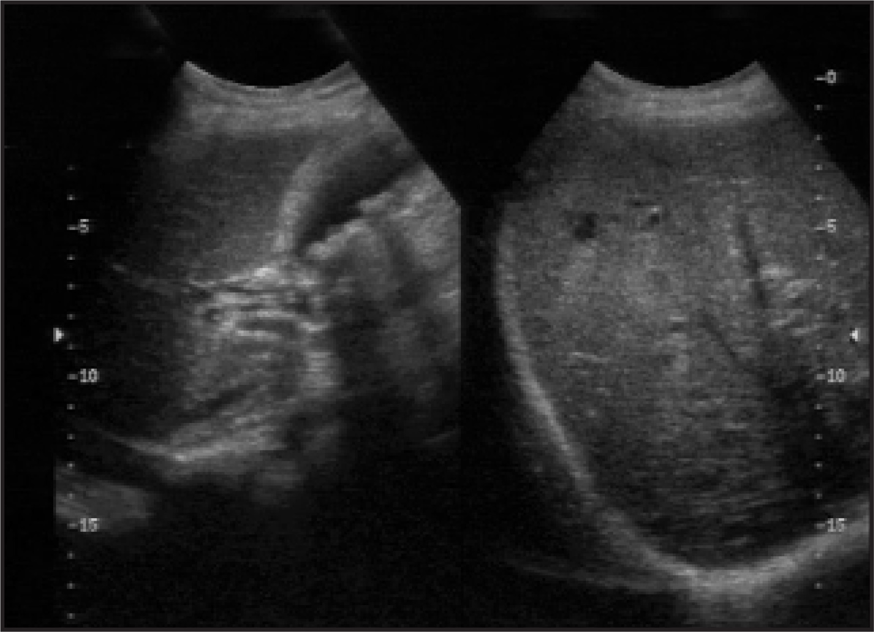
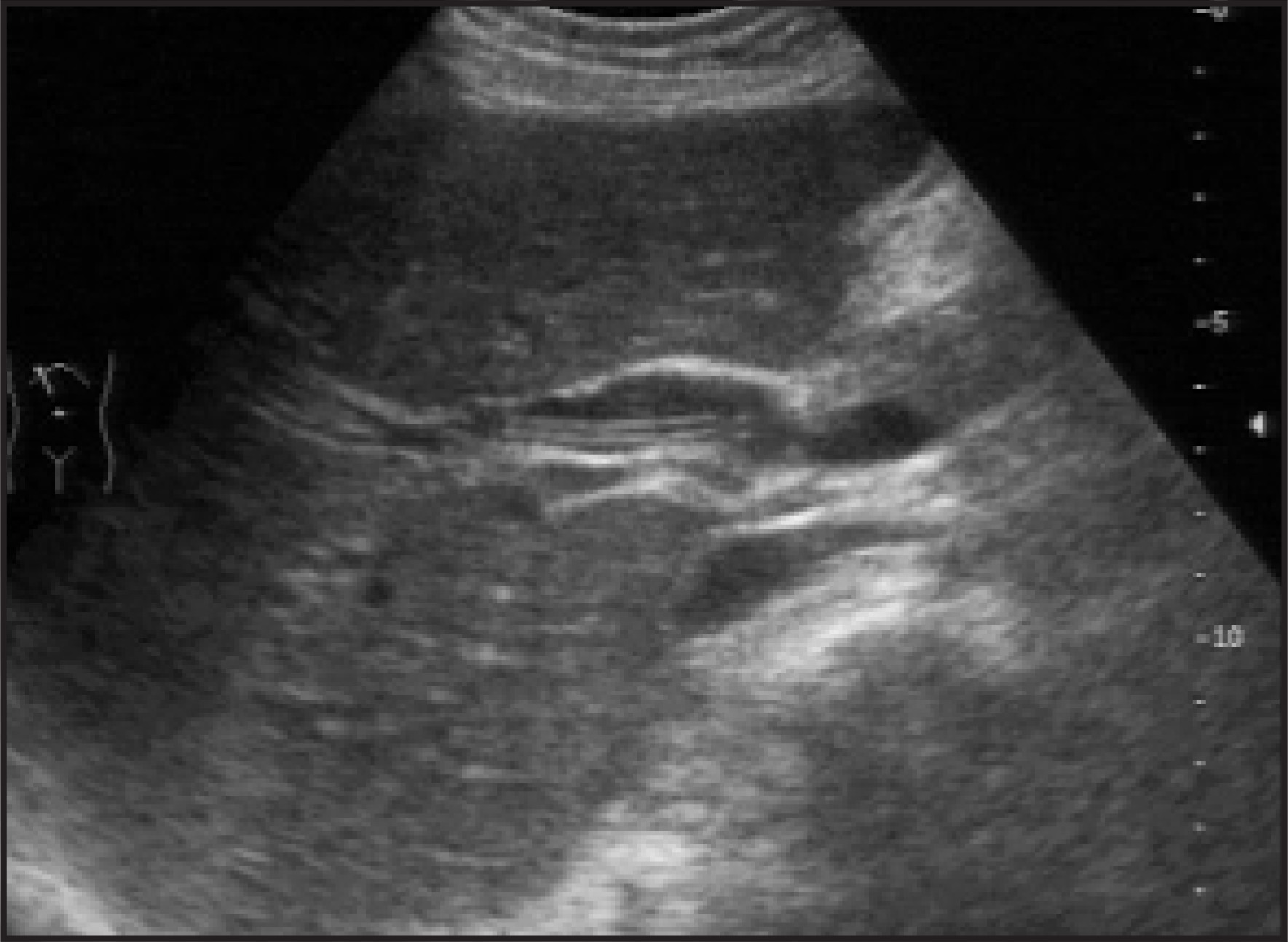
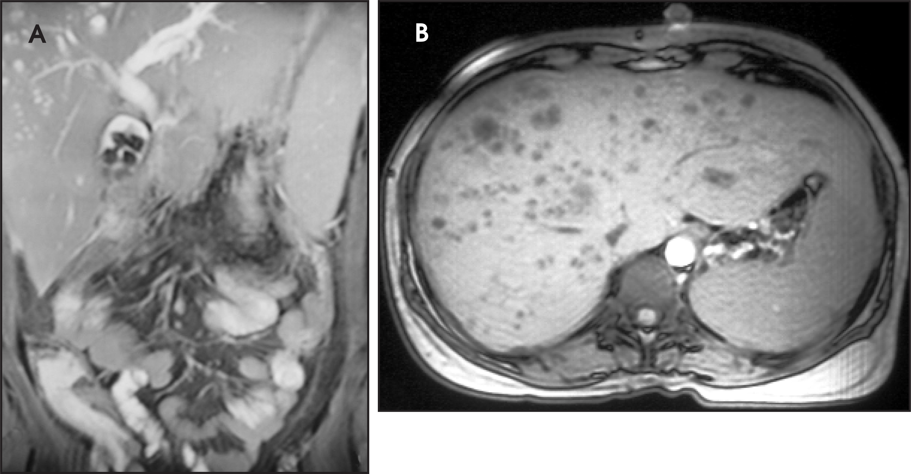
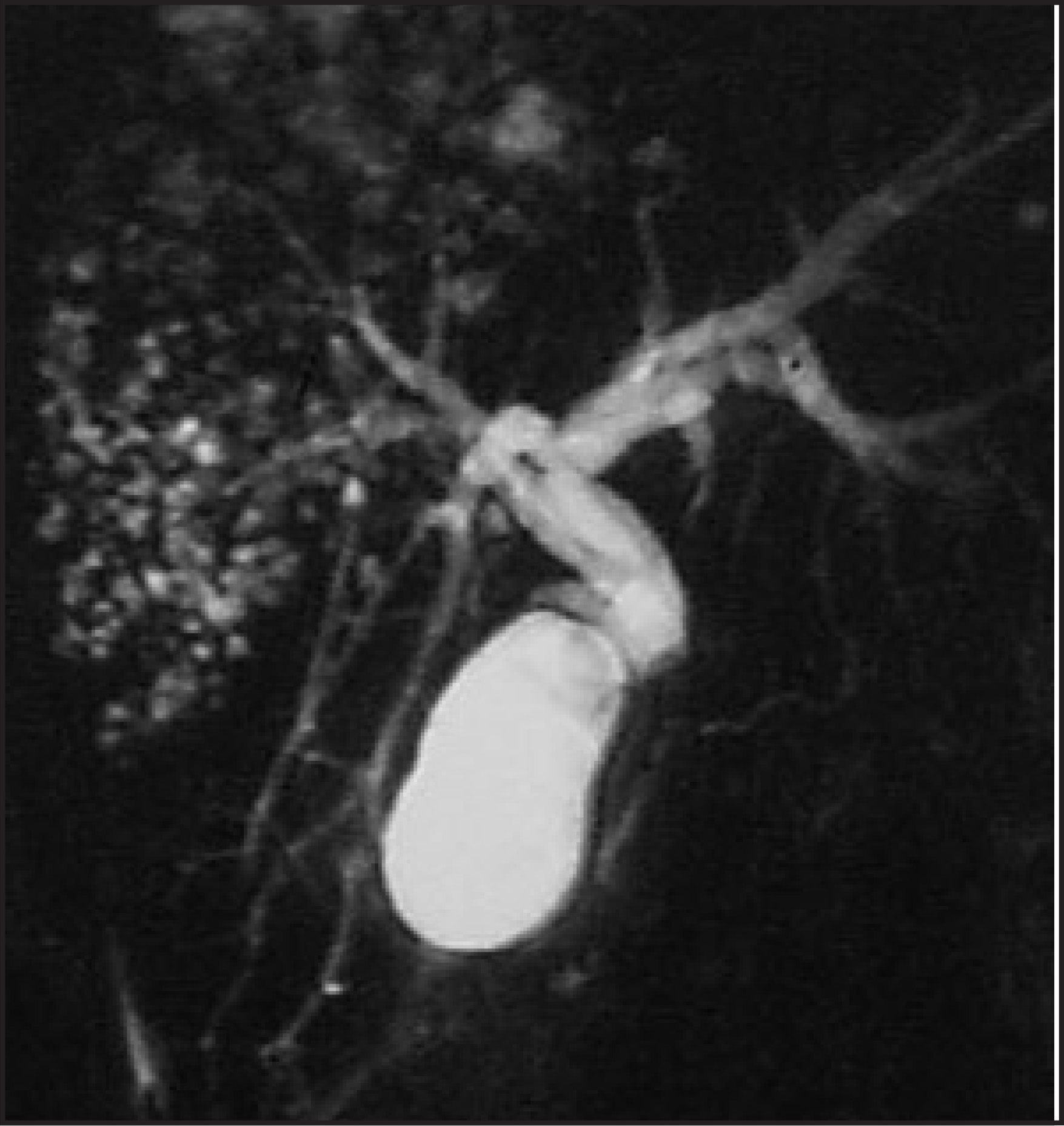
A 30-year-old woman presented with a 3-month history of recurrent, intermittent abdominal pain and 2 days of fever and vomiting. On examination, the patient had icterus and mild tenderness in the right hypochondrium. Laboratory work-up results at presentation revealed mild total hyperbilirubinemia and mildly elevated liver enzyme. Ultrasound of the abdomen (Figures 1 and 2) was followed by magnetic resonance imaging (MRI; Figure 3) and magnetic resonance cholangiopancreatography (MRCP; Figure 4) examinations.
IMAGING FINDINGS
An ultrasound scan revealed hepatomegaly with diffusely altered echotexture. Numerous small hypo-echoic lesions were present in the right lobe of the liver with a few foci showing coalescence, which was suggestive of small abscesses (Figure 1). The intrahepatic biliary ducts and the common bile duct (CBD) were dilated with intraluminal echogenic linear tubular structures, which were suggestive of roundworms (Figure 2). There were multiple gallstones, sludge, and a few nonshadowing echogenic structures that suggested the presence of worms in the gallbladder too (Figure 1). Other abdominal viscera were normal, and no ascites or enlarged lymph nodes were detected.
MRI and MRCP confirmed multiple intrahepatic abscesses, bilobar intrahepatic biliary dilation, and a dilated proximal CBD (Figures 3 and 4). A linear tubular structure with central hyperintensity was seen in the proximal CBD. On axial MRI, a “bull’s-eye” appearance suggestive of ascariasis was seen (Figure 3B). The right and left hepatic ducts also showed similar tubular linear defects. A markedly hypointense rounded focus with central hyperintensity was seen in the proximal CBD distal to the worm with a normal distal CBD, which was suggestive of choledocholithiasis (Figure 4). The gallbladder showed multiple markedly hypointense foci due to cholelithiasis, along with a few less hypointense foci caused by ascariasis (Figure 5).
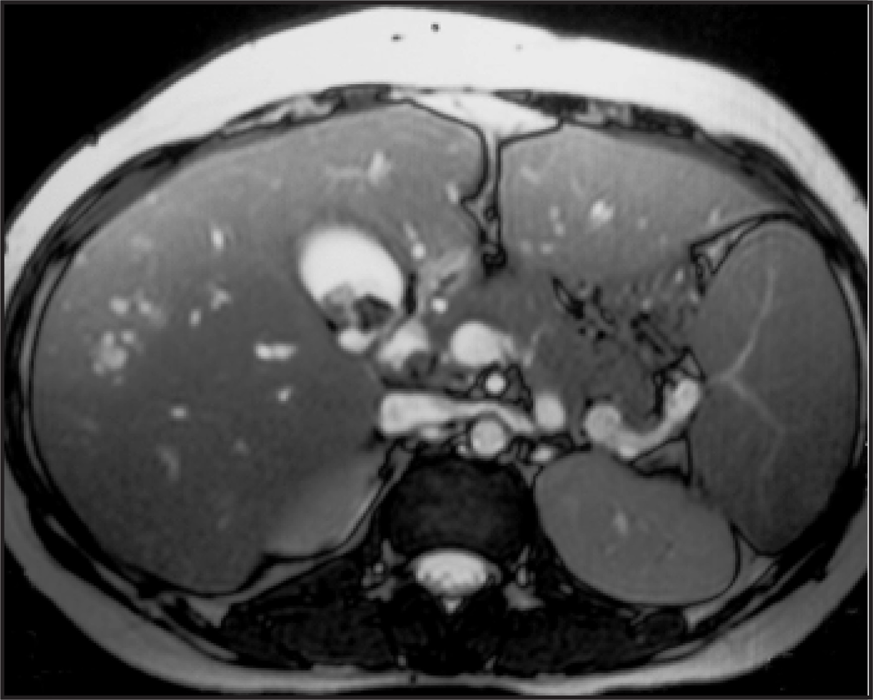
The patient’s condition improved after anti-helminthic therapy with albendazole and antibiotics was initiated.
DIAGNOSIS
Biliary ascariasis with cholelithiasis, choledocholithiasis, and cholangitic abscesses
DISCUSSION
The differential diagnosis for increased echogenicity within the CBD includes calculus, sludge, pus, thrombus, tumor, gas, foreign body, and parasites. Other biliary parasites are much smaller than Ascaris lumbricoides and, therefore, not part of the differential diagnosis in this case. On ultrasound, A lumbricoides in the biliary ducts usually manifests as an echogenic tubular structure, has a diameter of approximately 3 to 6 mm, and has a relatively hypoechoic center, with a more echogenic wall. This organism may exhibit slow movement. Ascarids typically lie parallel to the long axis of the bile duct. They may be coiled and, if multiple, may completely fill the bile duct, which produces either the “spaghetti sign,” or, if they are very densely packed in the bile ducts, may appear amorphous and manifest as hyperechoic pseudotumors. Other appearances described in the literature are the “strip sign,” “inner-tube sign,” and “bull’s-eye” appearance.1,2
Endoscopic retrograde cholangiopancreatography, a relatively invasive technique, can be used to image biliary ascariasis.2 On MRCP, the worm is seen as a tubular defect in the CBD with high signal intensity in the middle of the tubular defect caused by swallowed bile within the worm—this is the so-called “double-tube” appearance as was seen in this case. On transverse T2-weighted images, the worm is seen as “double bull’s-eye” or “eye-glass” appearance formed by the coiled portion of the Ascaris worm (2 high-density rings in the CBD).2
The human infection life cycle begins with ingestion of an egg, and then the larvae hatch in the small intestine. The larvae invade the small bowel mucosa, migrate through the circulatory system to the lungs, invade the alveoli, ascend the tracheobronchial tree, and are then swallowed into the small intestine, where they mature into adult worms. Intestinal infestation is often asymptomatic. Migration of worms into the biliary tree is a well-known complication, which may result in biliary colic, cholecystitis, cholangitis, intrahepatic abscesses, or pancreatitis.3
The hepatobiliary tree is one of the most common ectopic sites for ascariasis because of its easy accessibility, and this organism is a known cause of biliary tract disease, especially biliary colic, acute cholecystitis, cholangitis, and acute pancreatitis.4,5 The movement of the worm in the biliary tree is characterisitic on ultrasound examination. The diagnosis is established by means of microscopic identification of A lumbricoides eggs in fecal samples.
CONCLUSION
Biliary ascariasis is second only to cholelithiasis as a cause of acute biliary symptoms worldwide. Sonography is a well-established imaging modality for the diagnosis of hepatobiliary ascariasis. MRI and MRCP, although not the primary imaging modalities for hepatobiliary ascariasis, can reveal the extent of disease in the biliary tree, the status of the intrahepatic biliary radicals, and any other associated lesions (such as cholangitic abscesses) to better advantage and with more clarity than can other modalities.
References
- Anand R, Narula M, Gupta A, Vig K. Biliary ascariasis. Ind J Radiol Imag. 1999;9(1):23.
- Ng K, Wong H, Kong M. Biliary ascariasis CT, MR cholangiopancreatography, and navigator endoscopic appearance–Report of a case of acute biliary obstruction. Abdomin Imaging. 1999;24:470-472.
- Liu L, Weller P, Fauci A, Braunwald E, Isselbacher K. Harrison’s Principles of Internal Medicine. 1998:1208-1209.
- Danaci M, Belet U, Polat V, Incesu L. MR imaging features of biliary ascariasis. AJR Am J Roentgenol. 1999;173:503.
- Alper F, Kantarci M, Bozkurt M. Acute biliary obstruction caused by biliary ascariasis in pregnancy: MR cholangiography findings. Clin Radiol. 2003;58:896-898.
Citation
. RCOM RADIOLOGICAL. Applied Radiology. 2007;36(5):48A-48C. doi:10.37549/AR1511.