RCOM RADIOLOGICAL
Applied Radiology — Vol. 36 , Issue 5 , pp. 44 -44
DOI: 10.37549/AR1515
Published: May 1, 2007
Categories
CASE SUMMARY
A 47-year-old woman presented with an increasing mass over the left wrist. The patient underwent magnetic resonance imaging (MRI) examination for further evaluation. The patient had a similar abnormality in the flexor hallucis tendon sheath, which had previously been removed.
IMAGING FINDINGS
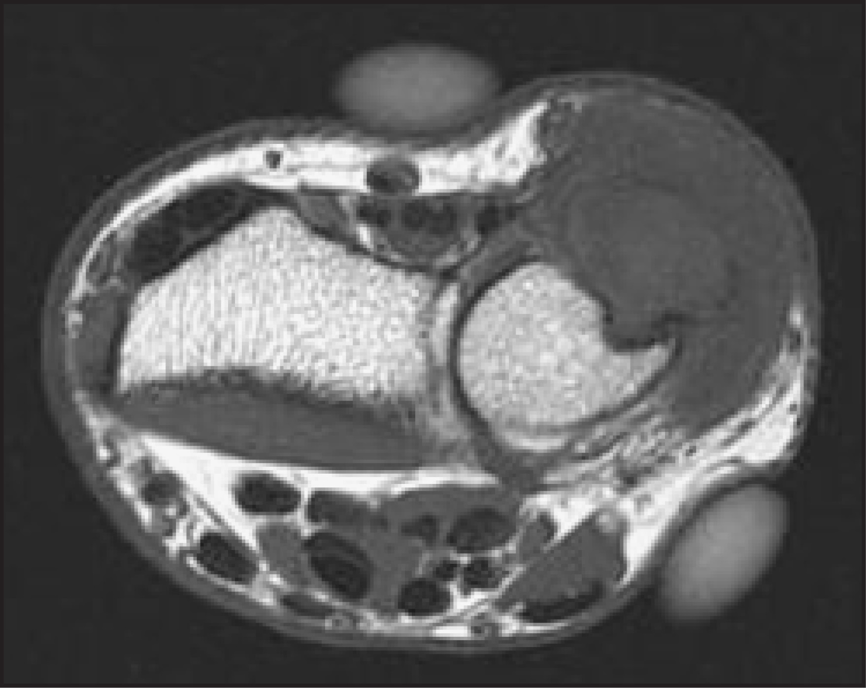
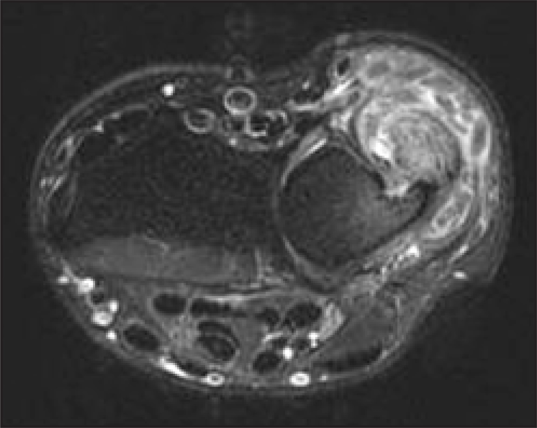
The MRI shows marked thickening of the extensor carpi ulnaris tendon associated with tenosynovitis (Figure 1). Also, within the tendon sheath are several oblong structures, which are predominantly low in signal on T2-weighted (T2W) images (Figure 2).
DIAGNOSIS
Rheumatoid tenosynovitis with rice-body formation
DISCUSSION
Rice-body formation is a nonspecific response to chronic synovial inflammation, which was first de scribed in association with tuberculous arthritis.1 Since then, rice-body formation has been associated with both rheumatoid arthritis and juvenile rheumatoid arthritis. The pathogenesis of rice-body formation remains controversial but may be related to microinfarction of the synovium2 with release of tissue into the joint or detachment of hypertrophied synovium.3
The MRI characteristics have been well documented with well-defined nodules of intermediate signal on T1-weighted (T1W) images that are relatively low in signal on T2W sequences.4 Although synovial osteochondromatosis and pigmented villonodular synovitis are arthropathies associated with low-signal structures around joints, tendons, and bursa, these 2 entities more commonly occur as monoarticular processes and are more often seen in larger joints, such as the knee or hip. Additionally, there is no evidence of hemosiderin deposition to suggest pigmented villonodular synovitis, and the cartilage components in synovial osteochondromatosis are high in signal on T2W images. The fact that the patient had a similar abnormality at a different site should suggest the possibility of a systemic disease.
CONCLUSION
Rice-body formation has a characteristic appearance on MRI, manifesting as masses of intermediate signal on T1W images and low signal on T2W images. Their presence should alert the radiologist to the possibility of an inflammatory arthropathy.
References
- Riese H. Die reiskorperchen in tuberculkoserkrankten synovalsacken. Deut Zeit Chirurg. 1895;42:1-99.
- Cheung H, Ryan L, Kozin F, McCarty D. Synovial origins of rice bodies in joint fluid. Arthritis Rheum. 1980;23:72-76.
- Berg E, Wainwright R, Barton B. On the nature of rheumatoid rice bodies: An immunologic, histochemical, and electron microscope study. Arthritis Rheum. 1977;20:1343-1349.
- Chung C, Coley B, Martin L. Rice bodies in juvenile rheumatoid arthritis. AJR Am J Roentgenol. 1998;170:698-700.
Citation
. RCOM RADIOLOGICAL. Applied Radiology. 2007;36(5):44-44. doi:10.37549/AR1515.