RCOM RADIOLOGICAL
Applied Radiology — Vol. 36 , Issue 6 , pp. 33 -36
DOI: 10.37549/AR1519
Published: June 1, 2007
Categories
CASE SUMMARY
A 29-year-old, right-hand–dominant man presented with an 8-month history of daily occipital headaches. On occasion, he reported problems with maintaining balance. Physical examination was entirely normal. Neurological examination was unremarkable except for impaired finger-nose coordination in both upper limbs.
IMAGING FINDINGS
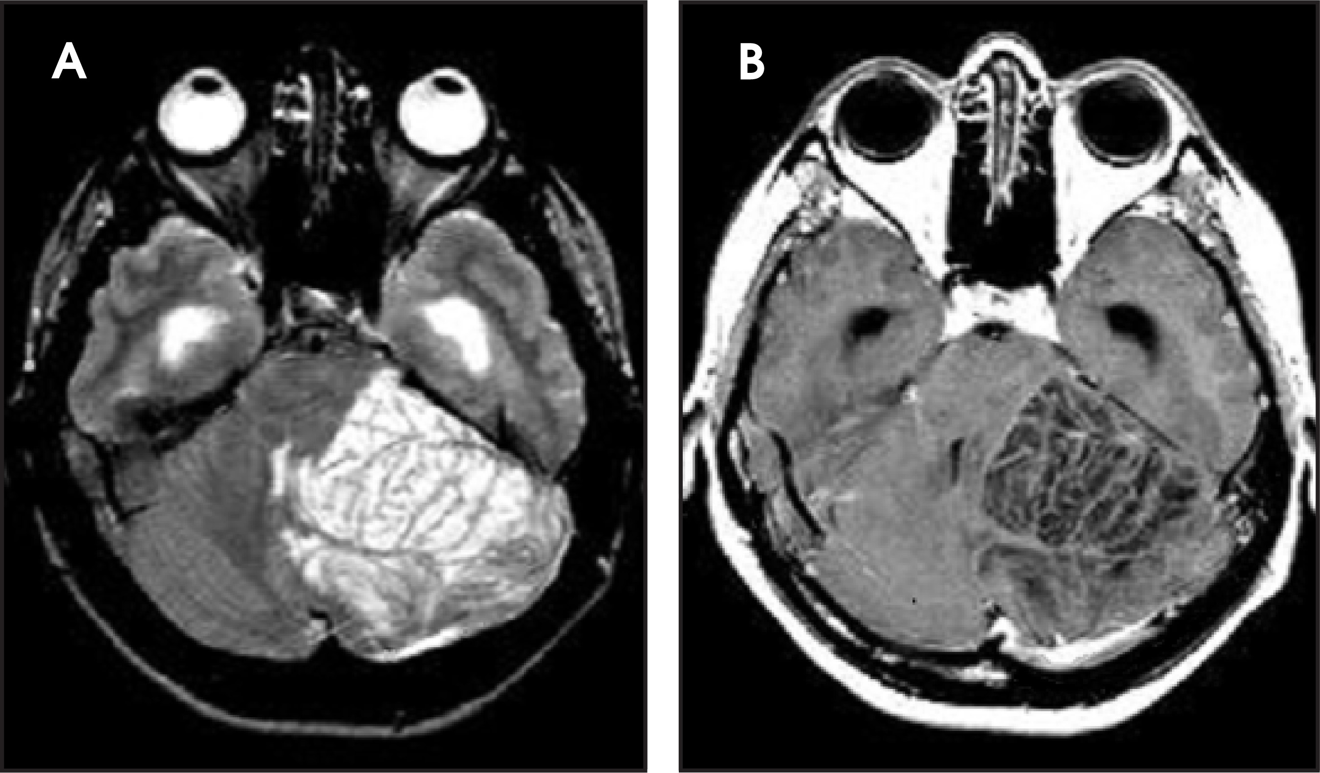
MR imaging (MRI) revealed a nonenhancing mass in the right cerebellar hemisphere, which was characterized by a striated pattern of hyperintensity on T2-weighted images (Figure 1A) and corresponding hypointensity on T1-weighted images (Figure 1B). The left cerebellar hemisphere was normal.
DIAGNOSIS
Lhermitte-Duclos disease (LDD)
DISCUSSION
Lhermitte-Duclos disease is a rare entity that was first reported in 1920.1 Histologically, it has been termed a dysplastic cerebellar gangliocytoma.1-3 This is because the fundamental nature of the lesion and, particularly, its pathogenesis remain unknown. Whether it represents a neoplastic, malformative, or hamartomatous lesion is still the subject of much debate.4
Macroscopically, it is typically a benign, slowly enlarging cerebellar mass. It seems to be more prevalent in young adults and shows no sexual prevalence. It may present with signs of cerebellar dysfunction or increased intracranial pressure secondary to obstructive hydrocephalus. However, cerebellar signs are minimal or absent in up to half of those with the lesion.5
MRI is the modality of choice in the radiologic evaluation of these lesions.6 The characteristic MRI features are of a nonenhancing posterior fossa mass with a “tiger-striped” or laminated appearance.7 This results from alternating bands of relative isointensity and hypointensity on T1-weighted sequences and relative isointensity and hyperintensity on T2-weighted sequences, as compared with normal neuronal tissue. Most other cerebellar masses destroy the folial pattern and show contrast enhancement. The nonenhancing striated MRI appearance of thickened folia in patients with LDD is distinctive and suggests this diagnosis, although atypical cases have been noted.8-10
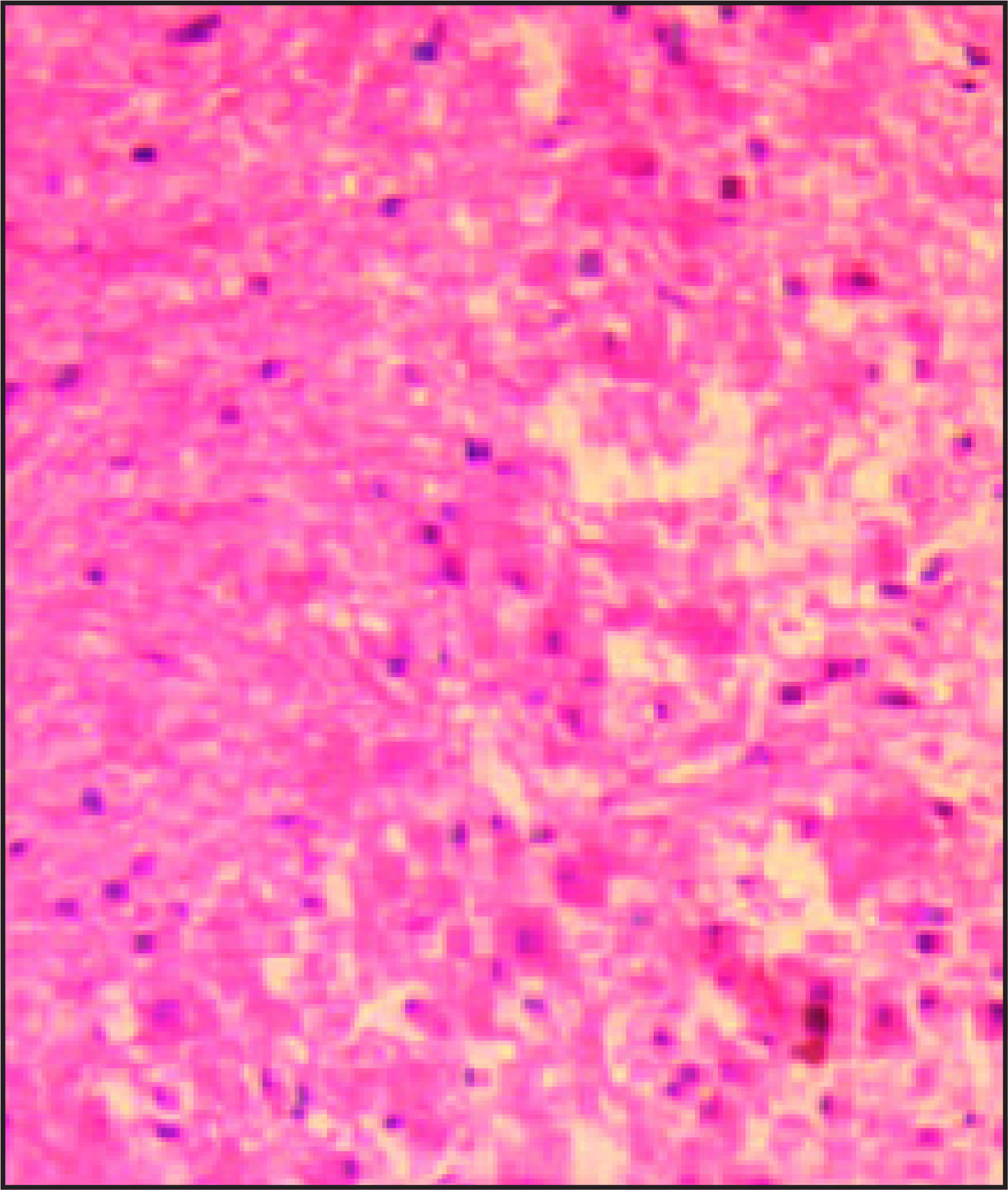
Normal cerebellar cortical structure consists of an outer molecular layer, a middle Purkinje layer, and an inner granular layer. In the disease, there is focal indolent growth of the cerebellar cortex in which there is folial enlargement because of a plethora of dysplastic cortical neurons and a thickening of the molecular layer (Figure 2). This results in the loss of Purkinje cells and the thinning of medullary white matter. Enlargement of the folia results in the loss of their secondary arborization and asymmetric expansion of the cerebellar hemisphere. On MRI, the abnormal T1 and T2 signals correspond to these changes.
Lhermitte-Duclos disease is closely associated with Cowden’s syndrome, a rare, autosomal-dominant, familial cancer syndrome characterized by multiple hamartomas and neoplasms of ectodermal, mesodermal, and endodermal origin. It may be found in up to 40% of cases of LDD.11 Other reported associations include gastrointestinal polyps, ovarian cysts, hepatic lesions, neuromas, and neurofibromas. There is also an increased predisposition to breast cancer and thyroid tumors.12,13
The treatment of the condition is usually surgical excision of the lesion and monitoring of any associated conditions. Long-term follow-up is strongly encouraged because of occasional symptomatic recurrence.14
CONCLUSION
Lhermitte-Duclos disease is a rare benign lesion of the cerebellum, the exact nature of which is still unknown. The lesion is hypointense on T1-weighted and hyperintense on T2-weighted MRI and produces a very typical appearance. The diagnostic images on MRI are unlikely to be confused with any other pathologic process in the cerebellum. This capability allows for management options independent of a biopsy. The disease should be considered when a young adult presents with clinical signs of cerebellar origin or progressive mass effect in the posterior fossa.
References
- Lhermitte J, Duclos P. Sur un ganglionuroma diffus du cortex du cervelet. Bull Assoc F Etude Cancer. 1920;9:99-107.
- Harding B, Adams J, Duchen L. Greenfields Neuropathology. 1992:592-594.
- Ambler M, Pogacar S, Sidman R. Lhermitte-Duclos disease (granule cell hypertrophy of the cerebellum) pathological analysis of the first familial cases. J Neuropathol Exp Neurol. 1969;28:622-647.
- Nowak D, Trost H. Lhermitte-Duclos disease (dysplastic cerebellar gangliocytoma): A malformation, hamartoma or neoplasm?. Acta Neurol Scand. 2002;105:137-145.
- Milbouw G, Born J, Martin D. Clinical and radiological aspects of dysplastic gangliocytoma (Lhermitte-Duclos disease): A report of two cases with review of the literature. Neurosurgery. 1988;22(1 Pt 1):124-128.
- Kulkantrakorn K, Awwad E, Levy B. MRI in Lhermitte-Duclos disease. Neurology. 1997;48:725-731.
- Smith R, Grossman R, Goldberg H. MR imaging of Lhermitte-Duclos disease: A case report. AJNR Am J Neuroradiol. 1989;10:187-189.
- Spaargaren L, Cras P, Bomhof M. Contrast enhancement in Lhermitte-Duclos disease of the cerebellum: Correlation of imaging with neuropathology in two cases. Neuroradiology. 2003;45:381-385.
- Ellis P. Case report. Lhermitte-Duclos disease: Enhancement following gadolinium-DPTA. Clin Radiol. 1996;51:222-224.
- Awwad E, Levy E, Martin D, Merenda G. Atypical MR appearance of Lhermitte-Duclos disease with contrast enhancement. AJNR Am J Neuroradiol. 1995;16:1719-1720.
- Murata J, Tada M, Sawamura Y. Dysplastic gangliocytoma (Lhermitte-Duclos disease) associated with Cowden disease: Report of a case and review of the literature for the genetic relationship between the two diseases. J Neurooncol. 1999;41:129-136.
- Albrecht S, Haber R, Goodman J, Duvic M. Cowden syndrome and Lhermitte-Duclos disease. Cancer. 1992;70:869-876.
- Liaw D, Marsh D, Li J. Germline mutations of the PTEN gene in Cowden disease, an inherited breast and thyroid cancer syndrome. Nat Genet. 1997;16:64-67.
- Kulkantrakorn K, Awwad E, Levy B. MRI in Lhermitte-Duclos disease. Neurology. 1997;48:725-731.
Citation
. RCOM RADIOLOGICAL. Applied Radiology. 2007;36(6):33-36. doi:10.37549/AR1519.