RCOM RADIOLOGICAL
Applied Radiology — Vol. 36 , Issue 6 , pp. 38 -41
DOI: 10.37549/AR1520
Published: June 1, 2007
Categories
CASE SUMMARY
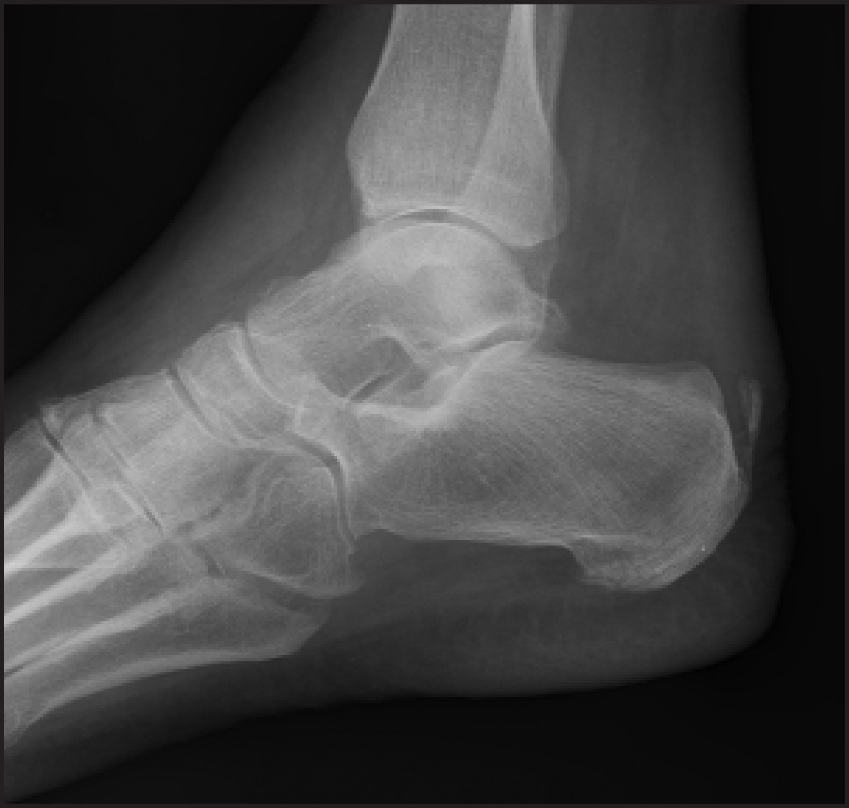
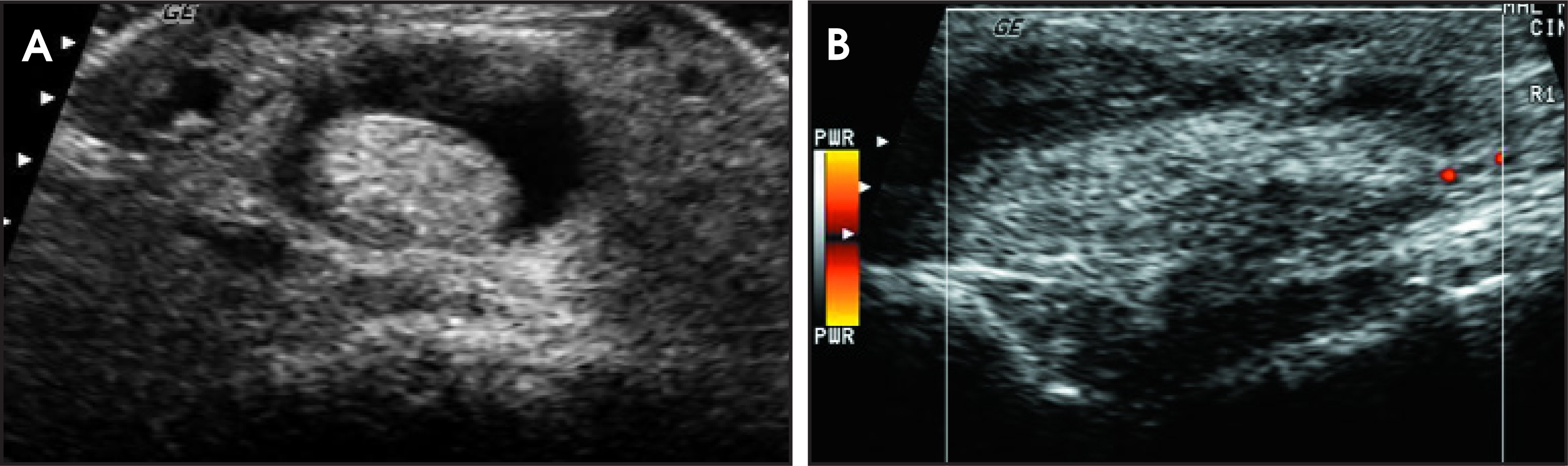
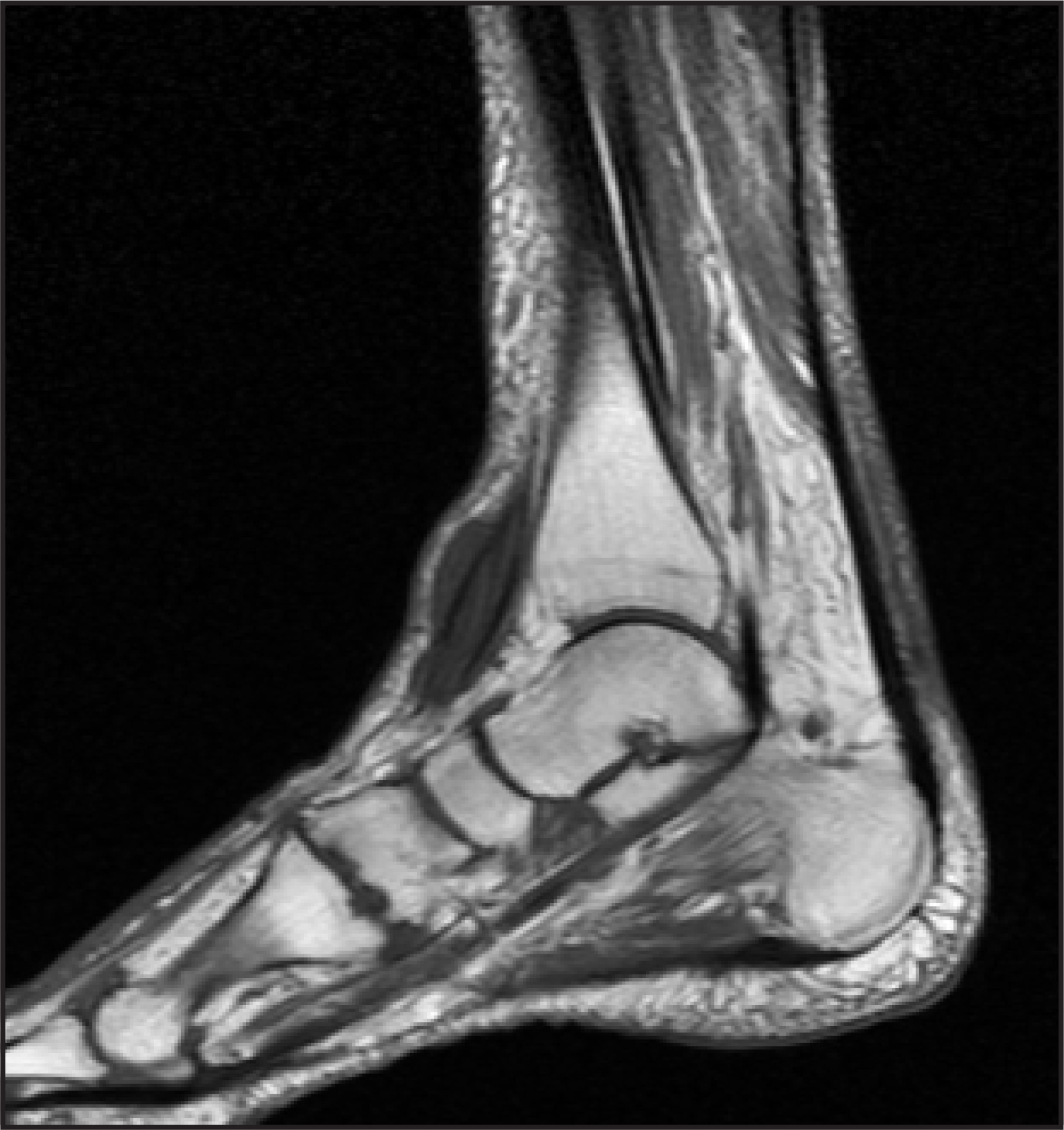
A 74-year-old white man with chronic bilateral ankle pain and bilateral lower extremity edema presented with a 4-month history of right ankle pain that resulted in difficulty walking. On physical examination, the patient was afebrile and there was bilateral lower extremity edema and a 2 × 3-cm firm, mobile, nontender bulge on the anterior aspect of his ankle. Plain-film radiography (Figure 1), ultrasound (US; Figure 2), and magnetic resonance imaging (MRI; Figure 3) studies were performed.
IMAGING FINDINGS
Right ankle radiographs showed bimalleolar ankle edema (not shown), as well as a 4.7 × 1.7-cm abnormal anterior ankle soft tissue opacity at the level of the tibiotalar joint (Figure 1). Ultrasound of the right ankle showed an abnormally thickened anterior tibialis tendon (ATT) at the level of the anterior aspect of the ankle (Figure 2). In the area of the palpable knot, there was a well-defined avascular hyperechoic mass, which was surrounded by fluid without any calcifications. It was unclear if any tendon fibers were still intact.
MRI of the right ankle showed focal enlargement of the ATT at the level of the tibiotalar joint, which was associated with intermediate T1 and high T2 signal. Some of the fibers of the ATT were still intact (Figure 3).
DIAGNOSIS
Partial tear of the anterior tibialis tendon
DISCUSSION
The ATT is the third most commonly ruptured tendon in the lower extremity.1 Tears of the ATT have been well described in the literature2 and can be classified as traumatic or spontaneous. This classification can also be thought of as acute or acute on chronic, respectively.3 Spontaneous ATT ruptures most commonly occur between the 6th and 8th decades of life, often with a history of forced plantar flexion.2 They can be associated with steroid injections, oral steroids, Marfan syndrome, hyperparathyroidism, diabetes, rheumatoid arthritis, Reiter syndrome, and psoriasis.1-3 Gallo et al3 analyzed the vascular supply of the ATT using immunohistochemical methods and injection techniques. Their study showed that the proximal portion of the tendon received blood from the anterior tibial artery, while the distal portion received blood from the medial tarsal artery. There was a 4.5- to 6.7-cm relatively avascular zone in the anterior portion of the tendon 0.5 to 3.0 cm proximal to its bony insertion that corresponded to the most frequently reported site of spontaneous ATT rupture.3 This region of avascularity also corresponded to the location of superior and inferior retinacula, which serve as pulleys for the ATT mechanism.1 The hypothesis is that the hypoxic state of the tissue in this critical zone and the chronic impingement by the retinacula render this region vulnerable to chronic degeneration and, eventually, to rupture. This hypothesis is also believed to pertain to the Achilles and posterior tibialis tendons.2
This case illustrates the importance of differentiating partial tendon tears from complete tendon tears,4-8 as the former are treated conservatively while the latter are often treated surgically. With US, there may be some overlap in the appearance of partial and complete tendon tears. Each may have focal tendon enlargement and heterogeneous signal as well as the presence or absence of intratendinous fluid. However, 1 study assessing the ability of US to differentiate partial from complete tears of the Achilles tendon showed 92% accuracy.5
The MRI findings of partial tears include increased T1 and T2 signal within a tendon that extends to a surface, focal or diffuse enlargement, focal or diffuse atrophy, partial disruption of tendon fibers, focal intratendinous fluid collections, and tendon enhancement. With MRI, there is also some overlap between the findings in complete and partial tendon tears. Complete tears are usually associated with discontinuity and separation of the torn ends of the tendon, and there usually is interposed edema or hemorrhage1,2,4,6 Clinically, the patient’s ability to dorsiflex the ankle is significantly impaired, though not necessarily absent, as the peroneal tendons are also able to dorsiflex the ankle.
CONCLUSION
Tendon tears are easily identified by MRI and US. It is of significant clinical importance to differentiate between complete and partial tears and, thus, all efforts should be made to make that distinction. Ultrasound can usually differentiate between these 2 diagnoses but in uncertain cases, MRI may be another useful imaging study.
References
- Jarvinen M, Jozsa L, Kannus P. Histopathological findings in chronic tendon disorders. Scand J Med Sci Sports. 1997;7(2):86-95.
- Schweitzer M, Karasick D. MR imaging of disorders of the Achilles tendon. AJR Am J Roentgenol. 2000;175:613-625.
- Gallo R, Kolman B, Daffner R. MRI of tibialis anterior tendon rupture. Skeletal Radiol. 2004;33:102-106.
- Premkumar A, Perry M, Dwyer A. Sonography and MR imaging of posterior tibial tendinopathy. AJR Am J Roentgenol. 2002;178:223-232.
- Hartgerink P, Fessell D, Jacobson J, van Holsbeeck M. Full-versus partial-thickness Achilles tendon tears: Sonographic accuracy and characterization in 26 cases with surgical correlation. Radiology. 2001;220:406-412.
- Aydingoz U, Aydingoz O. Spontaneous rupture of the tibialis anterior tendon in a patient with psoriasis. Clin Imaging. 2002;26:209-211.
- Chandnani V, Bradley Y. Achilles tendon and miscellaneous tendon lesions. Magn Reson Imaging Clin N Am. 1994;2:89-96.
- Bianchi S, Zwass A, Abdelwahab I, Zoccola C. Evaluation of tibialis anterior tendon rupture by ultrasonography. J Clin Ultrasound. 1994;22:564-566.
Citation
. RCOM RADIOLOGICAL. Applied Radiology. 2007;36(6):38-41. doi:10.37549/AR1520.