RCOM RADIOLOGICAL
Applied Radiology — Vol. 36 , Issue 7 , pp. 40 -41
DOI: 10.37549/AR1525
Published: July 1, 2007
Categories
CASE SUMMARY
A 22-year-old male university student presented to the emergency room with complaints of diffuse, lower abdominal pain of approximately 1 week’s duration. The patient described the pain as “waxing and waning.” He reported the pain as being 8 on a pain scale of 0 to 10. The pain varied from dull to sharp in character. He initially presented to his university’s health center for evaluation. He reported that an ultrasound was performed, which was unremarkable. The patient described worsening abdominal symptoms, which prompted further evaluation. He noted some initial diarrhea at the onset of symptoms, but no nausea or vomiting. Also, he reported fevers for the last week up to 103°F with sweats and myalgia. During the evening prior to presenting to the emergency room, the patient’s pain localized to the right lower quadrant. The patient was otherwise generally healthy except for some mild reflux symptoms and depression. At the time of presentation to the emergency room, the patient’s blood pressure was 126/78 mm Hg, his heart rate was 81 bpm, his respiratory rate was 18 breaths per minute, and his temperature was 36.4°C. A clinical examination revealed a soft, nontender, nondistended abdomen, with no hepatosplenomegaly. There was moderate tenderness to superficial and deep palpation as well as fullness in the right lower quadrant. There were no peritoneal signs. His stool was heme-negative. Laboratory results, including complete blood count, chemistries, and urinalysis were all negative.
IMAGING FINDINGS
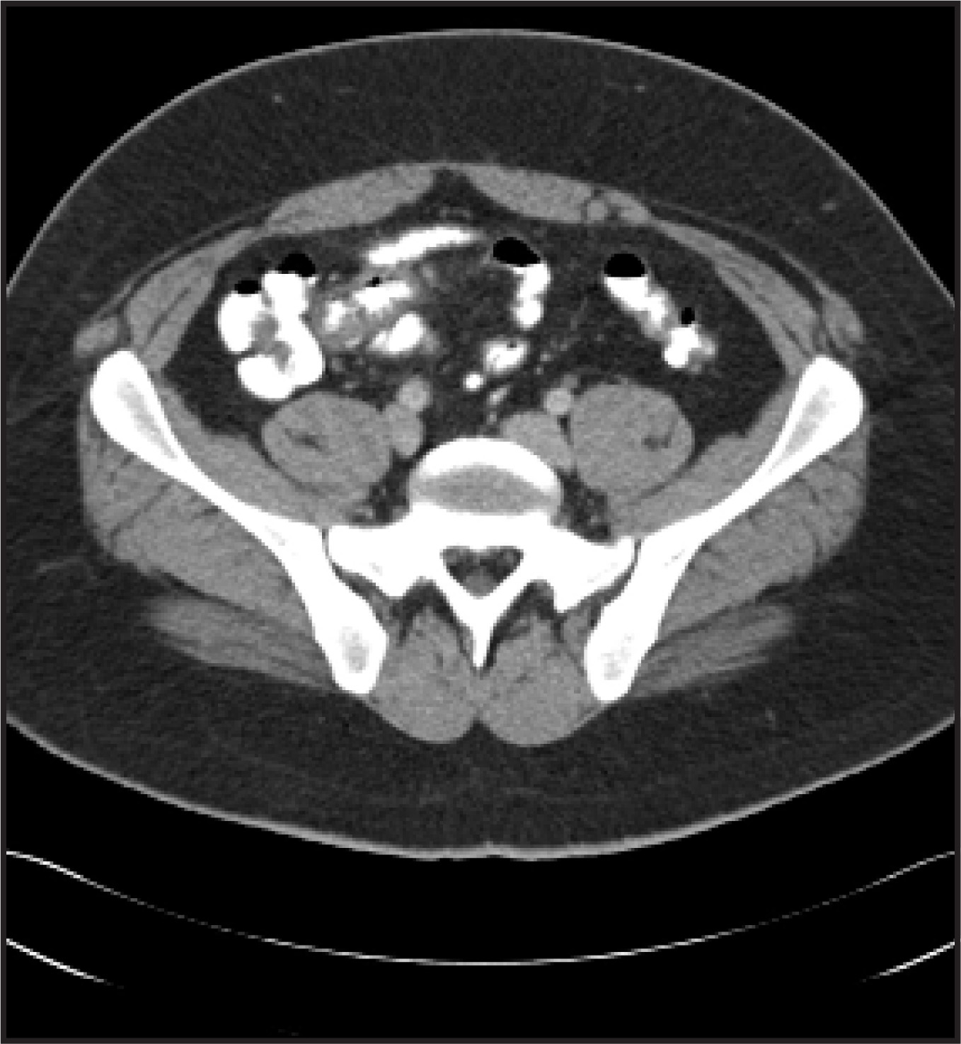
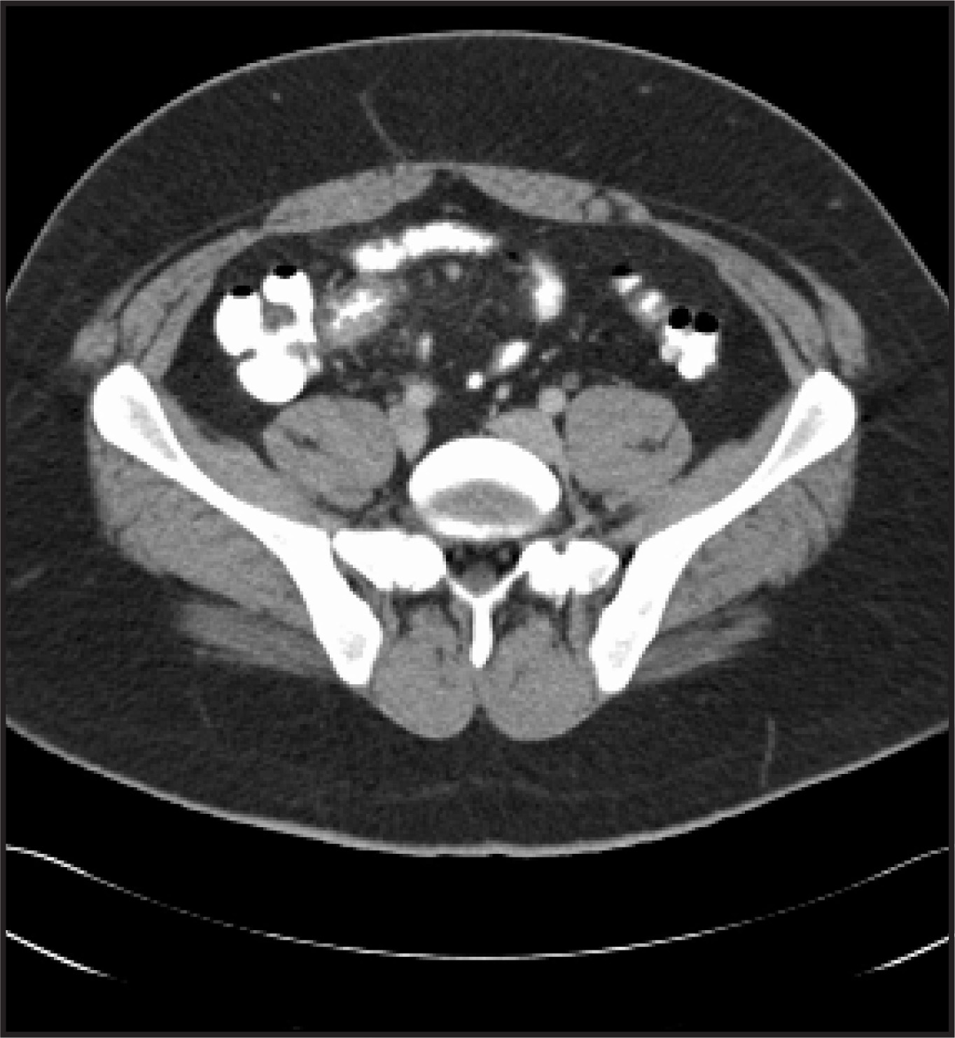
In the emergency room, CT scans of the abdomen and pelvis were ordered, which revealed inflammatory changes and diffuse thickening of the terminal ileum (Figures 1 and 2). The length of the involved segment of the terminal ileum measured approximately 10 cm. The cecum and appendix were normal in appearance. The diagnosis provided by the radiologist was Crohn’s disease and the differential diagnosis included infectious (Yersenia) and neoplastic (lymphoma) etiologies.
DIAGNOSIS
Inflammation of the terminal ileum secondary to an embedded plastic sword with ulceration in the ascending colon
DISCUSSION
The working diagnosis was Crohn’s disease, and the patient was admitted to the hospital. A gastroenterology consultation was requested. During this evaluation, it was discovered that the patient had been at a bar approximately 10 days before presentation to the emergency room. He recalled swallowing a spearlike mixed-drink plastic sword pick. The patient stated he was uncertain why he did this but reported symptoms of pain 3 to 4 days after ingestion.
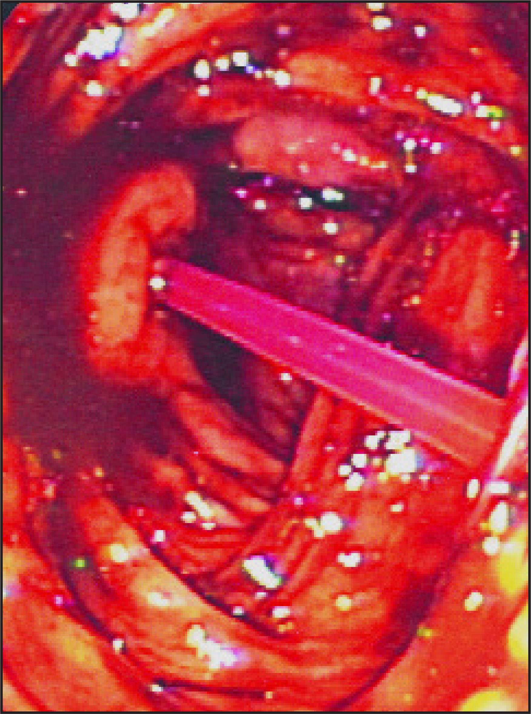
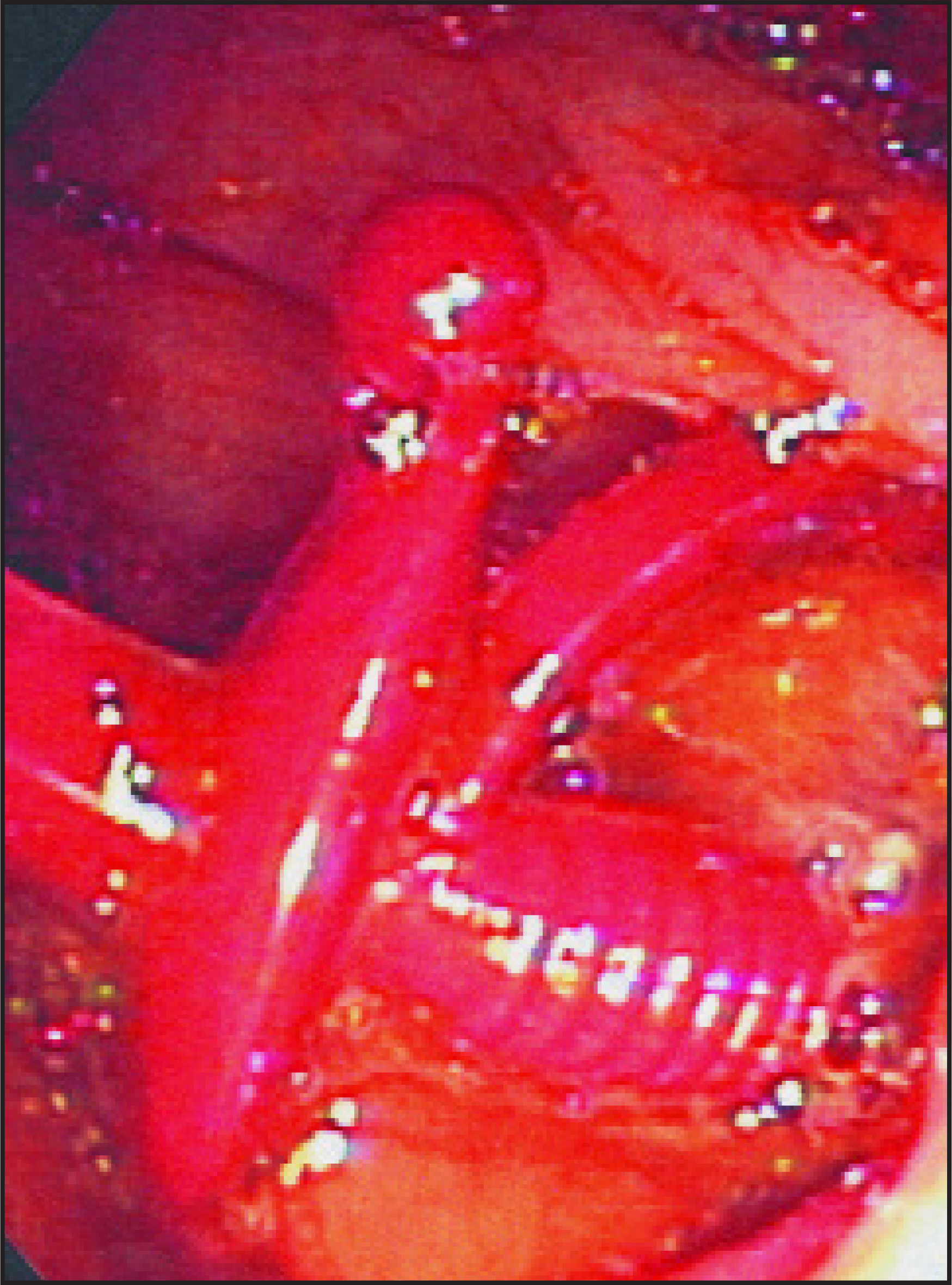
Based on the history, a colonoscopy was performed, which revealed a plastic sword pick with the tip and blade in the terminal ileum and the hilt embedded in the proximal ascending colon. The sword was removed cautiously during the colonoscopy. A localized area of mucosa in the terminal ileum was mildly erythematous and nodular. A single 6-mm ulcer was found in the proximal ascending colon at the embedded site.
CONCLUSION
The differential diagnosis for inflammation and wall thickening of the terminal ileum is extensive, including inflammatory, infectious, and neoplastic processes. In a young adult, the most common etiology is Crohn’s disease. However, with the right clinical history, foreign bodies should also be considered.
Citation
. RCOM RADIOLOGICAL. Applied Radiology. 2007;36(7):40-41. doi:10.37549/AR1525.