RCOM RADIOLOGICAL
Applied Radiology — Vol. 36 , Issue 7 , pp. 38 -39
DOI: 10.37549/AR1532
Published: July 1, 2007
Categories
CASE SUMMARY
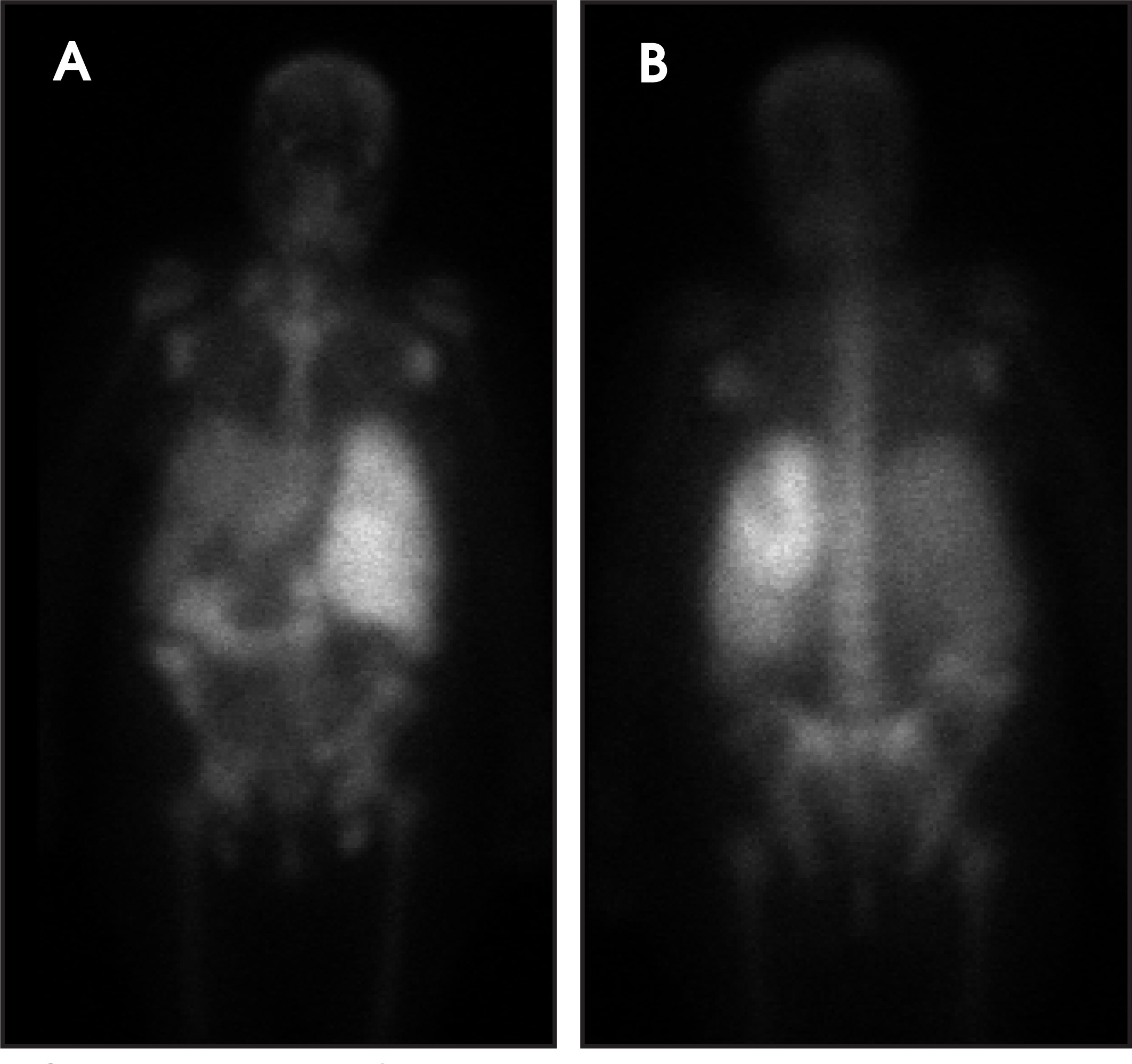
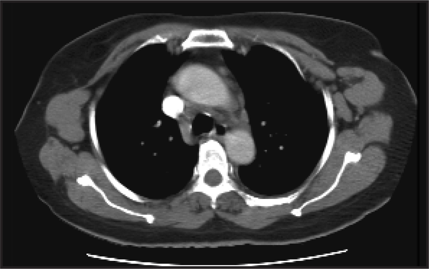
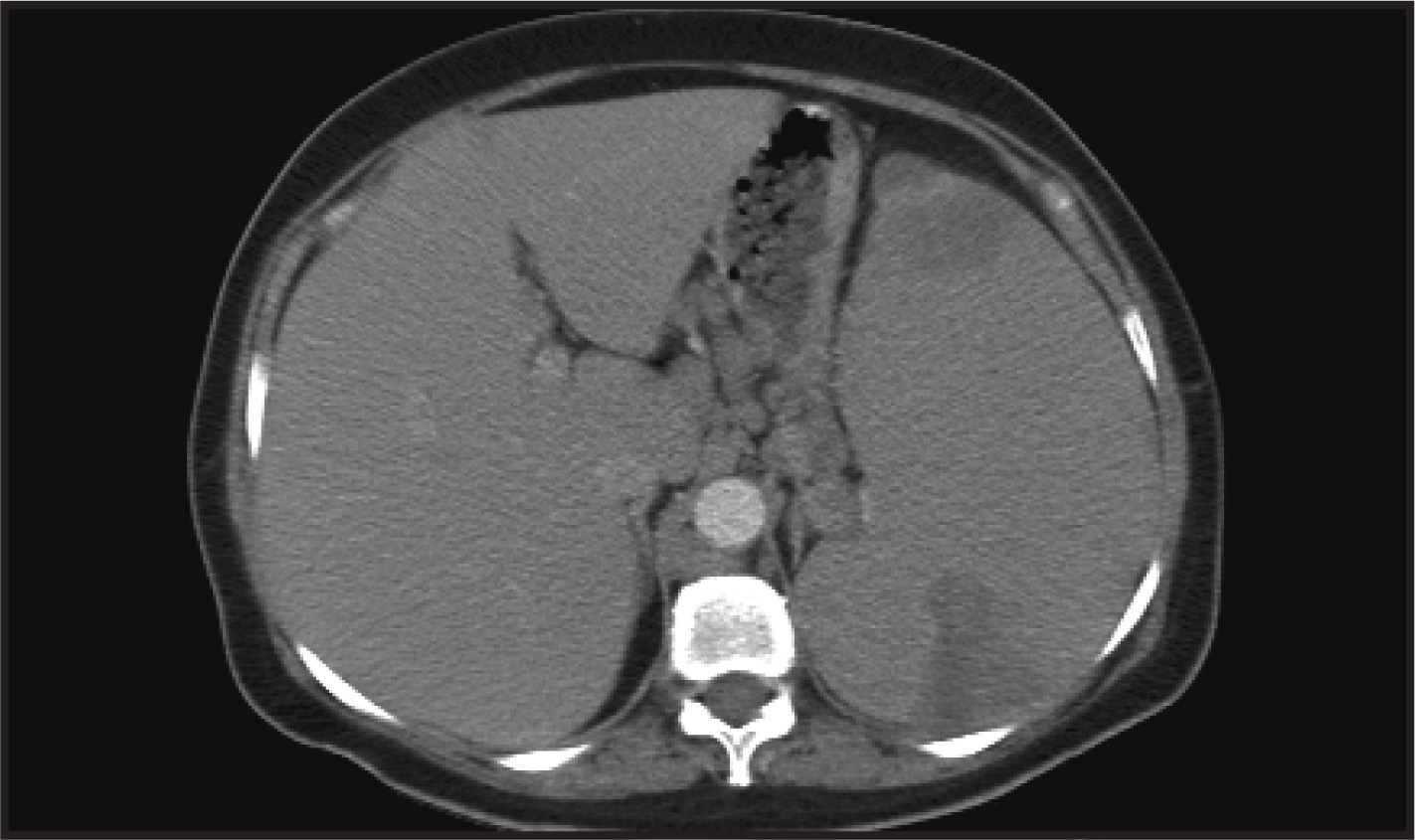
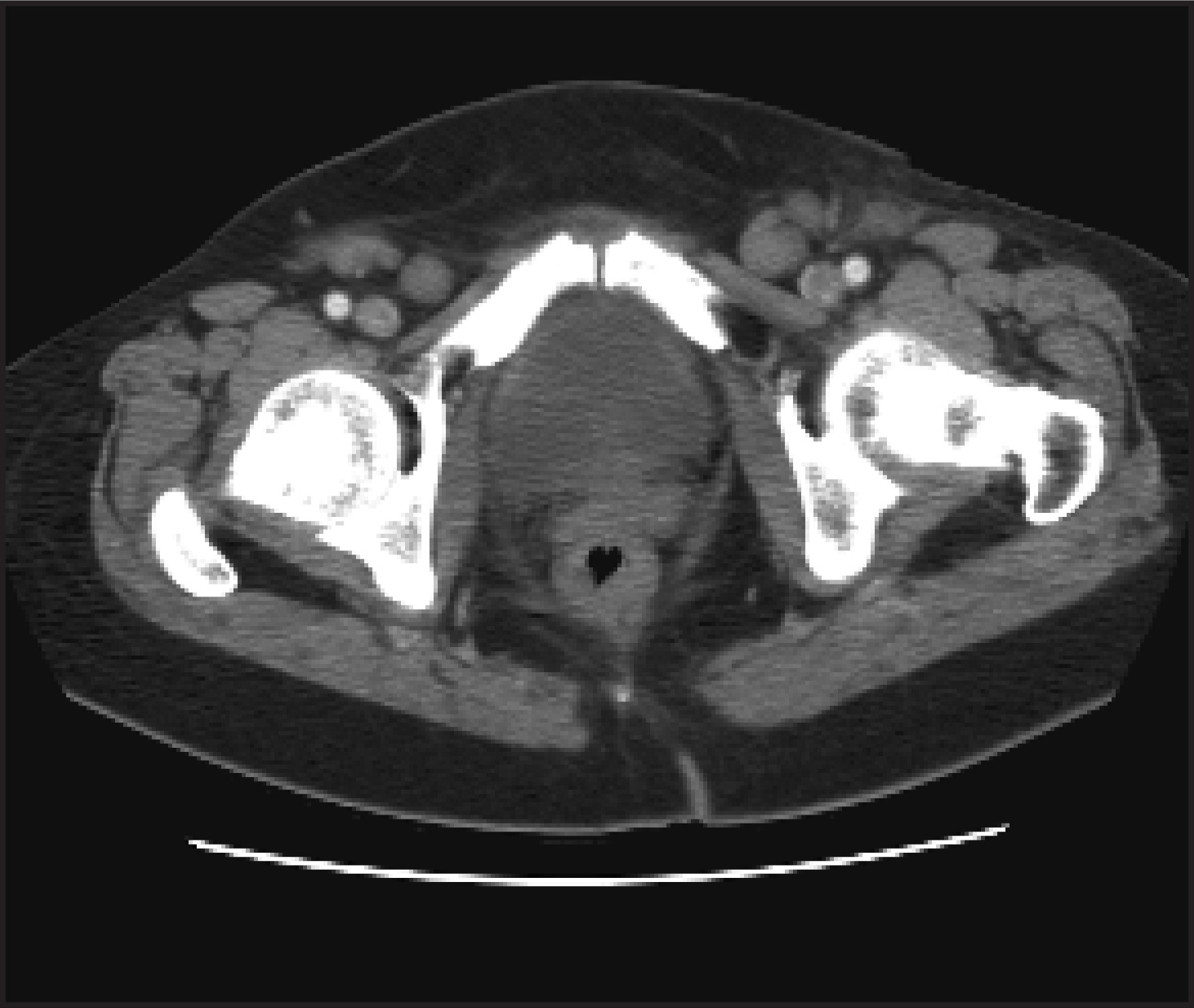
A 43-year-old woman with recently diagnosed stage IV mantle cell lymphoma (MCL) presented with symptoms of night sweats, weight loss, nausea, vomiting, and left-sided abdominal pain. Pertinent physical examination findings include marked splenomegaly. The patient underwent a gallium-67 citrate scintigraphy (Figure 1) as a baseline study prior to receiving chemotherapy. Computed tomography (CT) of the chest (Figure 2), abdomen (Figure 3), and pelvis (Figure 4) were obtained as a part of the staging work-up and revealed marked splenomegaly with a splenic infarct and multifocal lymphadenopathy attributed to the MCL. The patient received a first cycle of chemotherapy.
IMAGING FINDINGS
Anterior and posterior whole-body gallium-67 citrate scintigraphy performed 48 hours after intravenous injection of 10 mCi of gallium-67 citrate revealed marked splenomegaly. There was also a decreased uptake of radiotracer anteriorly in the upper part of the spleen and a large wedge-shaped defect posteriorly, which is indicative of an infarct. Uptake by MCL involving the right subclavicular, bilateral axillary, and bilateral inguinal lymph nodes were also noted (Figure 1). Contrast-enhanced transaxial CT of the chest and a transaxial CT image revealed a large bilateral axillary adenopathy (Figure 2). Contrast-enhanced transaxial CT of the abdomen and a transaxial image showed splenomegaly with hypodense anterior and large wedge-shaped defects posteriorly with hepatomegaly (Figure 3). Contrast-enhanced CT transaxial image of the pelvis shows bilateral inguinal lymphadenopathy (Figure 4).
DIAGNOSIS
Splenic infarct and massive splenomegaly in a patient with MCL
DISCUSSION
Mantle cell lymphoma is a malignant non-Hodgkin’s lymphoma of B-cell type.1 These tumors usually present with lymphoadenopathy and splenomegaly.2-5 In addition to the involvement of the lymph nodes and spleen, Waldeyer’s ring and the gastrointestinal tract are also involved. The disease is more commonly seen in elderly people, and it has a male predominance.
CT is the most common modality for staging lymphoma; however, its role in the evaluation of extranodal involvement is limited.6 Gallium-67 scintigraphy has been used in monitoring the response to therapy and in the follow-up of patients with MCL; however, gallium scintigraphy has a 50% false-negative rate. Recently, positron emission tomography (PET) using fluorodeoxyglucose (FDG) has been shown to be superior to gallium imaging. In addition, it may be more accurate and cost-effective than CT in the detection of extranodal lymphoma.7,8
Various forms of splenic involvement have been described in MCL, including spontaneous (pathologic) splenic rupture and splenic involvement by the blastic MCL, which mimicks the splenic marginal zone lymphoma.2-5 In this case, we describe another splenic finding associated with MCL, a splenic infarct on gallium-67 citrate scintigraphy.
CONCLUSION
An aggressive form of non-Hodgkin’s lymphoma, MCL has a poor prognosis. Various forms of splenic involvement are common with this disease. CT and gallium-67 citrate scintigraphy are used in the initial staging and follow-up of patients with MCL. However, FDG-PET appears to be a more sensitive imaging modality in the evaluation of MCL. Splenic infarct with massive splenomegaly associated with MCL is shown on gallium-67 citrate scintigraphy.
References
- Campo E, Raffeld M, Jaffe E. Mantle-cell lymphoma. Semin Hematol. 1999;36:115-127.
- Mollejo M, Lloret E, Solares J. Splenic involvement by blastic mantle cell lymphoma (large cell/anaplastic variant) mimicking splenic marginal zone lymphoma. Am J Hematol. 1999;62:242-246.
- Lunning M, Stetler-Stevenson M, Silberstein P. Spontaneous (pathological) splenic rupture in a blastic variant of mantle cell lymphoma: A case report and literature review. Clin Lymphoma. 2002;3:117-120.
- Strickland A, Marsden K, McArdle J, Lowenthal R. Pathologic splenic rupture as the presentation of mantle cell lymphoma. Leuk Lymphoma. 2001;41:197-201.
- Angelopoulou M, Siakantariz M, Vassilakopoulos T. The splenic form of mantle cell lymphoma. Eur J Haematol. 2002;68:12-21.
- Moog F, Bangerter M, Diederichs C. Extranodal malignant lymphoma: Detection with FDG PET versus CT. Radiology. 1998;206:475-481.
- Moog F, Bangerter M, Diederichs C. Lymphoma: Role of whole-body 2-deoxy-2-[F-18]fluoro-D-glucose (FDG) PET in nodal staging. Radiology. 1997;203:795-800.
- Schoder H, Meta J, Yap C. Effect of whole-body (18)F-FDG PET imaging on clinical staging and management of patients with malignant lymphoma. J Nucl Med. 2001;42:1139-1143.
Citation
. RCOM RADIOLOGICAL. Applied Radiology. 2007;36(7):38-39. doi:10.37549/AR1532.