RCOM RADIOLOGICAL
Applied Radiology — Vol. 36 , Issue 8 , pp. 11 -11
DOI: 10.37549/AR1535
Published: August 1, 2007
Categories
CASE SUMMARY
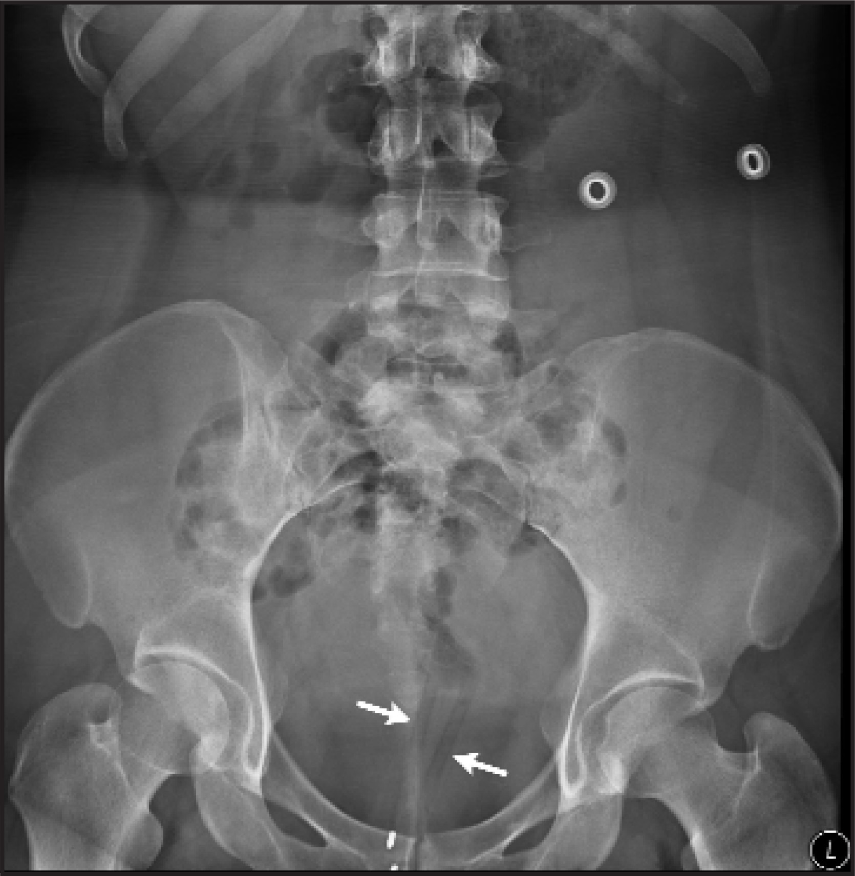
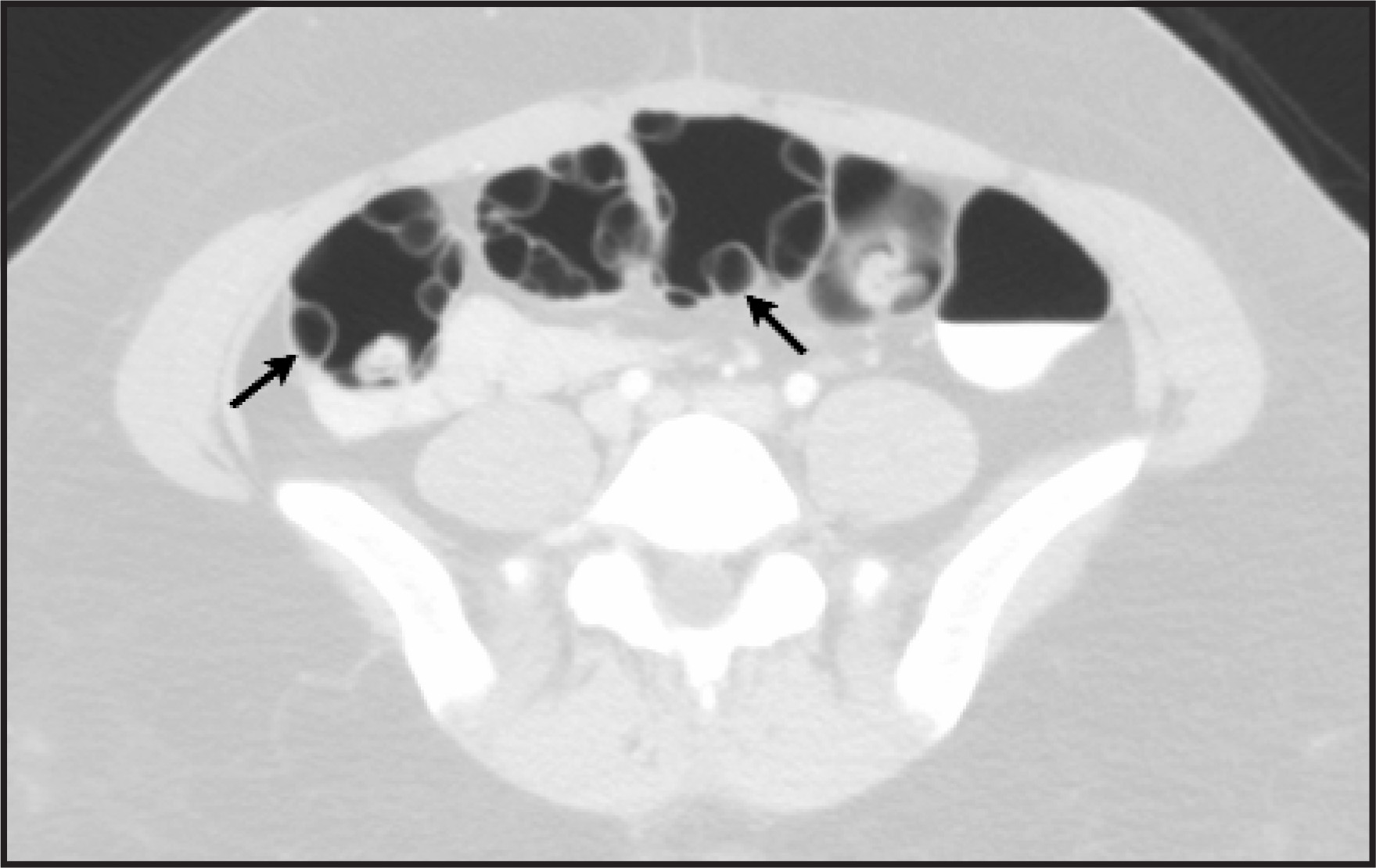
A mentally disabled 25-year-old woman with a history of repeated rectal injury secondary to the placement of foreign bodies (such as dresser screws, bottle caps, and metal springs per rectum) presented with foreign bodies (pens) in her rectum. An abdominal radiograph was performed (Figure 1) followed by a computed tomography (CT; Figure 2) to evaluate for rectal injury.
IMAGING FINDINGS
An abdominal radiograph was taken that revealed 2 linear foreign bodies (pens) in the rectosigmoid region (Figure 1). The pens were removed with the help of sigmoidoscopy. CT of the abdomen and pelvis was performed to evaluate for rectosigmoid injury. An axial CT image through the pelvis (in a lung window display) revealed the presence of multiple air-filled cystic areas arising from the inner wall of the sigmoid colon and protruding into the bowel lumen. These findings were suggestive of pneumatosis intestinalis (also known as pneumatosis cystoides) involving the rectosigmoid and distal sigmoid colon. (Figure 2).
DIAGNOSIS
Pneumatosis intestinalis of the sigmoid colon secondary to repetitive injury to the rectum from the insertion of foreign bodies
DISCUSSION
Pneumatosis (cystoides) intestinalis (PI) is defined as multiple gas-filled cysts in the gastrointestinal tract wall.1-3 The cysts may be located in the subserosa, submucosa, and, rarely, the muscularis layer.1,4 They may be single or multiple and vary in size from microscopic to several centimeters in diameter.4 They are usually lined by mixed inflammatory cells, macrophages, or foreign body giant cells1,3,4 with no communication between the air spaces and the bowel lumen.5,6 However, PI is a radiographic finding and not a diagnosis. PI is considered an ominous finding in ischemia, particularly if it is associated with portomesenteric venous gas.1,6 The majority of cases of PI occur in the jejunum and ileum, with 6% to 10% of cases involving the colon.6
Two forms of PI have been recognized: primary and secondary.4,5,7,8 Primary pneumatosis intestinalis (15% of cases) is a benign idiopathic condition, and patients are usually asymptomatic. These cysts are incidentally discovered on radiography or endoscopy. The secondary form (85% of cases) is associated with obstructive pulmonary disease, as well as obstructive and necrotic gastrointestinal diseases.6,8
Although the exact prevalence is unknown, PI is a rare condition. No sex or race predominance has been reported.6 The exact pathogenesis of PI is not known and many theories explaining the process have been put forth. The most prominent theories are mechanical, bacterial, and pulmonary mechanisms.7,8 More than 50 causative factors have been identified that result in PI.1,7 The breadth of pathologic conditions associated with PI formation suggests that its development is a multifaceted phenomenon.2 Common causes are summarized in Table 1.
Plain X-ray film findings of PI include air within the walls of the gastrointestinal tract. The patterns of the radiolucencies seen may be linear, curvilinear, small bubbles, or collections of larger cysts.5,7 Pneumoperitoneum or pneumoretroperitoneum can be seen secondary to cyst rupture.6-9
On barium enema, PI is visualized as a circumscribed attenuation pattern in the contrast column. When the cysts protrude into the lumen, they may mimic polyps or carcinomas on barium enema studies.2,3,7,8 Gas enters the bowel wall because of direct trauma. Enhanced gut permeability to gas can be induced by defects in the mucosa, the gut’s immune barrier (intramural lymphoid tissue), or both.2 The current case is interesting because it is the first published reported patient with PI after direct repetitive colon trauma.
On ultrasound, the appearance of PI includes circumferential, bright, echogenic foci in the bowel wall. Computed tomography (CT) with a wide lung parenchyma window is the best imaging modality for establishing the diagnosis of PI. It has greater sensitivity than plain film or ultrasound.6,7 CT can distinguish PI from intraluminal air or submucosal fat. A thickened bowel wall with contrast enhancement may suggest ischemia in the setting of PI. Dilated bowel loops and abnormal fluid levels suggest an obstructive cause.
When a foreign body causes PI, a careful history and physical examination should be followed by a biplanar radiograph of the abdomen to determine the exact position of the foreign body (or bodies) and to assess the presence of free air to exclude perforation.10 The most common reason for rectal foreign bodies is autoeroticism; other causes include criminal assault and medical diagnostic indications.10
CONCLUSION
Usually a benign condition, PI may be detected on various imaging modalities. It has a unique presentation when confined to the colon, with air cysts lining the inner wall. The presence of PI in the small bowel is considered an ominous finding in ischemia, particularly if it is associated with portomesenteric venous gas.
References
- Pear B. Pneumatosis Intestinalis: A review. Radiology. 1998;207:13-19.
- St. Peter S, Abbas M, Kelly K. The spectrum of pneumatosis Intestinalis. Arch Surg. 2003;138:68-75.
- Stein E. Anorectal and Colon Diseases. 2003:380-383.
- Jass J, Price A, Shepherd N. Morson and Dawson’s Gastrointestinal Pathology. 2003:402-403.
- Yamada T, Alpers D, Laine L. Textbook of Gastroenterology. 2003:1809-1811.
- Goyal S. Pneumatosis intestinalis. 2006.
- Feczko P, Mezwa D, Farah M, White B. Clinical significance of pneumatosis of the bowel wall. RadioGraphics. 1992;12:1069-1078.
- Margulis A, Burhenne H. Practical Alimentary Tract Radiology. 1993:44-45.
- Valdes-Dapena A, Stein G. Morphologic Pathology of the Alimentary Canal. 1970:679-680.
- Hicks T, Beck D, Opelka F, Timmeke A. Complications of Colon and Rectal Surgery. 1996:462-463.
Citation
. RCOM RADIOLOGICAL. Applied Radiology. 2007;36(8):11-11. doi:10.37549/AR1535.