RCOM RADIOLOGICAL
Applied Radiology — Vol. 36 , Issue 8 , pp. 11 -11
DOI: 10.37549/AR1537
Published: August 1, 2007
Categories
CASE SUMMARY
Several months after the cessation of chemotherapy for chronic lymphocytic leukemia (CLL), a 54-year-old man presented with enlarged axillary lymph nodes. Since the diagnosis of CLL 5 years earlier, the patient had experienced frequent night sweats but no fevers, chills, dyspnea, or cough. He had a 30 pack-year smoking history.
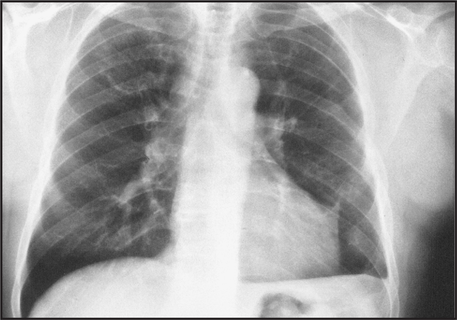
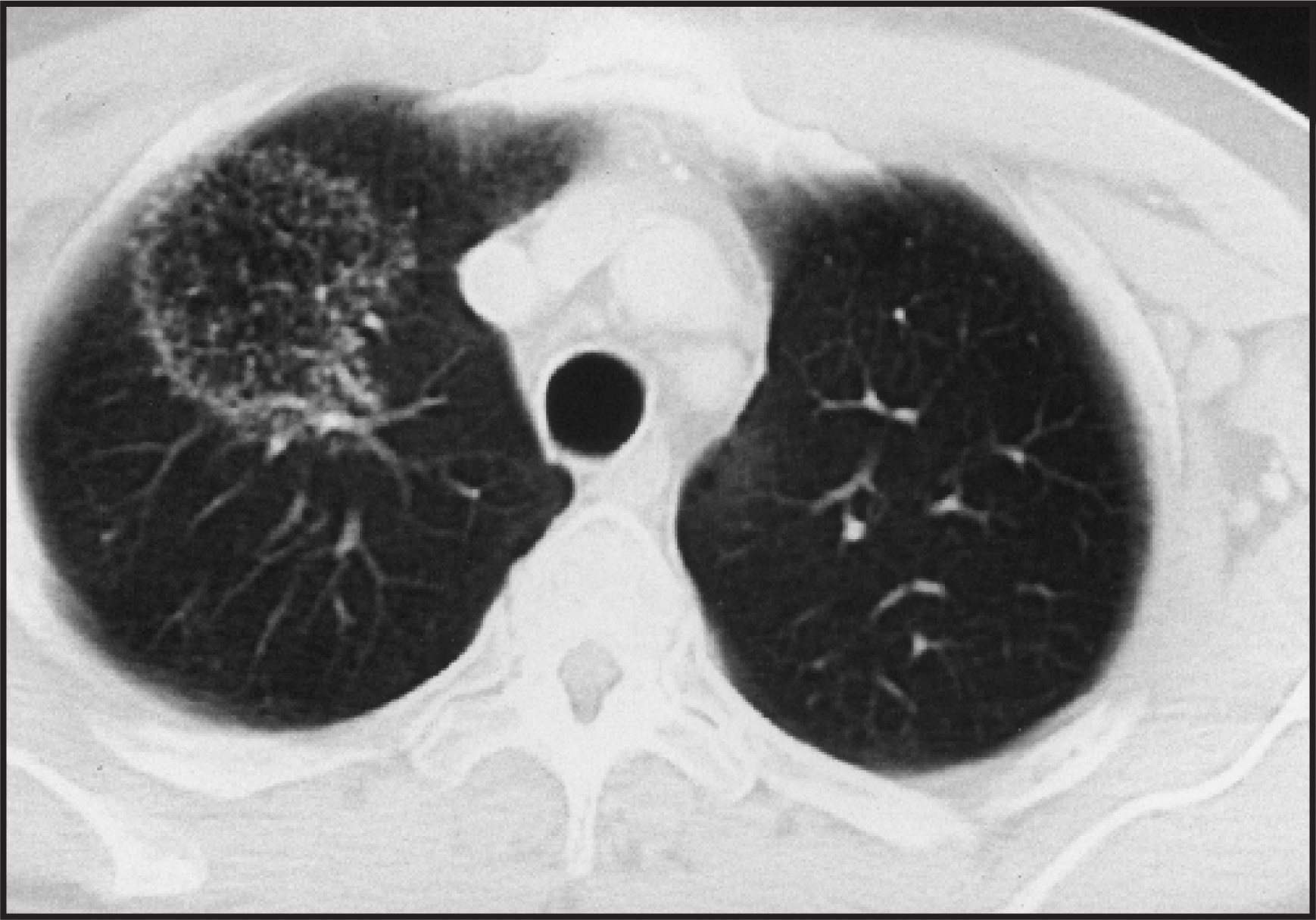
On physical examination, the patient’s vital signs were within normal limits. Bilateral, palpable lymph nodes were found in the neck, axilla, and inguinal areas. The breath sounds in the right upper lung were decreased. The laboratory results included an elevated white blood cell count of 15,000/mm3 (37.6% neutrophils, 52.9% lymphocytes, 2.8% basophils) and a high erythrocyte sedimentation rate of 33 mm/hr. Chest radiography (Figure 1) and chest computed tomography (CT) were performed (Figure 2).
IMAGING FINDINGS
A chest radiograph (Figure 1) showed pleural thickening in the region of the costophrenic sulcus, enlargement of the hilar and paratracheal lymph nodes, and a mass in the right upper lobe. A CT scan confirmed the mass and its solid peripheral margin and heterogeneous center (Figure 2). Bronchoscopy and fine-needle aspiration of the right-upper-lobe opacity revealed lymphoid cells.
Over the next 3 years, the patient underwent chemotherapy for CLL. A follow-up CT scan of the chest 3 years later showed the same right-upper-lobe lesion, which was unchanged in size and texture.
DIAGNOSIS
Focal organizing pneumonia with reversed halo sign
DISCUSSION
Organizing pneumonia occurs over a wide age range—most commonly in the fifth and sixth decades of life—without a gender preference. Patients have vague symptoms, usually a subacute presentation of fever, nonproductive cough, malaise, weight loss, and mild dyspnea over a time course of weeks to months.1 The physical examination often yields lung crackles in the affected areas, but the examination may be normal. On pulmonary function tests, a mild-to-moderate restrictive pattern, reduced carbon monoxide diffusion capacity, and mild hypoxemia are exhibited. Laboratory findings are consistent with an inflammatory process, including mild leukocytosis and elevations in C-reactive protein and erythrocyte sedimentation rate.
The histopathologic features of organizing pneumonia reveal intra-alveolar and intrabronchiolar polypoid granulation tissue, so-called Masson bodies, with fibroblasts, myofibroblasts, and loose connective tissue. Sometimes the granulation tissue contains inflammatory cells, eg, macrophages, plasma cells, and neutrophils with eosinophilia.1,2
Chest radiography commonly shows patchy, bilateral lower lobe areas of parenchymal opacity with a bias for the periphery of the lung. Air bronchograms can be present. On CT, several different manifestations of organizing pneumonia have been described. The most common appearance consists of bilateral, patchy, consolidative and ground-glass opacities, which are seen more often in the peripheral and lower lung regions. The dimensions range from a few centimeters to an entire lobe of the lung. Other presentations of organizing pneumonia include focal lesions, ringlike shadows, and nodular, bronchocentric, perilobular, and bandlike opacities.
Focal organizing pneumonia may be difficult to distinguish from malignancy. Diagnosis often requires biopsy or resection, which may yield a solitary mass with an irregular margin and spiculation. The mass tends to be flat or ovoid (rather than rounded) and may contain air bronchograms or may cavitate. It tends to be peripheral in location and may be in contact with the pleura.
A linear pattern of organizing pneumonia occurs in isolation or along with other patterns. It is seen as 2-cm or longer bands or arcs of opacity that measure at least 8 mm in width. Its margins may be smooth or irregular. Air bronchograms may be present. These bands may either extend toward the lung periphery in a radial fashion associated with bronchi or may appear peripheral without association with bronchi.
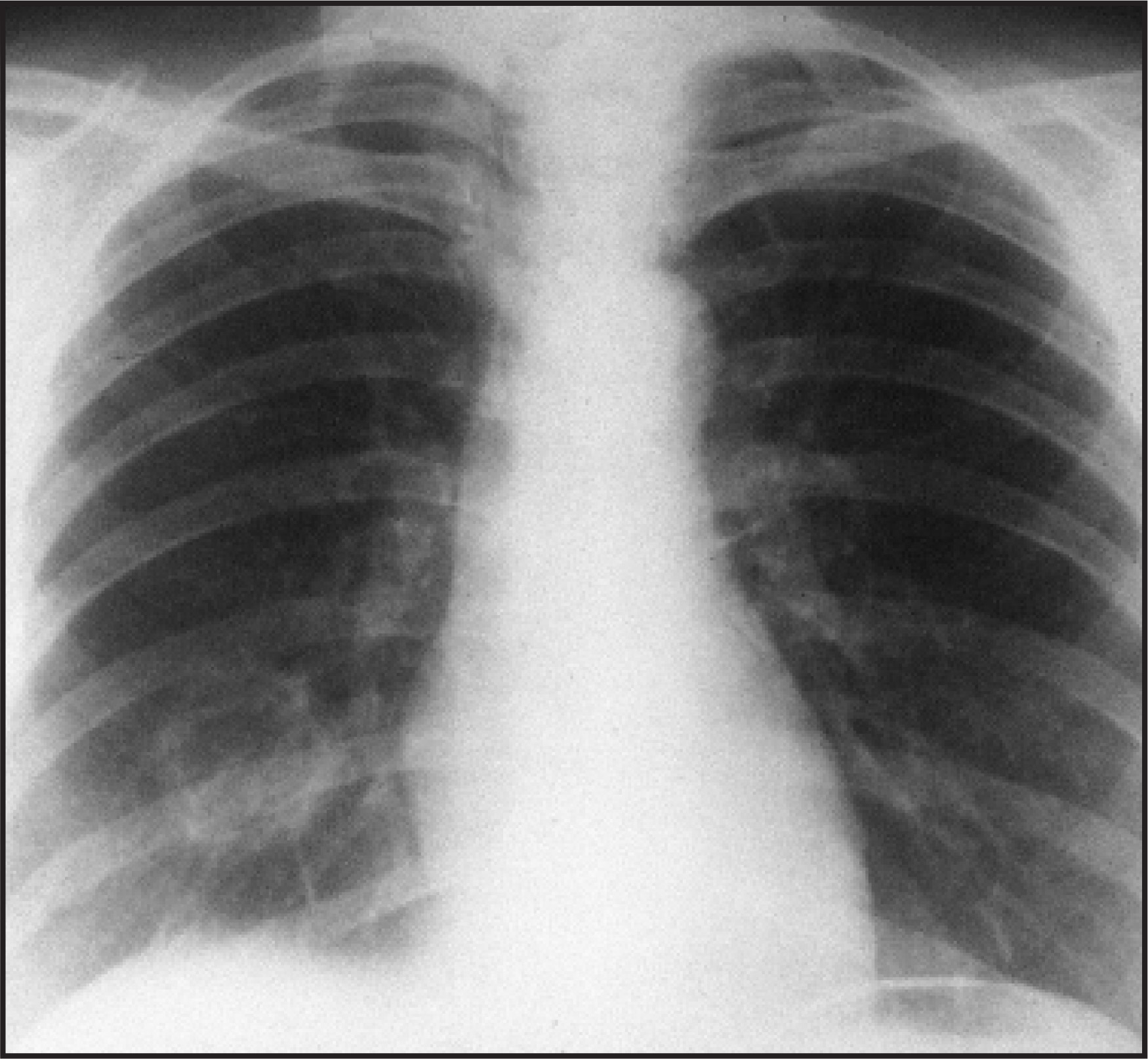
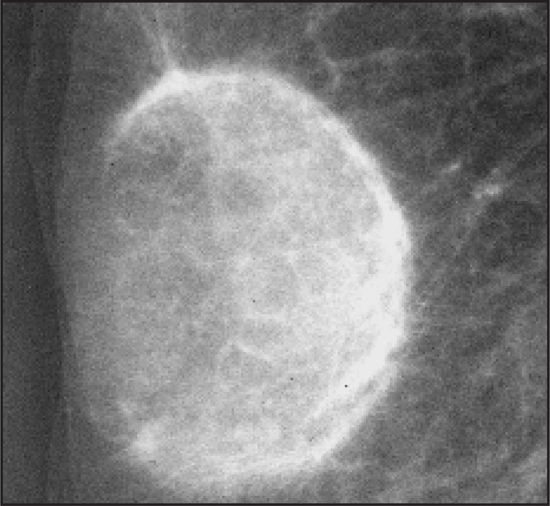
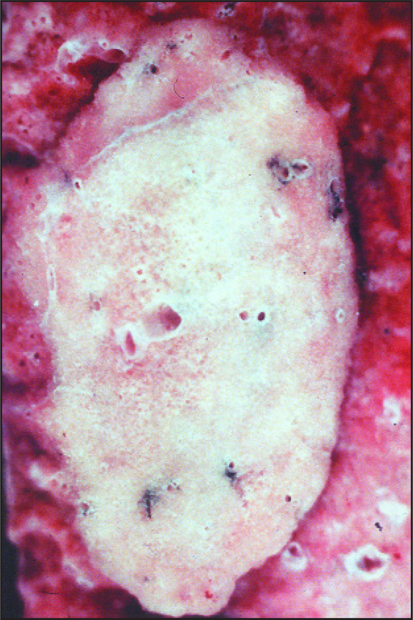
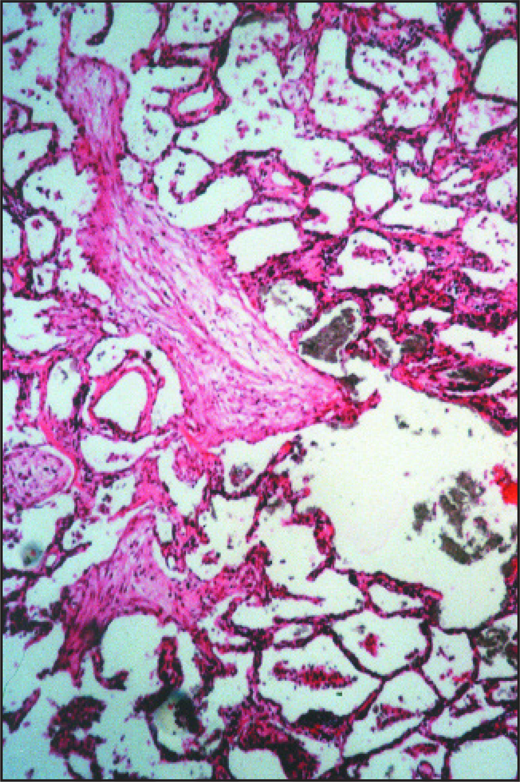
A recently described unusual appearance of organizing pneumonia consists of rings and crescents; initially 2 cases were described by Voloudaki et al.3 This appearance has also been dubbed the “reversed halo sign” (Figures 1 through 4). It consists of a central area of ground-glass opacity that is surrounded completely or partially by a ring or crescent of consolidation. Histologically, the central ground-glass area corresponds to alveolar septal inflammation and cellular debris, while the outside ring or crescent represents granulation tissue within alveoli and bronchioles (Figures 5 and 6).
Historical review of CT scans from patients with biopsy-proven organizing pneumonia by Kim et al4 revealed a ring or crescent pattern in 6 of 31 patients.4 A small comparison group of patients with biopsy-proven Wegener’s granulomatosis, bronchioloalveolar carcinoma, eosinophilic pneumonia, or Churg-Strauss syndrome failed to show any similar CT findings of a ring or crescent pattern. This suggests that the reversed halo, ring, or crescent patterns are more specific for organizing pneumonia than are the most commonly seen patchy ground-glass and consolidative patterns.
CONCLUSION
The reverse halo sign is an uncommon but relatively specific radiologic sign of cryptogenic organizing pneumonia. Radiologists should become familiar with this relatively new addition to the spectrum of imaging findings in organizing pneumonia.
References
- Cordier J. Organising pneumonia. Thorax. 2000;55:318-328.
- Oikonomou A, Hansell D. Organizing pneumonia: The many morphological faces. Eur Radiol. 2002;12:1486-1496.
- Voloudaki A, Bouros D, Froudarakis M. Crescentic and ring-shaped opacities. CT features in two cases of bronchiolitis obliterans organizing pneumonia (BOOP). Acta Radiol. 1996;37:889-892.
- Kim S, Lee K, Ryu Y. Reversed halo sign on high-resolution CT of cryptogenic organizing pneumonia: Diagnostic implications. AJR Am J Roentgenol. 2003;180:1251-1254.
Citation
. RCOM RADIOLOGICAL. Applied Radiology. 2007;36(8):11-11. doi:10.37549/AR1537.