RCOM RADIOLOGICAL CASE OF THE MONTH
Applied Radiology — Vol. 30 , Issue 6 , pp. 43 -45
DOI: 10.37549/AR1011
Published: June 1, 2001
Categories
CASE SUMMARY
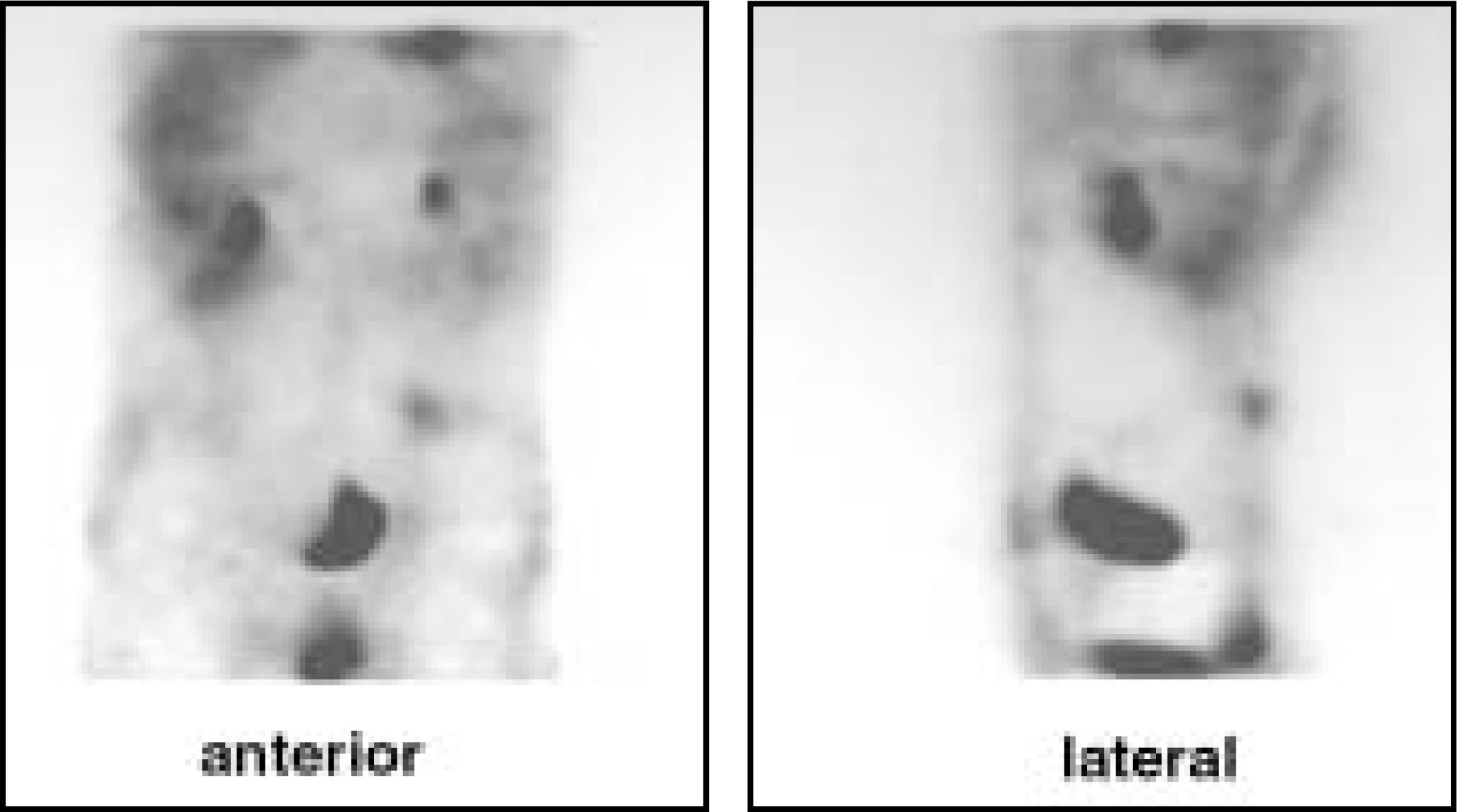
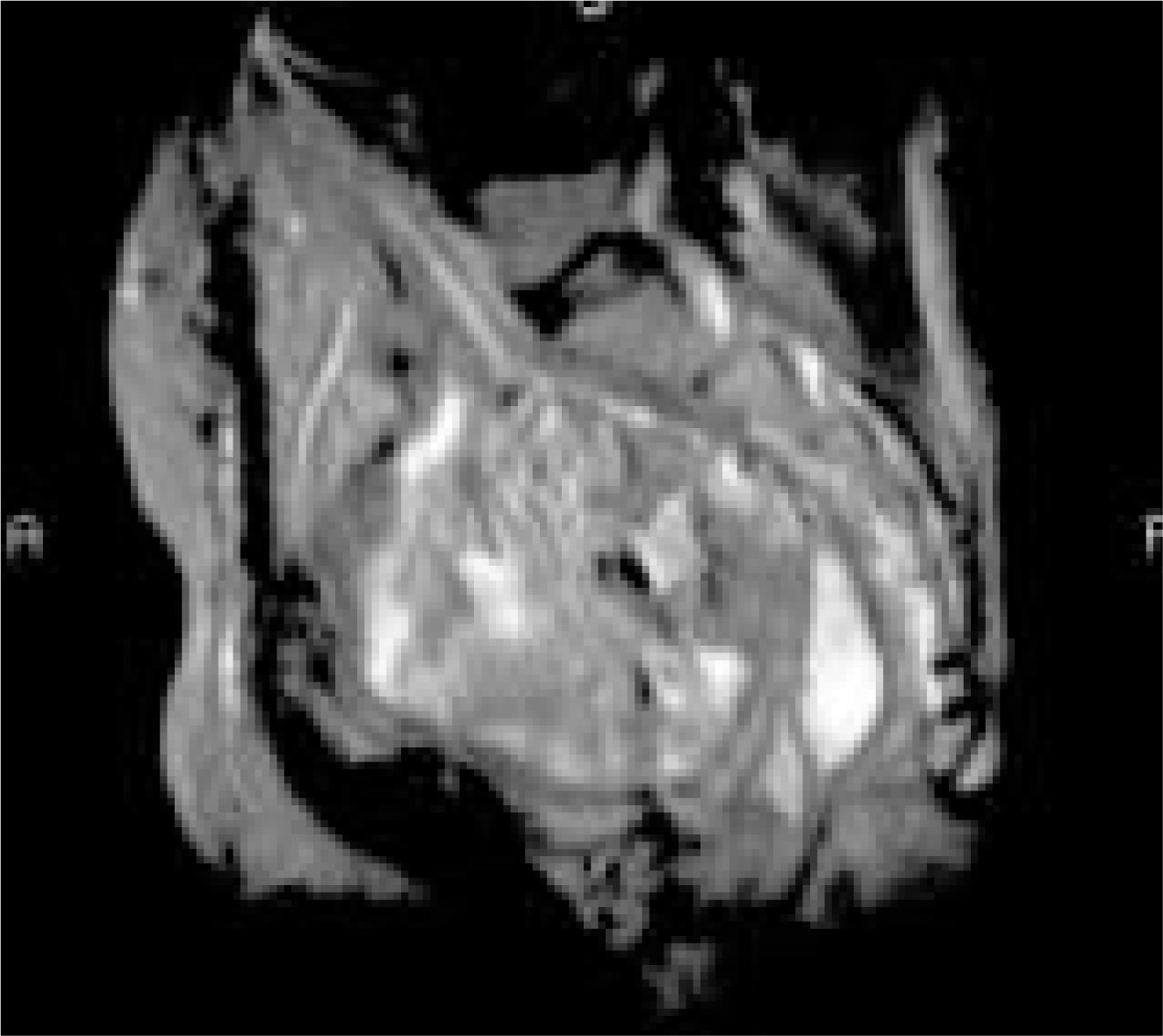
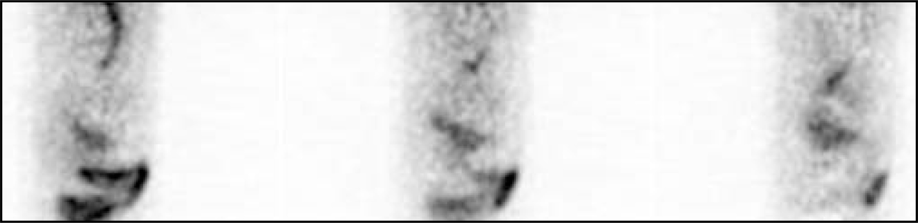
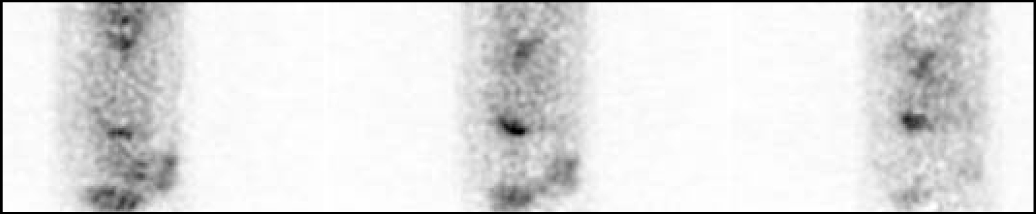
The patient is a 64-year-old Hispanic man who underwent resection of mucinous, moderately well-differentiated adenocarcinoma of the rectosigmoid (Stage T3N1M0; Duke’s C) in July 1996. At the time of surgery, his serum carcinoembryonic antigen (CEA) level was 3.5 ng/mL. In December 1997, he had a second surgery for recurrence, after which he completed a course of remote after-loading high-dose-rate irradiation via intraoperative interstitial implant. During a left herniorrhaphy procedure in August 1998, a deep-needle biopsy of the presacral area was performed, revealing mucinous, metastatic colonic adenocarcinoma. Although serum CEA assay results were within normal limits (2.0 ng/mL); the tumor tissues were CEA-membrane positive. In the months immediately preceding and following the biopsy, an F-18 fluorodeoxyglucose (FDG) positron emission tomography (PET) scan, magnetic resonance imaging (MRI) study, and a nuclear imaging study with arcitumomab were performed (figures 1 through 3).
DIAGNOSIS
Recurrence of colorectal cancer, mucinous type
IMAGING FINDINGS
In this case, the MRI and PET study were unrevealing, whereas the arcitumomab study (CEA-Scan, Immunomedics, Inc., Morris Plains, NJ) confirmed the extent of the lesion.
DISCUSSION
Approximately 600,000 people in the United States are under postsurgical care for colorectal cancer. These patients need regular follow-up visits to detect recurrence and complications arising from treatment. Surveillance guidelines for the postsurgical colorectal cancer patient, such as the National Comprehensive Cancer Network (NCCN)1 and others, include serum CEA and imaging (usually CT) at the core of the recommended follow-up protocol. Unfortunately, many patients present with no symptoms or unclear signs of recurrence, and the usual work-up is not sufficient to detect new disease serum using CEA, CT, or MR alone.
After primary surgery, many patients develop subtle thickening, which is difficult to biopsy, or stable masses, which on biopsy are found to contain tumor. Furthermore, there may also be recurrence with no radiological signs; the serum CEA levels may remain stable or within normal limits even though histologically, the tumor has CEA expressed on the cell membrane.2 There are also patients who do not show any overt clinical signs of recurrence and are more difficult to evaluate.
At our institution, we prefer to use MRI to evaluate rectal lesions. In our experience, MRI can also often differentiate mucinous and nonmucinous rectal tumors,3 because mucinous tumors show high signal intensity on T2-weighted fast spin-echo images. Additionally, recent studies have shown that MR is superior to CT in the evaluation of the presacral space in postsurgical patients.4
In our experience, two nuclear medicine procedures, F-18 FDG PET and Tc-99m-labeled arcitumomab, when combined with the results of CT or MR, enhance the early detection of recurrence of colorectal cancer. Through the correlation of functional and morphological information, one can enhance the staging or detection of recurrence in the patient with colorectal cancer. Studies have shown that arcitumomab in conjunction with CT or MR is a reliable means of detecting metastases or recurrence of colorectal cancer,5 regardless of serum CEA levels.6 F-18 FDG PET also has a role in the work-up to detect colorectal cancer recurrence in the patient with rising serum CEA.7
Nevertheless, false-positive F-18 FDG studies in the evaluation of the oncology patient is a well-known phenomenon.8 The reasons for false-negative studies are not as well documented, but recently it has been shown that as may as 41% of mucinous tumors do not have avid uptake of F-18 FDG and, as such, may not show with PET imaging.9 This case study may be such an example.
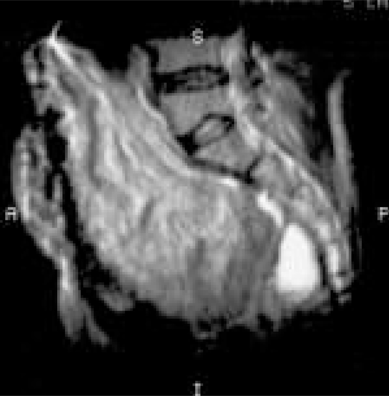
Due to the extent of the disease, external beam radiation was considered over surgery for this patient. In January 1999, he completed 25 sessions of external beam radiation therapy over the course of 44 days (total radiation 50 Gray). The MRI study revealed no progression of disease (figure 4), serum CEA levels in March 1999 were 3.9 ng/mL (normal <5.0 ng/mL) and an arcitumomab study in April 1999 confirmed response of the tumor to the radiation (figure 5).
CONCLUSION
Through June 2000, several MRI studies, a PET scan, a methylene diphosphonate (MDP) bone scan, and arcitumomab studies continue to show no evidence of disease. This patient continues under our care. Further follow-up using serum CEA, MRI, arcitumomab studies, and PET are being employed as needed. To date this patient has had 4 arcitumomab studies with no reactions to the arcitumomab murine antibody fragment. Recent studies indicate that multiple arcitumomab injections are safe. Additionally, serial arcitumomab nuclear imaging has the potential to detect recurrence sooner and improve tumor-free survival.10,11
We believe that if nuclear medicine imaging is being considered in the management of a colorectal cancer patient, it is advantageous to know the histology of the tumor before selecting the nuclear medicine procedure. In cases in which the patient does not have rising serum CEA or the tumor is mucinous type, then a nuclear imaging study with arcitumomab should be the study of choice.
References
- NCCN Colorectal cancer practice guidelines. Oncology. 1996;10(11; suppl):140-175.
- Guadagni F, Roselli M, Cosimelli M. Quantitative analysis of CEA expression in colorectal adenocarcinoma and serum: lack of correlation. Int J Cancer. 1997;72:949-954.
- Hussain S, Outwater E, Siegelman E. Mucinous versus nonmucinous rectal carcinomas: Differentiation with MR imaging. Radiology. 1999;213:79-85.
- Pema P, Bennett W, Bova J, Warman P. CT vs. MRI in diagnosis of recurrent rectosigmoid carcinoma. J Comput Assist Tomogr. 1994;18(2):256-261.
- Hughes K, Pinsky C, Petrelli N. Use of carcinoembryonic antigen radioimmunodetection and computed tomography for predicting the resectability of recurrent colorectal cancer. Ann Surg. 1997;226:621-631.
- Erb D, Nabi H. Clinical and technical considerations for imaging colorectal cancers with technetium-99m-labeled AntiCea fab' fragment. J Nuc Med Tech. 2000;28(1):12-18.
- Strauss L, Clorius J, Sclag P. Recurrence of colorectal tumors: PET evaluation. Radiology. 1999;170:329-332.
- Strauss L. Fluorine-18 deoxyglucose and false-positive results: a major problem in the diagnostics of oncological patients. Eur J Nucl Med. 1996;23:1409-1415.
- Whiteford M, Whiteford H, Yee L. Usefulness of FDG-PET scan in the assessment of suspected metastatic or recurrent adenocarcinoma of the colon and rectum. Dis Colon Rectum. 2000;43:759-767.
- Wegener W, Petrelli N, Serafini A, Goldenberg D. Safety and efficacy of arcitumomab imaging in colorectal cancer after repeated administration. J Nucl Med. 2000;41:1016-1020.
- Lechner P, Lind P, Goldenberg D. Can postoperative surveillance with serial CEA immunoscintigraphy detect resectable rectal cancer recurrence and potentially improve tumor-free survival?. J Am Coll Surg. 2000;191:511-518.
Citation
. RCOM RADIOLOGICAL CASE OF THE MONTH. Applied Radiology. 2001;30(6):43-45. doi:10.37549/AR1011.