RCOM RADIOLOGICAL CASE OF THE MONTH
Applied Radiology — Vol. 30 , Issue 6 , pp. 40 -42
DOI: 10.37549/AR1010
Published: June 1, 2001
Categories
CASE SUMMARY
A 37-year-old woman with non-insulin-dependent diabetes mellitus (NIDDM) presented to the hospital with severe abdominal pain and vomiting. Over the previous year, she had developed numerous intermittent episodes of diarrhea, nausea, and vomiting. Physical examination revealed a soft abdomen with a palpable mass in the right lower quadrant. Laboratory test results were significant for hemoglobin of 9.7 g/dL and blood glucose of 189 g/dL. Her medical history was significant for Wilms’ tumor at 3 months old. The tumor was treated by left nephrectomy with adrenalectomy, adjuvant vincristine chemotherapy, and external beam radiation.
DIAGNOSIS
Leiomyosarcoma in a patient with Wilms’ tumor; a secondary primary tumor
IMAGING FINDINGS
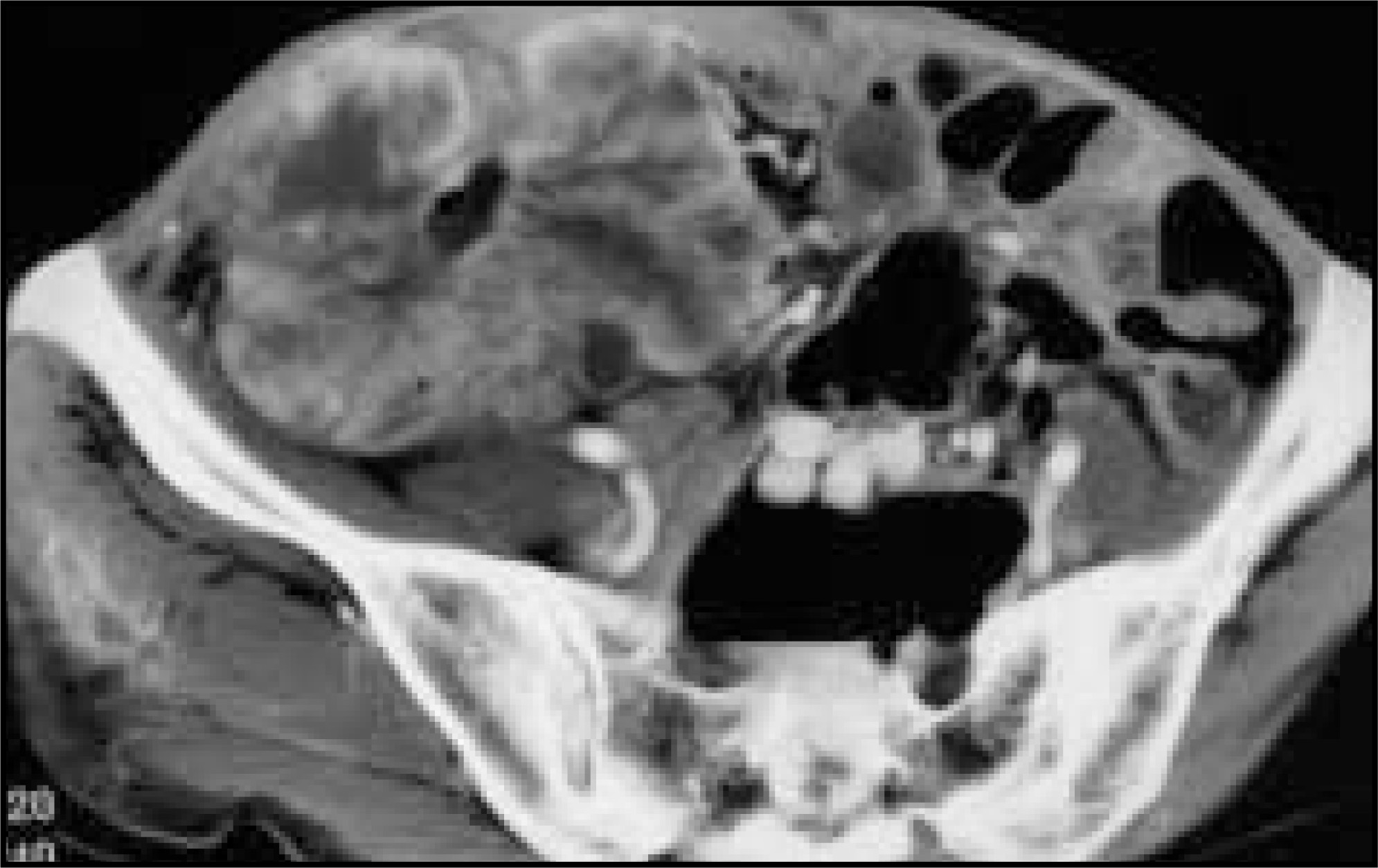
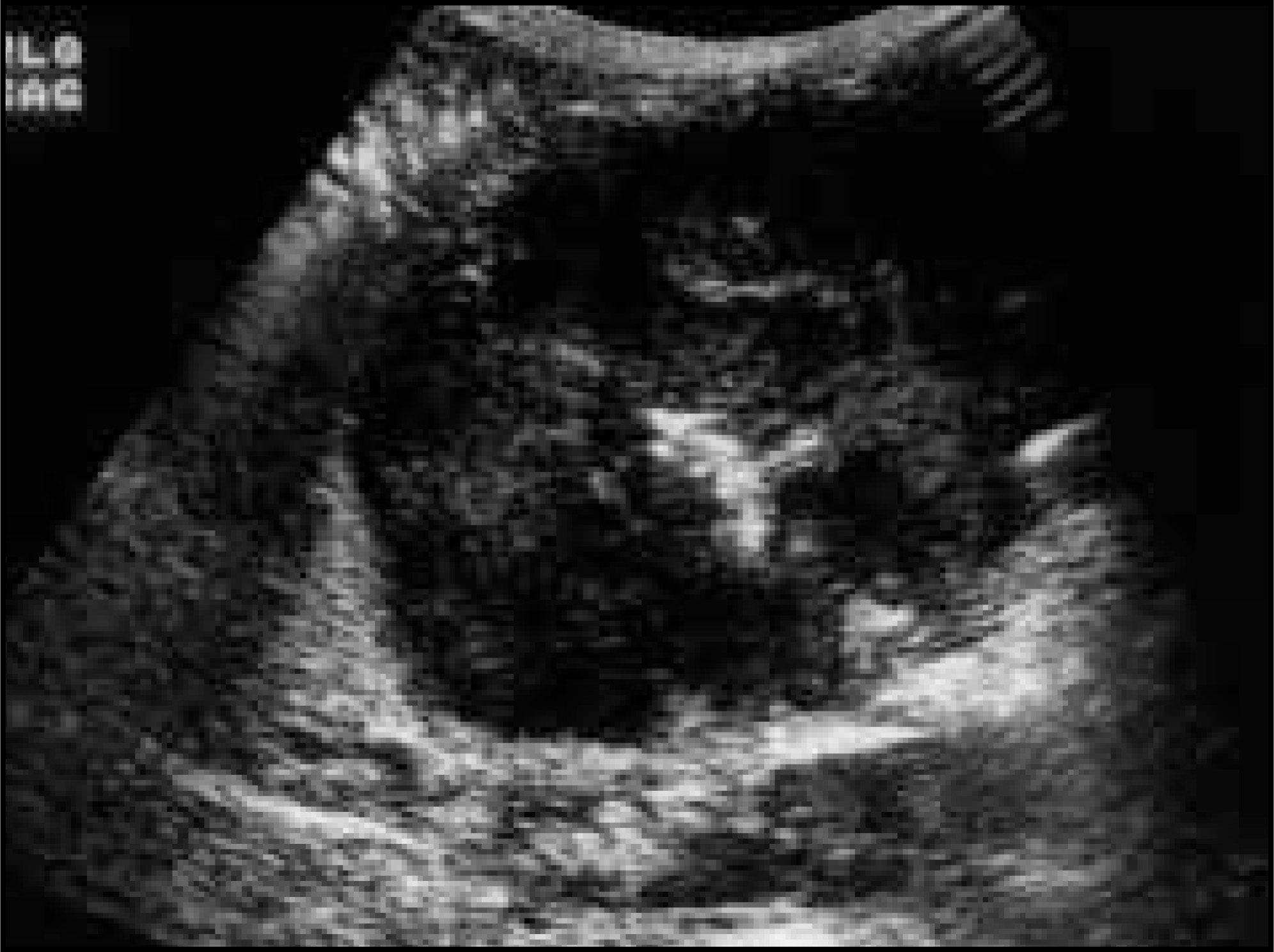
Computed tomography of the abdomen and pelvis was performed with oral and intravenous contrast. The study showed a 10-cm mass in the right lower quadrant involving the small bowel (figure 1). Double-contrast barium evaluation demonstrated medial displacement of the hepatic flexure. Ultrasound evaluation confirmed a 9.7 × 8.9 × 6.3-cm complex, predominantly solid mass in the right pelvis (figure 2). Ultrasound-guided needle biopsy was performed.
DISCUSSION
The incidence of second primary tumors (SPT) among childhood cancer survivors has been reported as six-fold greater than the general population.1,2 The risk following radiotherapy alone is reported as four-fold greater than the general population; radiotherapy in combination with cyclophosphamide increases the risk nine-fold. Second primary tumors have been reported in patients treated for central nervous system tumors (particularly retinoblastoma), Hodgkin's disease, and Wilms’ tumor, as in this patient.
Wilms’ tumor is the most common intra-abdominal malignancy in childhood.3 Advances in treatment strategies over the past 30 years have increased 5-year survival from 20% to 95% in those with the more favorable prognosis.4 Radiation therapy, once commonly utilized, is now utilized only in patients with metastases, lymph node involvement, or unfavorable histology.
The national Wilms’ tumor study group reviewed the incidence of SPT following treatment of Wilms’ tumor.2 The risk of development of SPT after treatment of Wilms’ tumor is 8.5 times higher than the cancer incidence for the general population.2 The present case is one of a leiomyosarcoma. Abdominal irradiation, as therapy for Wilms’ tumor, is associated with 1.43 cases of new cancer per 10 Gy of radiation dose administered during the therapy for Wilms’ tumor. The relative risk for thyroid, bone, connective tissue, gastrointestinal (GI), and central nervous system tumors and leukemia increase in patients treated for Wilms’ tumor.2
The dose-limiting structure for radiation therapy to the abdomen and pelvis is usually the small bowel.5 The clinical signs and symptoms of acute small bowel toxicity secondary to radiation therapy include nausea, vomiting, diarrhea, and malabsorption. Late complications include stricture formation, fistula, bowel obstruction, and secondary malignancies. Radiological findings include bowel wall thickening, stricture, and dilatation related to obstruction.
In the general population, small bowel neoplasms account for 3% to 6% of all GI tumors.6,7 Malignant small bowel tumors are more common than benign small bowel tumors. Endocrine cell tumors (carcinoids) and lymphomas are the most common cancers of the small bowel. Adenocarcinoma and leiomyosarcoma are the third and fourth most common malignancies of small bowel.
Leiomyosarcoma represents 20% of all small intestine malignancies.7,8 Patients with leiomyosarcoma usually present in the fifth and six decades of life with a male to female ratio of 3:1. Leiomyosarcoma occurs most commonly in the jejunum and ileum and is the most common neoplasm of Meckel’s diverticula. The presenting symptoms for leiomyosarcoma of the large or small bowel vary by site of origin. Patients with involvement of the duodenum and ileum present most commonly with blood loss.4 Patients with involvement of the colon present most commonly with pain. Gastrointestinal leiomyosarcoma is usually radioresistant; therefore, surgery is the primary treatment. Complete surgical resection, including en bloc resection of adjacent structures, improves overall survival. Leiomyosarcoma spreads through local extension to adjacent organs, peritoneal surfaces, and omental seeding. Hematogenous spread to the liver, lungs, adrenal glands, spleen, brain, and bone has been reported.
The radiological findings of GI leiomyosarcoma have been described previously.7,9 Plain radiographs may demonstrate a large soft-tissue mass that contains air, representing communication of the lumen of the tumor with the lumen of bowel. Deformity/displacement of small bowel segments, ulcerations, and tracts and cavities that may fill with contrast may be observed on barium studies. Computed tomography may demonstrate a bulky, eccentrically growing, enhancing, soft-tissue mass. Central areas of necrosis may also be detected.
References
- Hawkins M, Draper G, Kingston J. Incidence of second primary tumours among childhood cancer survivors. Br J Cancer. 1987;56:339-347.
- Breslow N, Takashima J, Whitton J. Second malignant neoplasms following treatment for Wilms’ tumor: A report from the National Wilms’ Tumor Study Group. J Clin Oncol. 1995;13:1851-1859.
- Webber B, Parham D, Drake L, Wilimas J. Renal tumors in childhood. Pathol Annu. 1992;27(Pt 1):191-232.
- Johnson M, Gibbs D, Gouldman J. Leiomyosarcoma of the colon: A second malignant neoplasm after treatment for a Wilms’ tumor. Am Surg. 1999;65(1):6-10.
- Hong A, Stevens G, Stephen M. Protection of the small bowel during abdominal radiation therapy with a tissue expander prosthesis. Aust N Z J Surg. 2000;70:690-692.
- Miettinen M, Sarlomo-Rikala M, Lasota J. Gastrointestinal stromal tumors: Recent advances in understanding of their biology. Hum Pathol. 1999;30:1213-1220.
- Ashley S, Wells S. Tumors of the small intestine. Semin Oncol. 1988;15:116-128.
- Akwari O, Dozois R, Weiland L, Beahrs O. Leiomyosarcoma of the small and large bowel. Cancer. 1978;42:1375-1384.
- Maglinte D, Gore R, Levine M, Laufer I. Textbook of Gastrointestinal Radiology. 1994.
Citation
. RCOM RADIOLOGICAL CASE OF THE MONTH. Applied Radiology. 2001;30(6):40-42. doi:10.37549/AR1010.