RCOM RADIOLOGICAL CASE OF THE MONTH
Applied Radiology — Vol. 30 , Issue 7 , pp. 40 -41
DOI: 10.37549/AR1020
Published: July 1, 2001
Categories
CASE SUMMARY
A 28-year-old man presented with brisk gastrointestinal (GI) bleeding requiring transfusion.
DIAGNOSIS
Leiomyoma (GI stromal tumor) of the jejunum
IMAGING FINDINGS
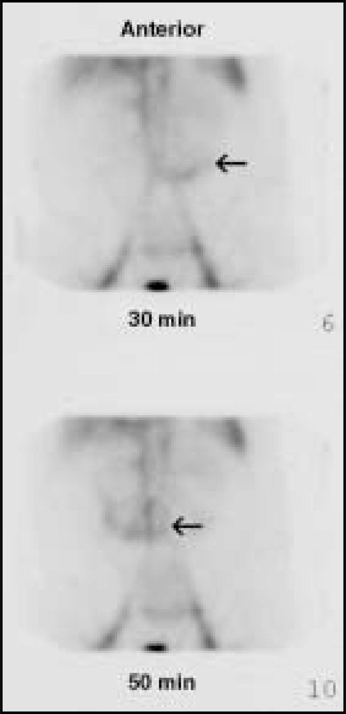
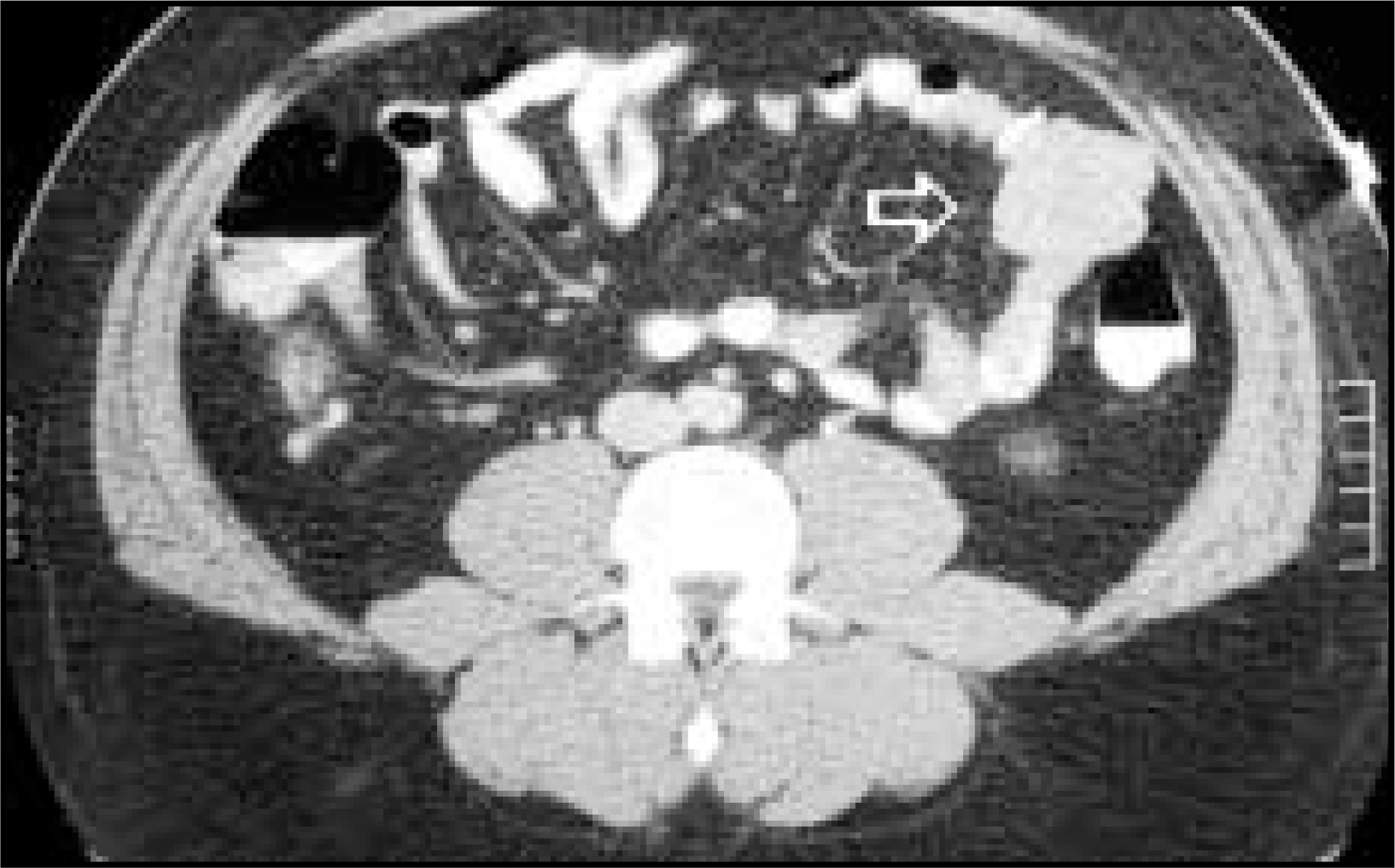
The diagnosis of small-bowel stromal neoplasms can be made by small-bowel examination (figure 1), but as they are typically extraluminal, only a small portion of the lesion may be recognized. Angiography can demonstrate the hypervascularity of stromal tumors, as well as active bleeding should it be brisk, but is invasive. A scintigraphic bleeding scan (figure 2) is only of benefit in lesions with active bleeding and is nonspecific as to the source. It has been shown that CT (figure 3) detects more than 90% of smooth muscle tumors of the small bowel and is an excellent imaging modality with which to identify these lesions preoperatively.1,4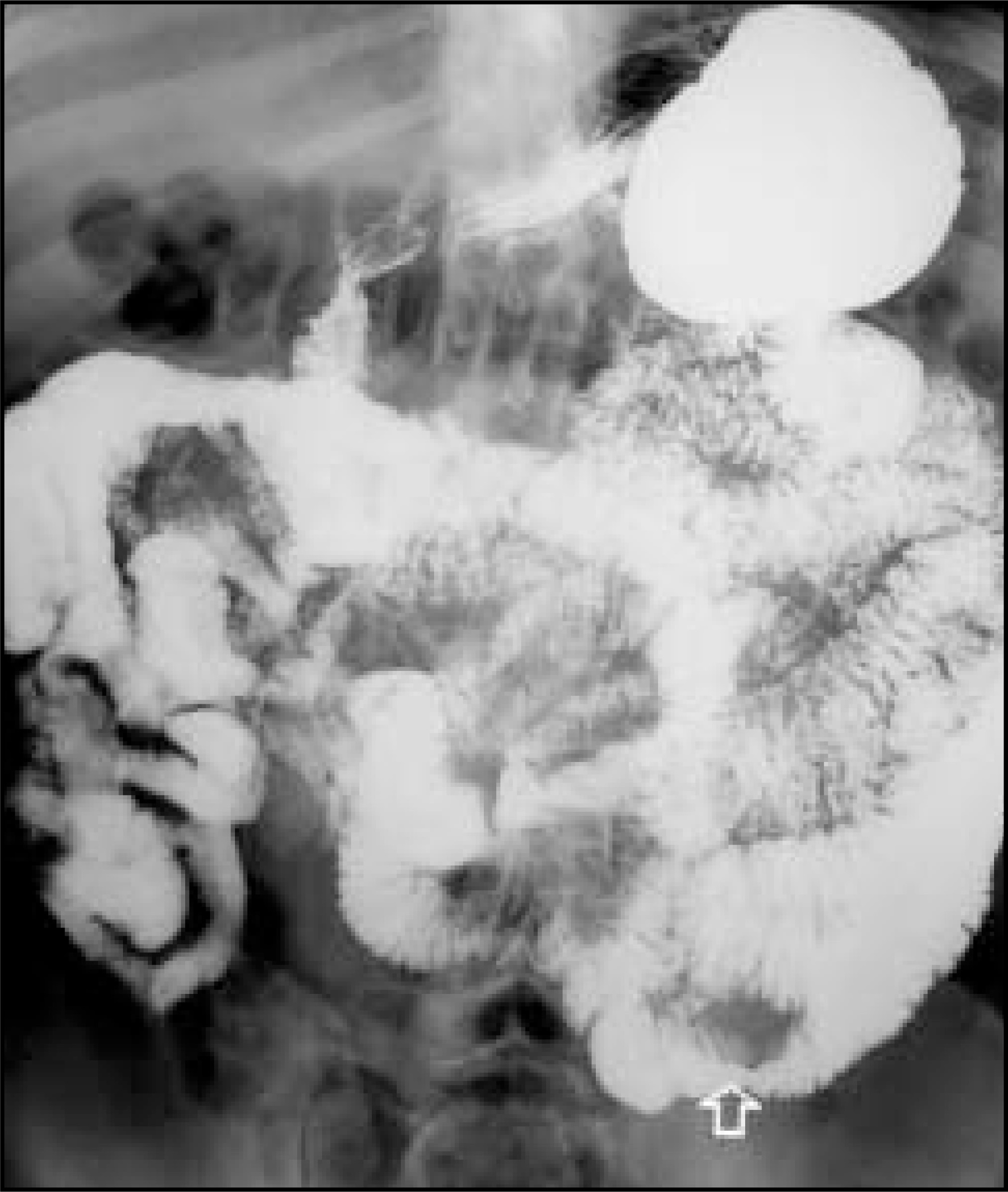
DISCUSSION
Leiomyoma is the most common benign tumor of small bowel. Approximately 50% of cases are found in the jejunum. Almost one-half of all lesions are <5 cm. It is said that for every 14 stomach tumors and 46 colonic tumors, there is a lesion of the small intestine, despite the fact that the small bowel represents 75% of the total length of the gastrointestinal tract. Of all smooth muscle tumors of the small bowel, only 50% are symptomatic.1,2
As most benign and malignant stromal tumors of the small bowel are exophytic, they can become quite large before they are palpated or cause obstruction. Bleeding is the most common presenting sign. Patients younger than 50 years old who present with GI bleeding commonly bleed from an identifiable lesion, such as leiomyoma, Meckel’s diverticulum, or other small-bowel tumors; whereas older patients bleed from more difficult to identify sources such as angiodysplasia, which may be multiple.3
Stromal tumors may be subserosal, intraluminal, or submucosal. They are hypervascular and can either ulcerate superficially (figure 4) or demonstrate central necrosis with possible communication to the bowel lumen by way of a fistula. Leiomyoma is 2- to 3-times more common than leiomyosarcoma. Leiomyosarcoma is typically larger than the benign leiomyoma and more frequently demonstrates aggressive behavior, such as central necrosis. Still, imaging cannot reliably differentiate between the two, which is even difficult histologically.1,2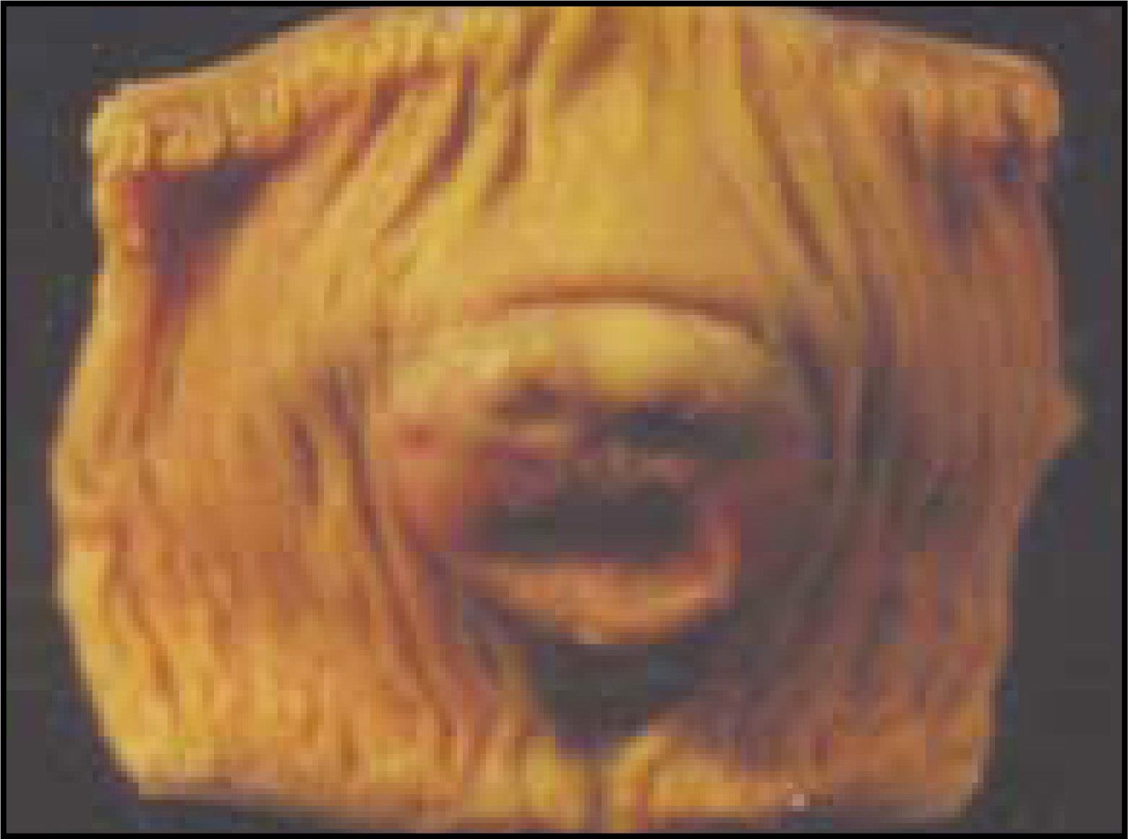
References
- Blanchard D, Budde J. Tumors of the small bowel. World J Surg. 2000;24:421-429.
- Buckley J, Fishman E. CT Evaluation of small bowel neoplasms: Spectrum of disease. RadioGraphics. 1988;18:379-392.
- Katz L. Role of surgery in gastrointestinal bleeding. Semin Gastrointest Dis. 1999;10(2):78-81.
- Yoshikawa K, Yamaguti T. The role of dual-phase enhanced helical computed tomography in difficult intestinal bleeding. J Clin Gastroenterol. 2000;31:83-84.
Citation
. RCOM RADIOLOGICAL CASE OF THE MONTH. Applied Radiology. 2001;30(7):40-41. doi:10.37549/AR1020.