RCOM RADIOLOGICAL CASE OF THE MONTH
Applied Radiology — Vol. 30 , Issue 11 , pp. 52 -53
DOI: 10.37549/AR1049
Published: November 1, 2001
Categories
CASE SUMMARY
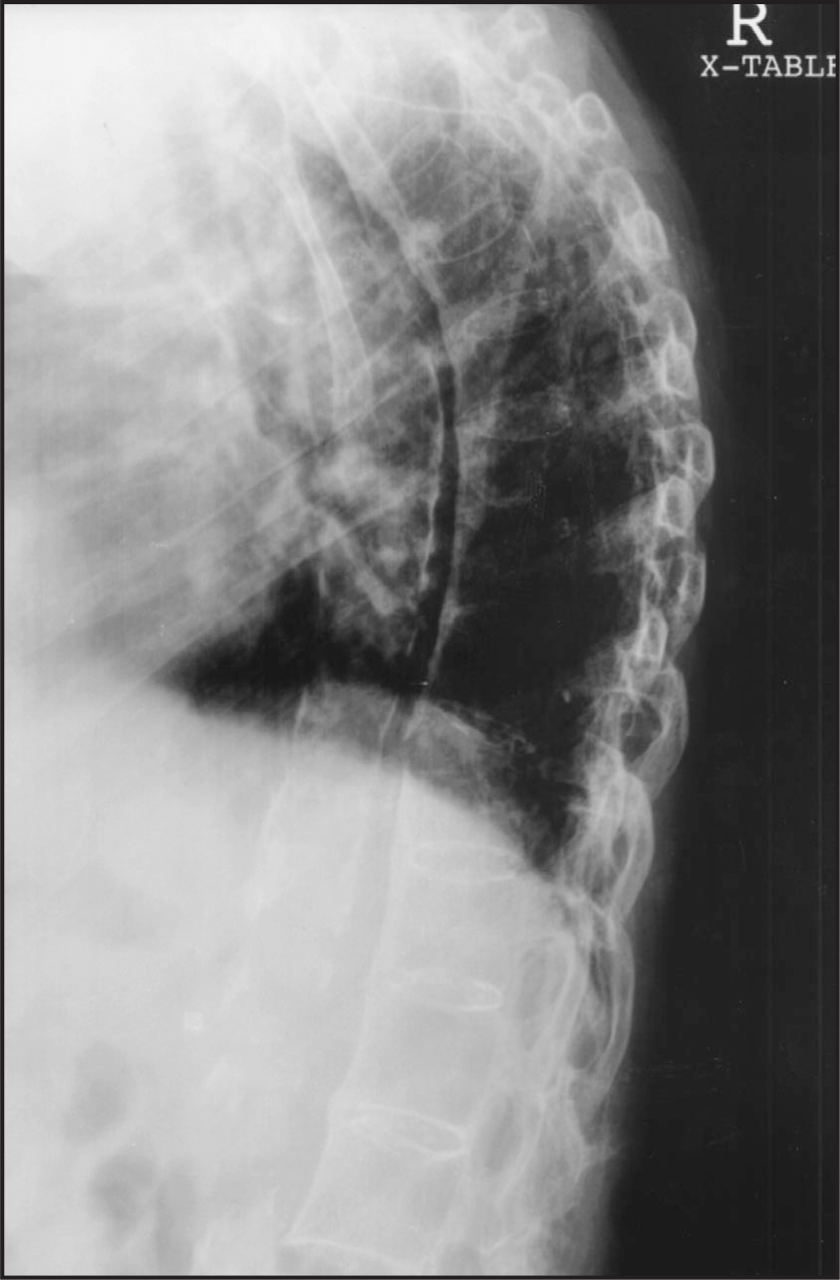
A 74-year-old female inpatient was referred for magnetic resonance imaging (MRI) of the thoracolumbar spine after complaints of chronic, unrelenting back pain (figure 1). The exact pain history was sketchy, as the patient demonstrated some signs of dementia. The patient’s daughter reported no known trauma, and there was no history of previous back surgery. Two days after the MRI was obtained, the neurological surgery service requested a myelogram of the thoracic spine (figure 2) for further elucidation of the lesion that was demonstrated on MR imaging.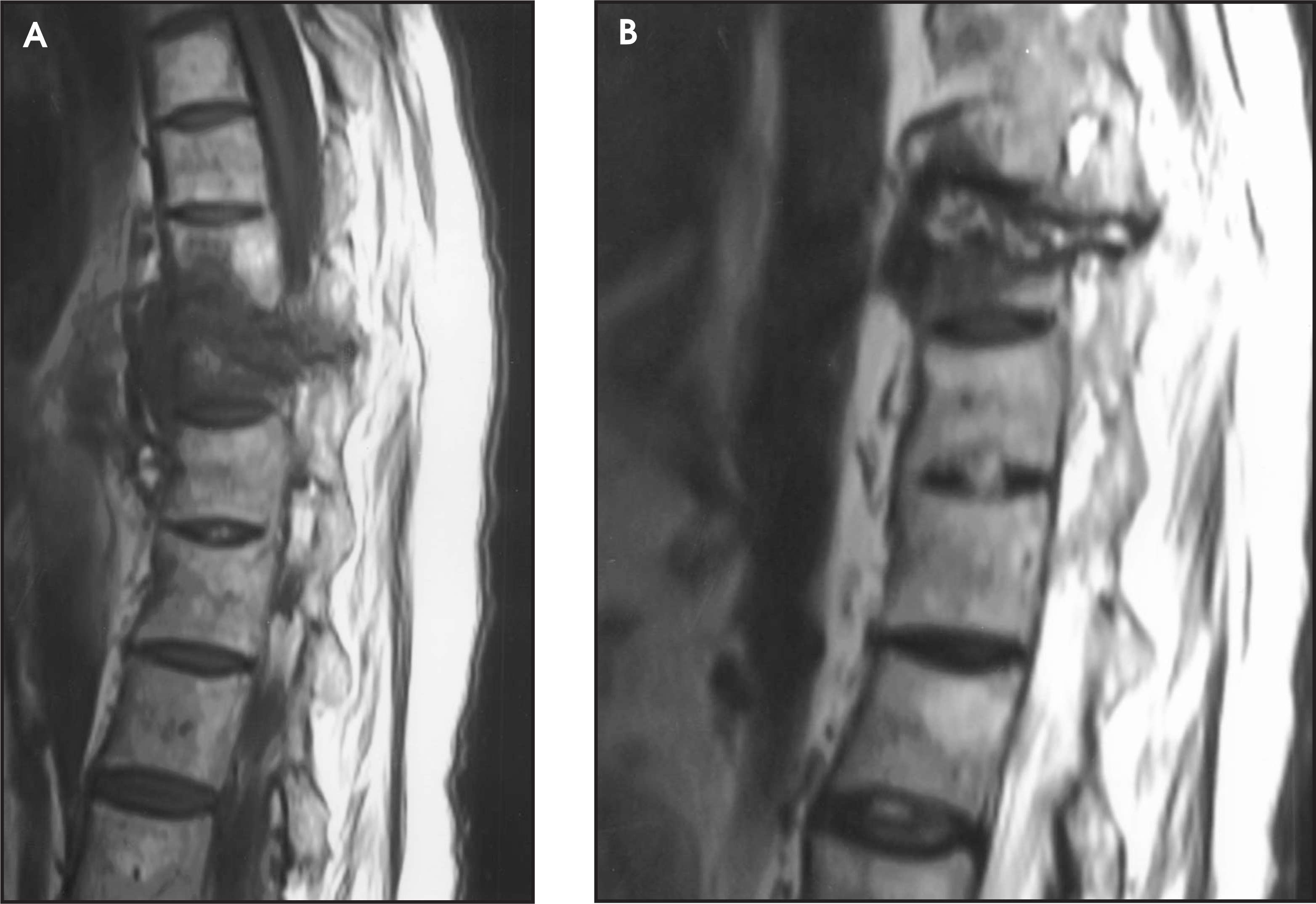
DIAGNOSIS
Ankylosing spondylitis with pseudarthrosis of the thoracic spine
IMAGING FINDINGS
T1- and T2-weighted sagittal images of the thoracolumbar spine are shown in figure 1. A cross-table lateral plain radiograph obtained as a myelogram scout image is shown in figure 2.
The MR imaging appearance of pseudarthrosis is characteristic. In our case, the T1-weighted sagittal image (figure 1A) shows an area of decreased signal intensity in the midthoracic spine involving both the vertebral body and associated posterior elements. Additional low signal intensity anterior to the affected body is likely hematoma. Note that the low signal intensity area is contiguous—apparent “bridging” of anterior and posterior spinal elements. The T2-weighted sagittal image (figure 1b), shows an area of low-intensity signal surrounding a central, “bridging” high-intensity signal (representing the actual pseudarthrosis site).
The radiograph obtained as a scout image for a myelogram also demonstrates typical bony ankylosis and exaggerated kyphosis (figure 2). Note the well-defined midthoracic fracture, evident anteriorly just superior to the diaphragm. This finding in a clinical setting of chronic/subacute back pain would suggest possible pseudarthrosis even if a trauma history were not relayed.
DISCUSSION
Pseudarthrosis is a potential complication of long-standing ankylosing spondylitis. Thought to be an insufficiency lesion, pseudarthrosis often causes back pain without known trauma or several days after minimal trauma. It occurs most frequently at the thoracolumbar junction, and the most common mechanism of injury appears to be torsional strain. The pseudarthrosis represents subacute or chronic fracture of both the vertebral body and adjacent posterior elements of a given spinal segment. Sequelae from this lesion can include neurologic deficit.
In patients with chronic ankylosing spondylitis with long-standing back pain, infection must also be included in the differential diagnosis. MRI can be useful in distinguishing pseudarthrosis from infection. In infection, MR imaging shows contrast enhancement in the adjacent discs and soft tissues, while pseudarthrosis does not demonstrate increased enhancement of the involved discs. In this case, notice the anterior bony changes characteristic of ankylosing spondylitis in both MR views (figure 1).
References
- Petersson T, Laasonen L, Leirsalo-repo M, Tervahartiala P. Spinal pseudarthrosis complicating ankylosing spondylitis: A report of two patients. Br J Rheumatol.. 1996;34:1319-1323.
- Peh W, Ho T, Chan F. Case report: Pseudoarthrosis complicating ankylosing spondylitis—Appearances on magnetic resonance imaging. Clin Radiol.. 1993;47:359-361.
Citation
. RCOM RADIOLOGICAL CASE OF THE MONTH. Applied Radiology. 2001;30(11):52-53. doi:10.37549/AR1049.