RCOM RADIOLOGICAL CASE OF THE MONTH
Applied Radiology — Vol. 31 , Issue 1 , pp. 37 -38
DOI: 10.37549/AR1068
Published: January 1, 2002
Categories
CASE SUMMARY
A 76-year-old man was admitted to the hospital because of a persistent fever. His past medical history was remarkable for his having undergone a splenectomy for treatment of a gunshot wound during World War II.
DIAGNOSIS
Thoracic splenosis
IMAGING FINDINGS
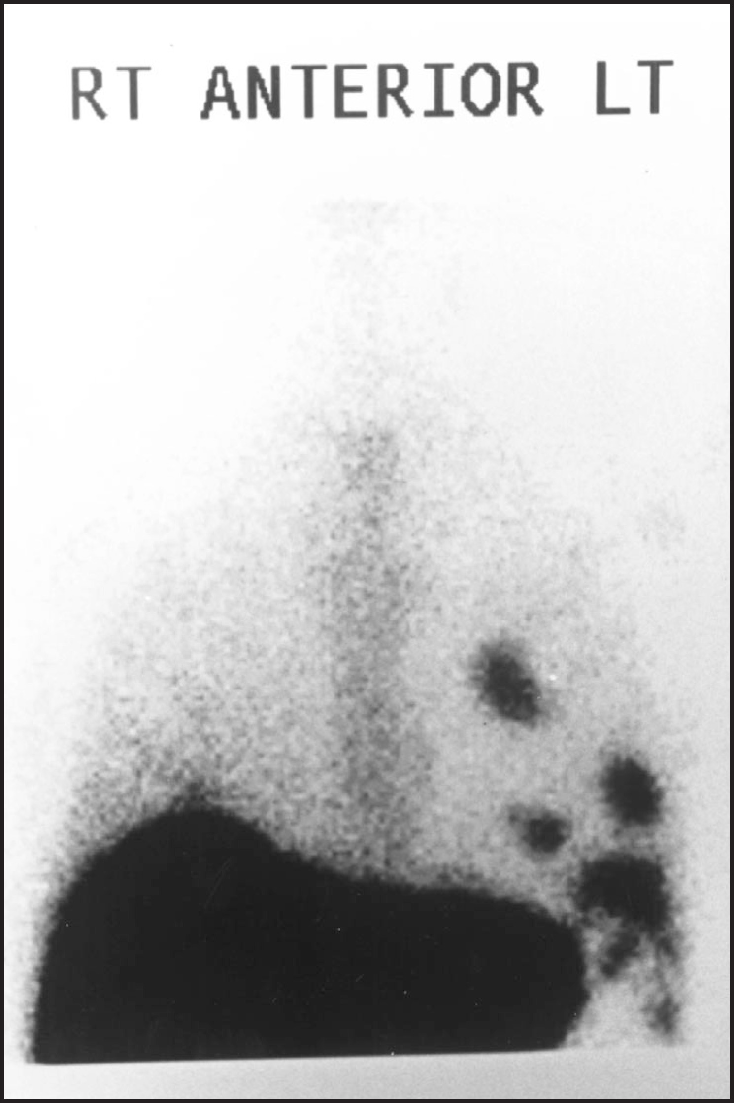
A chest x-ray revealed left-sided pleural effusion with a possible pleural-based mass (not shown). A subsequent computed tomography (CT) examination demonstrated multiple high-attenuation, pleural-based nodules in the left hemithorax and the absence of normal splenic tissue in the left upper quadrant of the abdomen (figure 1). Technetium-99m–labeled sulfur colloid scan demonstrated absence of normal splenic uptake in the left upper quadrant and multiple areas of abnormal radiopharmaceutical uptake in the left hemithorax (figure 2).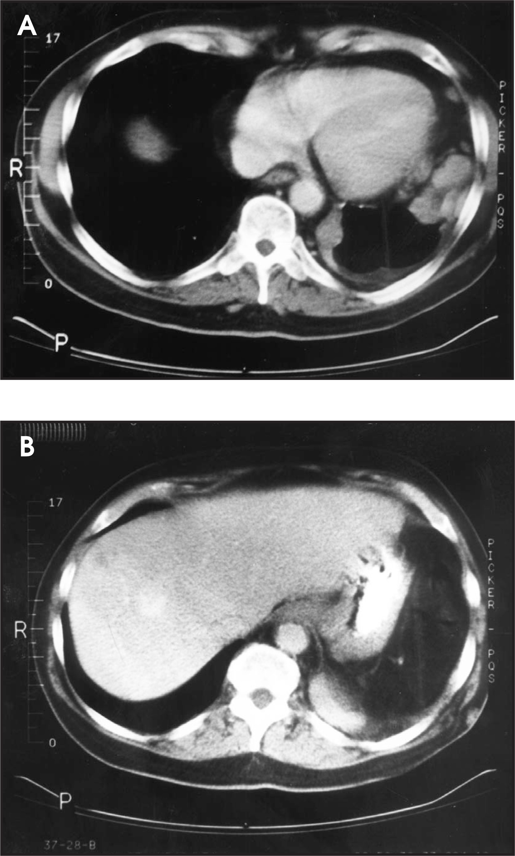
DISCUSSION
Splenosis is the autoimplantation of splenic tissue from the left upper quadrant to other sites, usually occurring after splenic injury. Abdominal splenosis typically presents with multiple nodules within the mesentery, peritoneum, and omentum. Thoracic splenosis is rare and is seen in patients who have sustained splenic trauma in association with traumatic rupture or tearing of the diaphragm. This entity was first described by Shaw and Shafi1 in 1937 as an autopsy finding in a patient who had undergone previous posttraumatic splenectomy. In a 1993 review of 17 cases of posttraumatic splenosis, the mean interval between trauma and imaging studies was 5.1 years with a range of 2.7 to 7.9 years.2 More recently, the diagnosis has been reported within 1 year of the traumatic episode.3
Most patients with thoracic splenosis are asymptomatic, although a patient presenting with hemoptysis has been reported.4 This is in contrast to abdominal splenosis, which may present with abdominal pain or small-bowel obstruction.4,5 Peripheral blood smears may demonstrate findings typically seen in asplenic individuals, specifically absence of Howell-Jolly bodies as well as the presence of pitted erythrocytes and siderocytes.5
Chest radiographic findings in thoracic splenosis demonstrate multiple pleural-based nodules in the left hemithorax. On CT, the pleural-based nodules demonstrate attenuation characteristics similar to the spleen. The nodules have been reported to range in size from <1 cm to 6 cm.2,3 On MRI, the nodules demonstrate signal intensity and enhancement characteristics identical to normal splenic tissue.6 However, none of the radiographic, CT, or MRI findings are pathognomonic for thoracic splenosis. Therefore, other differential diagnostic considerations include metastatic pleural disease (including lung and breast cancer) and lymphoma. If only a solitary pleural nodule is present, mesothelioma and benign fibrous tumor of the pleura should be included in the differential diagnosis.3
Historically, the diagnosis of thoracic splenosis has been made at thoracotomy; typical findings include multiple reddish-brown pleural-based nodules ranging in size from 3 mm to 6 cm.5,7 The diagnosis can also be confirmed noninvasively with radionuclide studies. Several authors have documented the diagnosis of thoracic splenosis using radionuclides that localize to the spleen.3,8-10 Commonly used agents include Technetium-99m sulfur colloid, Technetium-99m–labeled heat-damaged red blood cells, and Indium 111 platelets. Typical findings, as in this case, include absence of the normal splenic uptake of tracer with multiple areas of abnormal uptake in the left hemithorax.
SUMMARY
The diagnosis of thoracic splenosis should be considered in patients who present with multiple pleural-based masses on chest x-ray or CT in which a history of significant trauma (usually requiring splenectomy) can be elicited. The diagnosis can be confirmed with noninvasive radionuclide studies, thereby obviating the need for additional diagnostic studies, including open thoracotomy and biopsy.
References
- Shaw A, Shafi A. Traumatic autoplastic transplantation of splenic tissue in man with observations on late results of splenectomy in 6 cases. J Pathol Bacteriol.. 1937;45:215-235.
- Normand J, Rioux M, Dumant M. Thoracic splenosis after blunt trauma: Frequency and imaging findings. AJR Am J Roentgenol. 1993;161:739-741.
- White C, Meyer C. General case of the day. Thoracic splenosis. RadioGraphics.. 1998;18:255-257.
- Cordier J, Gamondes J, Marx P. Thoracic splenosis presenting with hemoptysis. Chest.. 1992;101:626-627.
- Roucos S, Tabet G, Jebara V. Thoracic splenosis. J Thorac Cardiovasc Surg.. 1990;99:361-363.
- Bordlee R, Eshaghi N, Oz O. Thoracic splenosis: MR demonstration. J Thorac Imaging.. 1995;10:146-149.
- O'Connor J, Brown C, Thomas J. Thoracic splenosis. Ann Thorac Surg.. 1998;66:552-553.
- Kwan A, Drum D, Ahn C, Tow D. Intrathoracic splenosis mimicking metastatic lung cancer. Clin Nucl Med.. 1994;19:93-95.
- Hardin V, Morgan M. Thoracic splenosis. Clin Nucl Med. 1994;19:438-440.
- Naylor M, Karstaedt N, Finck S, Burnett O. Noninvasive methods of diagnosing thoracic splenosis. Ann Thorac Surg.. 1999;68:243-244.
Citation
. RCOM RADIOLOGICAL CASE OF THE MONTH. Applied Radiology. 2002;31(1):37-38. doi:10.37549/AR1068.