RCOM RADIOLOGICAL CASE OF THE MONTH
Applied Radiology — Vol. 31 , Issue 4 , pp. 36 -37
DOI: 10.37549/AR1081
Published: April 1, 2002
Categories
CASE SUMMARY
A 76-year-old woman presented with a pelvic mass on physical examination. Her medical history was unremarkable. An ultrasound examination was performed.
DIAGNOSIS
Struma ovarii
IMAGING FINDINGS
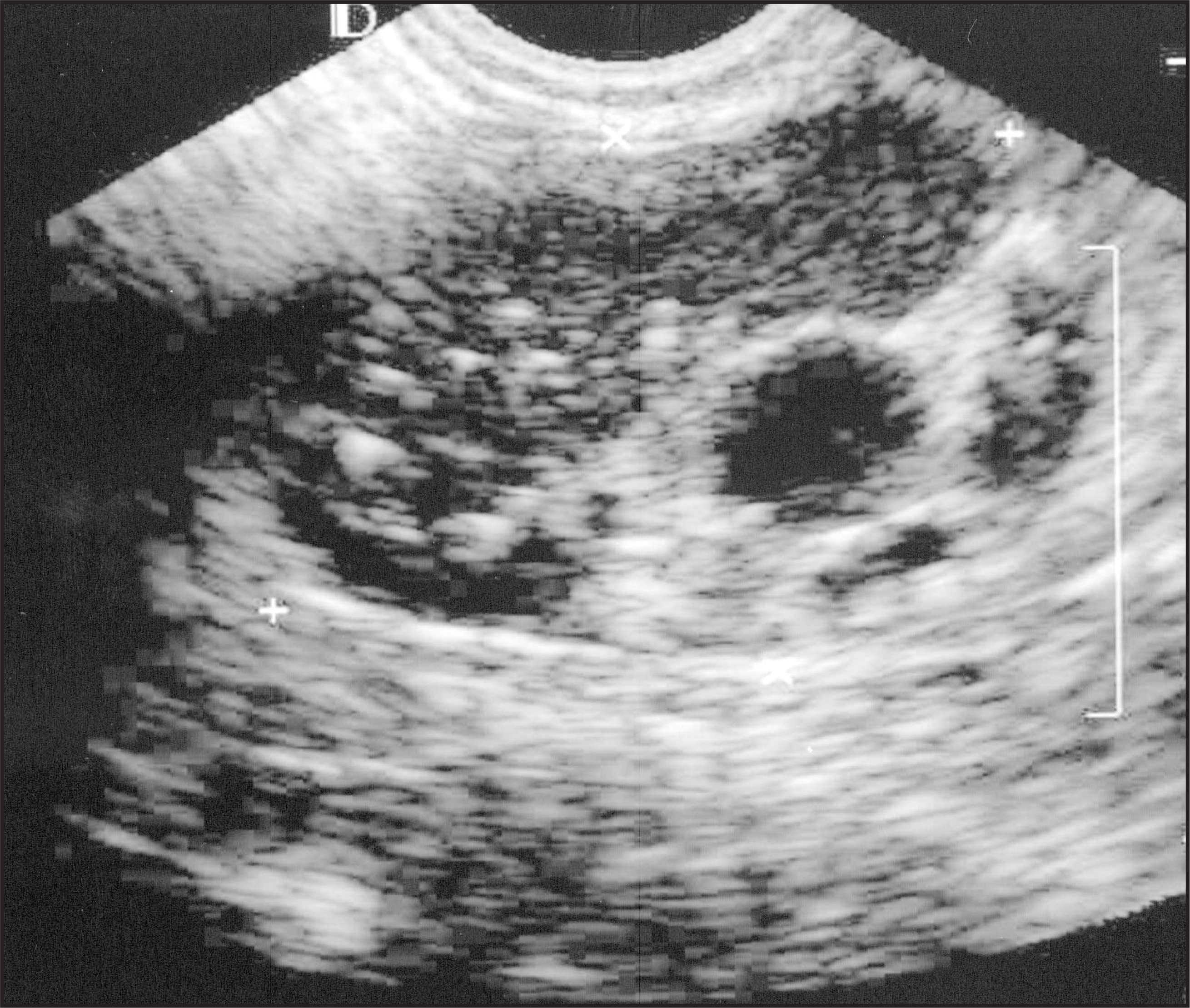
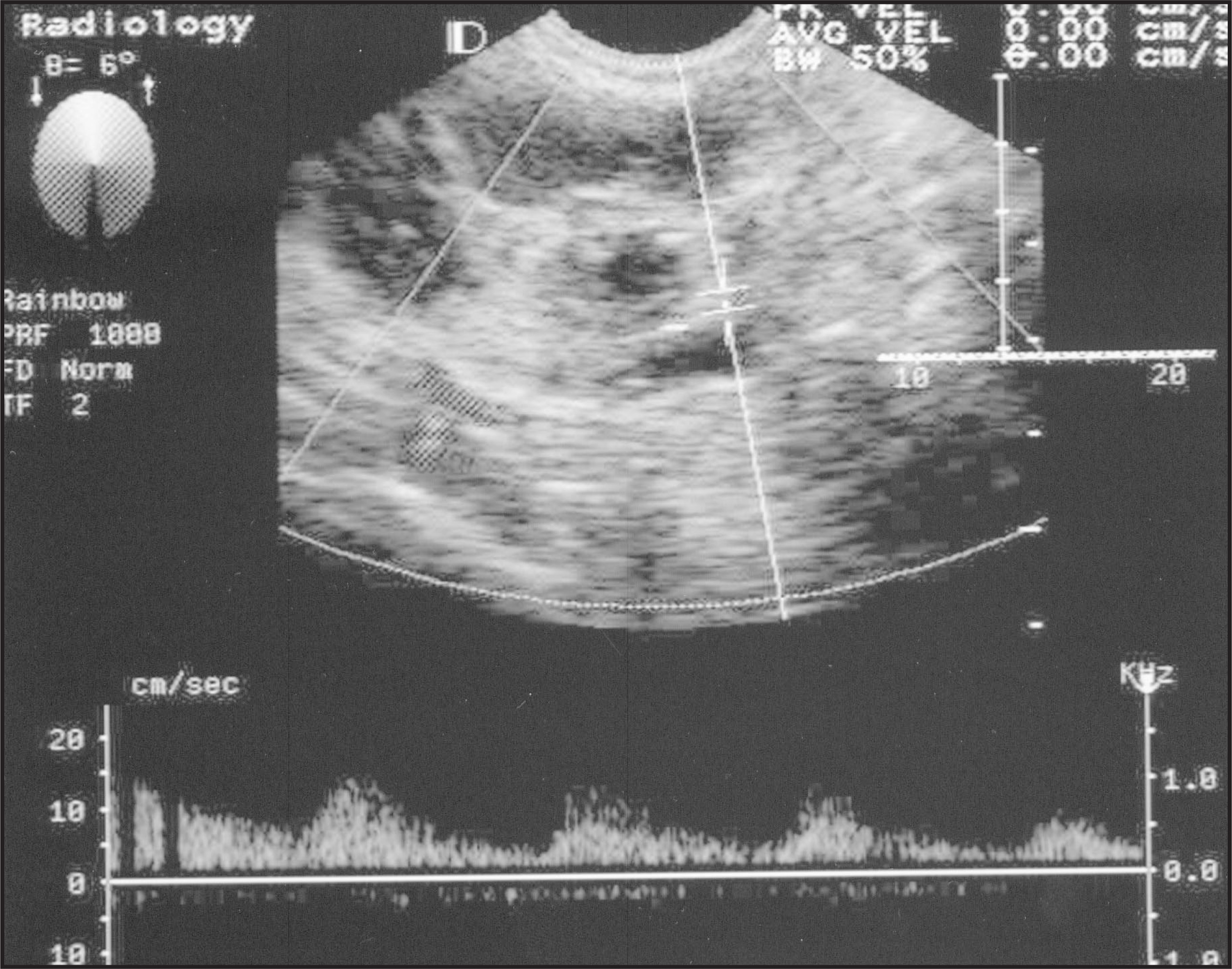
Ultrasound of the pelvis was performed with a 3-Mhz transducer. A well-defined heterogeneous mass was noted in the right adnexa measuring 4.7 × 5.0 × 3.5 cm (figure 1). The mass was primarily hypoechoic with echogenic foci scattered throughout. Eccentrically located within the mass was a 1.5-cm heterogeneous nodule with a sonolucent center. Doppler evaluation showed arterial flow (figure 2). No ascities or adenopathy was noted. No normal right ovarian tissue was seen. The left ovary was normal in appearance. The uterus was absent surgically. Because of the possibility of malignancy, the right adnexal mass was removed surgically and pathologically proven to be struma ovarii.
DISCUSSION
Struma ovarii is a rare germ-cell tumor of the ovary, consisting mainly of thyroid tissue. Ninety-five percent of these tumors are benign. Five percent of these tumors contain a carcinoid component considered malignant transformation.1 Only 5% of these malignant tumors metastasize. Struma ovarii tumors are generally asymptomatic and are usually discovered incidentally during a routine physical examination. Seldom, patients may complain of a palpable pelvic mass. However, in a few cases, these tumors can secrete thyroid hormones and the patients present with symptoms seen in hyperthyroidism such as thyroid enlargement and tachycardia.2
Elevated cancer antigen 125 (CA-125) levels are rarely seen with struma ovarii, usually in the setting of ascites.3 In order to make the correct diagnosis, histologic examination of the mass ovary must show thyroid follicles.4 Immunohistochemical staining for thyroglobulin may be done in problematic cases.
For benign pure struma ovarii, surgical resection is usually curative with complete resolution of the patient’s symptoms.
References
- Zalel Y. Sonographic and clinical characteristics of struma ovarii. J Ultrasound Med. 2000;19:857-860.
- Thomas R. Metastatic malignant struma ovarii. Clin Nucl Med. 1992;17:577-578.
- Bethune M. Struma ovarii presenting as acute pseudo: Meigs syndrome with an elevated CA-125 level. Aust NZ J Obstet Gynecol. 1996;36:372-373.
- Szyfelbein W. Struma ovarii stimulating ovarian tumors of other types. Am J Surg Pathol. 1995;19:21-29.
Citation
. RCOM RADIOLOGICAL CASE OF THE MONTH. Applied Radiology. 2002;31(4):36-37. doi:10.37549/AR1081.