RCOM RADIOLOGICAL CASE OF THE MONTH
Applied Radiology — Vol. 31 , Issue 5 , pp. 43 -45
DOI: 10.37549/AR1087
Published: May 1, 2002
Categories
CASE SUMMARY
A 46-year-old male-to-female transsexual with bilateral breast augmentations on hormone therapy presented for evaluation of left breast tenderness. No breast masses or other focal abnormalities could be identified on physical examination.
IMAGING FINDINGS
Bilateral mammography with standard and pushback views revealed bilateral wedge-shaped densities radiating from the nipples, slightly larger on the left (figure 1A). The densities blended gradually into surrounding lucent adipose tissue and had strand-like extensions. A 6-mm focal asymmetric density was visible in the outer left breast in the standard and pushback craniocaudal views. This resolved on a craniocaudal magnification compression view consistent with mammary stroma (figure 1B). Implants were visible bilaterally. The pectoral muscles could be seen deep to the implants.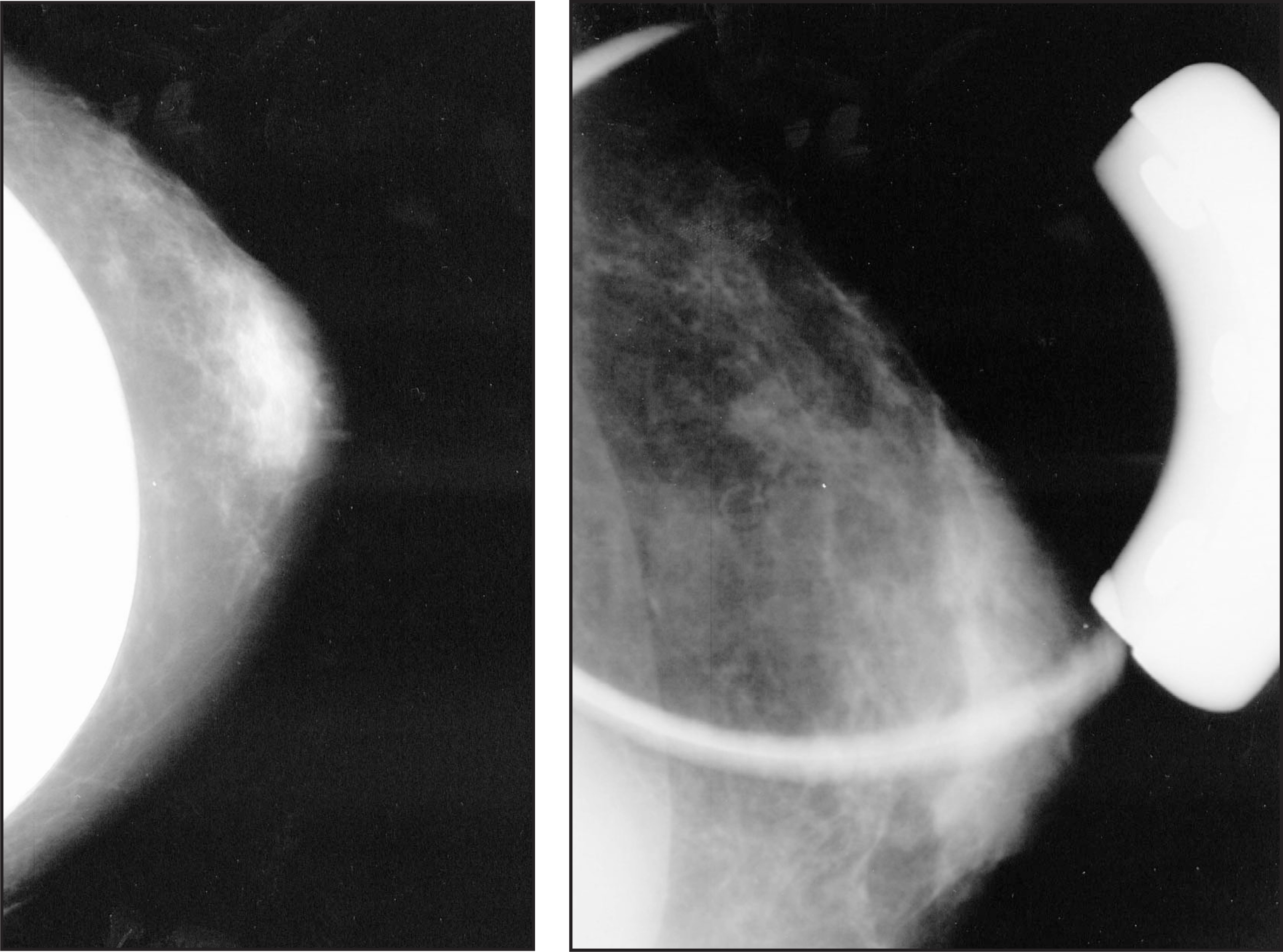
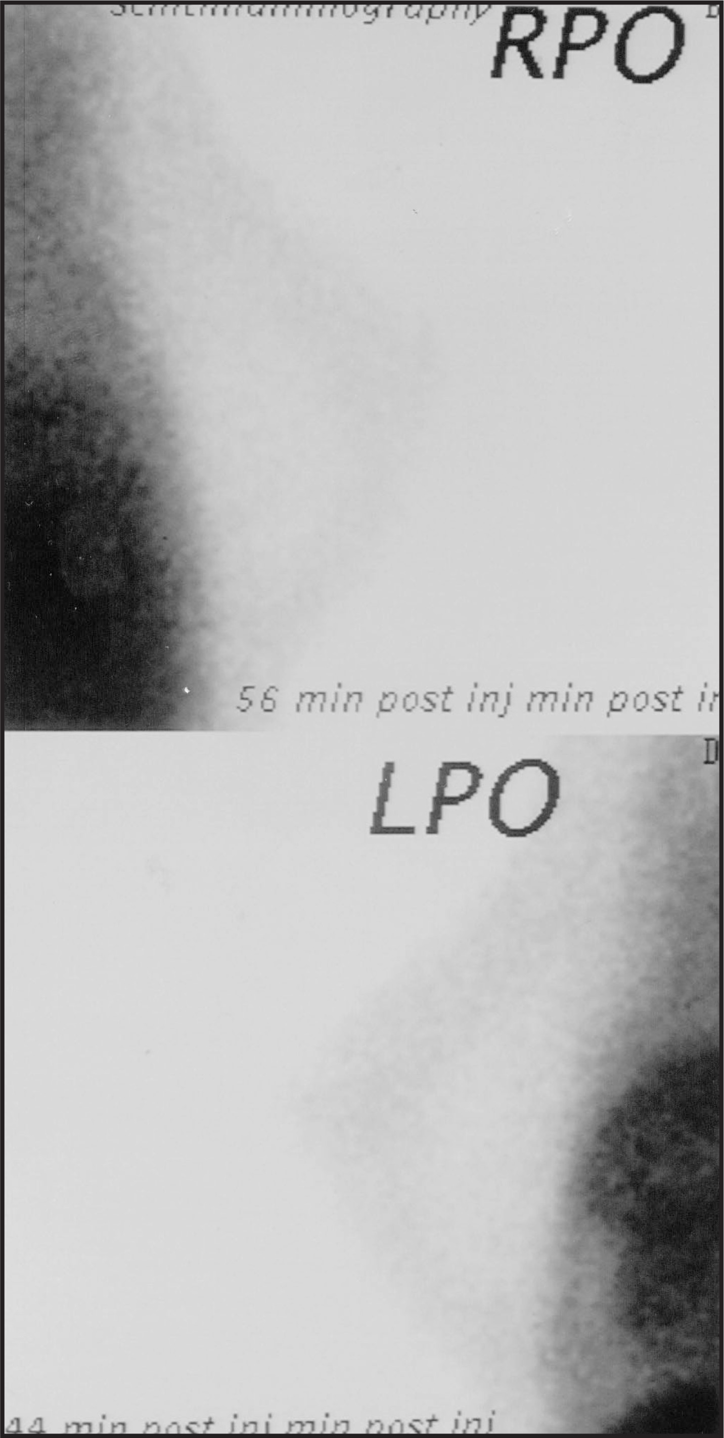
Scintigraphy of the breasts with Tc-99m sestamibi revealed activity at the surface of the breasts where skin and subcutaneous tissues were viewed tangentially with mild diffuse increases at the sites of gynecomastia in the retroareolar areas (figure 2). There were photopenic defects deep to the foci of gynecomastia corresponding to the breast implants.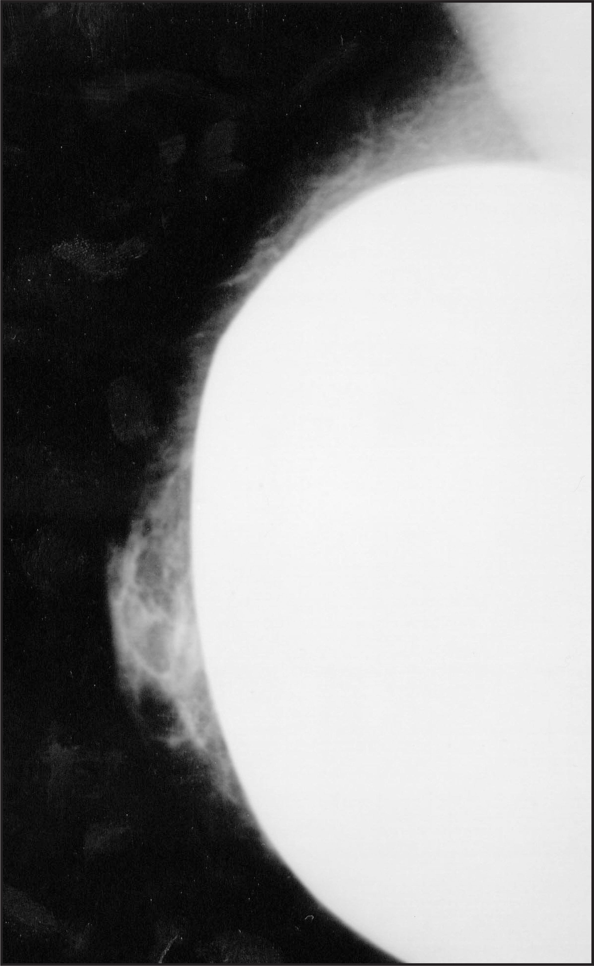
DIAGNOSIS
Breasts of a male-to-female transsexual
DISCUSSION
Transsexualism is defined as “the desire to live and be accepted as a member of the opposite sex, usually accompanied by the wish to make his or her body as congruent as possible with the preferred sex through surgery and hormone therapy.”1 Breast development may result if male-to-female transsexuals undergo hormone replacement therapy or sex reassignment surgery.1-3 Breast augmentation is frequently performed in these patients.1,3 Although transsexualism is a well-known entity, I can find no previously published reports of the mammographic or scintimammographic appearance of the breasts of male-to-female transsexuals.
Hormone therapy and surgical castration in male-to-female transsexuals result in hypertrophy of vestigial breast tissue, ie, gynecomastia.2 The wedged-shape, retroareolar location, bilaterality, lack of a discrete edge, and strand-like extensions seen in the mammogram in this case are typical of gynecomastia.4,5 Subpectoral implants have a greater risk of cranial and lateral migration in male-to-female transsexuals than in women. Subglandular implants are, therefore, preferred.3 Identification of the pectoral muscles deep to the implants demonstrates the subglandular location of the implants. Hence, the mammographic appearance of the breasts seen in this case is what would be expected in a transsexual; gynecomastia directly overlying breast implants.
Incidence of transsexualism is estimated at 1 in 11,900 males and 1 in 30,400 females. Not all patients with gender identity disorders are candidates for sex reassignment surgery. Selection of patients for surgery is a lengthy process involving careful diagnostic assessment followed by a real-life experience in the desired gender role and hormone therapy. Psychotherapy is now optional, since it is usually ineffective for this condition. Breast augmentation may be performed if breast enlargement is not sufficient for comfort in the social gender role after 18 months of hormone therapy.1 Approximately two-thirds to three-fourths of male-to-female transsexuals request augmentation.3 Augmentation mammoplasty may be performed preceding, simultaneously with, or subsequent to genital surgery. Some believe that it is safer to perform augmentation mammoplasty first as it is reversible and the effects on the patient’s adjustment can be observed before performing irreversible penovaginal surgery and castration.6
SUMMARY
This case demonstrates the mammographic and scintimammographic appearance of the breasts of a male-to-female transsexual with subglandular implants (the most common type of augmentation mammoplasty in transsexuals). As expected, the mammogram has the same appearance of gynecomastia as seen in other genetic males,4,5 but with an implant directly beneath it. The increased activity in the retroareolar areas and the photopenic defects below them on scintimammography correspond to the mammographic findings.
References
- Levine S, Brown G, Coleman E. Harry Benjamin International Gender Dysphoria Association’s standards of care for gender identity disorders.. Int J Transgender. 1998.
- Kanhai R, Hage J, van Diest P. Short-term and long-term histologic effects of castration and estrogen treatment on breast tissue of 14 male-to-female transsexuals in comparison with two chemically castrated men.. Am J Surg Pathol.. 2000;24((1)):74-80.
- Kanhai R, Hage J, Asscheman H. Augmentation mammoplasty in male-to-female transsexuals.. Plast Reconstr Surg. 1999;104:542-549.
- Appelbaum A, Evans G, Levy K. Mammographic appearance of male breast disease.. Radiographics. 1999;19:559-568.
- Cooper R. Mammography in men.. Radiology. 1994;191:651-656.
- Edgerton M. The surgical treatment of male transsexuals.. Clin Plast Surg. 1974;1:285-323.
Citation
. RCOM RADIOLOGICAL CASE OF THE MONTH. Applied Radiology. 2002;31(5):43-45. doi:10.37549/AR1087.